Fundamentals of Anatomy Physiology Eleventh Edition Chapter 29


- Slides: 145

Fundamentals of Anatomy & Physiology Eleventh Edition Chapter 29 Development and Inheritance Lecture Presentation by Deborah A. Hutchinson Seattle University © 2018 Pearson Education, Inc.

Learning Outcomes 29 -1 Explain the relationship between differentiation and development, and specify the various stages of development. 29 -2 Describe the process of fertilization, and explain how developmental processes are regulated. 29 -3 List the three stages of prenatal development, and describe the major events of each. 29 -4 Explain how the three germ layers are involved in forming the extra-embryonic membranes, and discuss the importance of the placenta as an endocrine organ. 29 -5 List several fetal changes that occur during the second and third trimesters. 2 © 2018 Pearson Education, Inc.

Learning Outcomes 29 -6 Describe the interplay between the maternal organ systems and the developing fetus, and discuss the structural and functional changes in the uterus during gestation. 29 -7 List and discuss the events that occur during labor and delivery. 29 -8 Identify the features and physiological changes of the postnatal stages of life. 29 -9 Relate basic principles of genetics to the inheritance of human traits. 3 © 2018 Pearson Education, Inc.

Introduction to Development and Inheritance § Development – Gradual modification of anatomical structures and physiological characteristics – From fertilization to maturity 4 © 2018 Pearson Education, Inc.

29 -1 Development § Prenatal development – Embryonic and fetal developmental stages § Postnatal development – Begins at birth – Continues to maturity • State of full development or completed growth 5 © 2018 Pearson Education, Inc.

29 -1 Development § Pre-embryonic development – Processes that occur in first 2 weeks after fertilization – Produces embryo § Embryonic development – Occurs during third through eighth weeks – Embryology—study of these events § Fetal development – Development of fetus – Begins at start of ninth week, continues until birth 6 © 2018 Pearson Education, Inc.

29 -1 Development § Differentiation – Creation of different cell types during development – Occurs through selective changes in genetic activity • Some genes are turned off, others are turned on § Inheritance (heredity) – Transfer of genetically determined characteristics from generation to generation § Genetics – Study of mechanisms responsible for inheritance 7 © 2018 Pearson Education, Inc.

29 -2 Fertilization § Fertilization – Fusion of two haploid gametes, each containing 23 chromosomes – Produces zygote containing 46 chromosomes § Sperm – Delivers paternal chromosomes to fertilization site – Small, efficient, and highly streamlined § Oocyte – Contains everything needed to support development of embryo for nearly a week 8 © 2018 Pearson Education, Inc.

Figure 29– 1 a Fertilization. × 300 a A secondary oocyte and numerous sperm at the time of fertilization. Notice the difference in size between the gametes. 9 © 2018 Pearson Education, Inc.

29 -2 Fertilization § Secondary oocyte – Released from ovary at ovulation – Surrounded by zona pellucida • Thick, glycoprotein-rich envelope – Corona radiata • Protective layer of follicle cells outside zona pellucida 10 © 2018 Pearson Education, Inc.

29 -2 Fertilization § Fertilization – Occurs in uterine tube within a day after ovulation • Secondary oocyte travels a few centimeters • Sperm must cover distance between vagina and ampulla of uterine tube § Capacitation – Must occur before sperm can fertilize secondary oocyte • Contact with secretions of seminal glands • Exposure to conditions in female reproductive tract 11 © 2018 Pearson Education, Inc.

29 -2 Fertilization § Acrosome reaction – Acrosome releases protein-digesting enzymes to penetrate corona radiata and reach oocyte surface • Hyaluronidase • Acrosin § Oocyte activation – Triggered by fusion of sperm and oocyte membranes 12 © 2018 Pearson Education, Inc.

29 -2 Fertilization § Oocyte activation – Sodium ions enter oocyte and depolarize membrane – Calcium ions are released from smooth endoplasmic reticulum, causing • Exocytosis of vesicles adjacent to oocyte membrane (cortical reaction) • Completion of meiosis II and formation of second polar body • Activation of enzymes that increase metabolic rate 13 © 2018 Pearson Education, Inc.

29 -2 Fertilization § Cortical reaction – Releases enzymes that • Inactivate sperm receptors • Harden zona pellucida – Results in block to polyspermy • Prevention of fertilization by more than one sperm § After oocyte completes meiosis II, it becomes mature ovum 14 © 2018 Pearson Education, Inc.

29 -2 Fertilization § Female pronucleus – Reorganized nuclear material remaining in ovum after completion of meiosis – Moves toward male pronucleus § Male pronucleus – Swollen nucleus of sperm – Moves toward female pronucleus 15 © 2018 Pearson Education, Inc.

29 -2 Fertilization § Amphimixis – Fusion of female pronucleus and male pronucleus – “Moment of conception” – Cell becomes a zygote with 46 chromosomes – Fertilization is complete § First cleavage – Produces two daughter cells (blastomeres) 16 © 2018 Pearson Education, Inc.

Figure 29– 1 b Fertilization (Part 1 of 6). Oocyte at Ovulation releases a secondary oocyte and the first polar body; both are surrounded by the corona radiata. The oocyte is suspended in metaphase of meiosis II. Corona radiata First polar body Zona pellucida 17 © 2018 Pearson Education, Inc.

Figure 29– 1 b Fertilization (Part 2 of 6). 1 Fertilization and Oocyte Activation Acrosomal enzymes from multiple sperm create gaps in the corona radiata. A single sperm then makes contact with the oocyte membrane, and membrane fusion occurs, triggering oocyte activation and the completion of meiosis. Fertilizing sperm Second polar body 18 © 2018 Pearson Education, Inc.

Figure 29– 1 b Fertilization (Part 3 of 6). 2 Pronuclei Develop and DNA Synthesis Occurs The sperm is absorbed into the cytoplasm, and the female pronucleus develops. As the male pronucleus develops, most remaining sperm break down. Nucleus of fertilizing sperm Female pronucleus 19 © 2018 Pearson Education, Inc.

Figure 29– 1 b Fertilization (Part 4 of 6). 3 Spindle Formation Begins Pronuclei chromatin condenses into chromosomes, and spindle fibers appear in preparation for the first cell division. Centrioles Male pronucleus Female pronucleus 20 © 2018 Pearson Education, Inc.

Figure 29– 1 b Fertilization (Part 5 of 6). 4 Amphimixis Occurs and Cleavage Begins Maternal and paternal chromosomes align on metaphase plate 21 © 2018 Pearson Education, Inc.

Figure 29– 1 b Fertilization (Part 6 of 6). 5 First Cleavage Forms Two Blastomeres The first cleavage division nears completion about 30 hours after fertilization. Blastomeres 22 © 2018 Pearson Education, Inc.

29 -3 Gestation § Gestation – Time spent in prenatal development – Consists of three integrated trimesters, each 3 months long 23 © 2018 Pearson Education, Inc.

29 -3 Gestation § First trimester – Pre-embryonic through early fetal development – Rudiments of all major organ systems appear § Second trimester – Development of organs and organ systems – Body shape and proportions change § Third trimester – Rapid fetal growth and deposition of adipose tissue – Most major organ systems are fully functional 24 © 2018 Pearson Education, Inc.

29 -4 The First Trimester § First trimester – Involves four general processes • Cleavage • Implantation • Placentation • Embryogenesis 25 © 2018 Pearson Education, Inc.

29 -4 The First Trimester § Cleavage – Sequence of cell divisions that begins immediately after fertilization – Zygote becomes a pre-embryo, which develops into multicellular blastocyst – Ends when blastocyst contacts uterine wall § Implantation – Begins with attachment of blastocyst to endometrium of uterus – Sets stage formation of embryonic structures 26 © 2018 Pearson Education, Inc.

29 -4 The First Trimester § Placentation – Occurs as blood vessels form around periphery of blastocyst – Placenta develops to permit exchange between maternal and embryonic blood § Embryogenesis – Formation of viable embryo – Establishes foundations for all major organ systems 27 © 2018 Pearson Education, Inc.

29 -4 The First Trimester § First trimester – Most dangerous prenatal stage – Only about 40 percent of conceptions produce embryos that survive first trimester 28 © 2018 Pearson Education, Inc.

29 -4 The First Trimester § Cleavage and blastocyst formation – Blastomeres • Identical cells produced by cleavage – Morula • Stage after 3 days of cleavage • Pre-embryo is a solid ball of cells resembling mulberry • Reaches uterus on day 4 or 5 29 © 2018 Pearson Education, Inc.

29 -4 The First Trimester § Cleavage and blastocyst formation – Blastocyst • Forms from morula • Hollow ball • Inner cavity is called a blastocoele • Blastomeres are no longer identical – Trophoblast • Outer layer of cells of blastocyst • Provides nutrients to developing embryo 30 © 2018 Pearson Education, Inc.

29 -4 The First Trimester § Cleavage and blastocyst formation – Inner cell mass • Clustered at one end of blastocyst • Exposed only to blastocoele • Insulated from contact with outside uterine environment by trophoblast • Will form embryo 31 © 2018 Pearson Education, Inc.

Figure 29– 2 Cleavage and Blastocyst Formation (Part 1 of 2). Blastomeres Polar bodies 2 -cell stage DAY 1 First cleavage division 4 -cell stage DAY 2 DAY 0: Fertilization 32 © 2018 Pearson Education, Inc.

Figure 29– 2 Cleavage and Blastocyst Formation (Part 2 of 2). Zona pellucida Early morula DAY 3 Advanced morula DAY 4 Hatching Inner cell mass DAY 6 Blastocoele Days 7– 10: Implantation in uterine wall © 2018 Pearson Education, Inc. Trophoblast Blastocyst 33

29 -4 The First Trimester § Implantation – Occurs 7– 10 days after fertilization – Blastocyst adheres to uterine lining – Trophoblast cells divide rapidly, creating layers • Cytotrophoblast – Cells closest to interior of blastocyst • Syncytiotrophoblast – Outer layer – Erodes path through uterine epithelium by secreting hyaluronidase 34 © 2018 Pearson Education, Inc.

Figure 29– 3 Stages in Implantation (Part 1 of 2). DAY 6 FUNCTIONAL LAYER OF ENDOMETRIUM Uterine glands DAY 7 UTERINE CAVITY Blastocyst Trophoblast Blastocoele Inner cell mass 35 © 2018 Pearson Education, Inc.

Figure 29– 3 Stages in Implantation (Part 2 of 2). DAY 8 Endometrial capillary Syncytiotrophoblast Cytotrophoblast DAY 9 Developing villi Amniotic cavity Lacuna 36 © 2018 Pearson Education, Inc.

29 -4 The First Trimester § Lacunae – Trophoblastic channels carrying maternal blood § Villi – Extend away from trophoblast into endometrium – Increase in size and complexity until day 21 § Amniotic cavity – Fluid-filled chamber created as inner cell mass separates from trophoblast – Next to superficial layer of inner cell mass • Deeper layer is exposed to blastocoele 37 © 2018 Pearson Education, Inc.

29 -4 The First Trimester § Ectopic pregnancy – Implantation occurs somewhere other than within uterus – 0. 6 percent of pregnancies – Does not produce viable embryo – Can be life threatening to the mother 38 © 2018 Pearson Education, Inc.

29 -4 The First Trimester § Embryonic period – Implantation to week 9 – Differentiation • Involves changes in genetic activity of some cells but not others – Induction • Cells release chemical substances that affect differentiation of other embryonic cells • Can control highly complex processes 39 © 2018 Pearson Education, Inc.

29 -4 The First Trimester § Early embryogenesis – Gastrulation • Forms third layer of cells in embryo • Through cell migration – Three germ layers • Ectoderm • Mesoderm • Endoderm 40 © 2018 Pearson Education, Inc.

Figure 29– 4 The Inner Cell Mass and Gastrulation (Part 2 of 2). Day 15: Gastrulation By day 15, superficial cells of the two-layered embryonic disc have begun to migrate toward a central line known as the primitive streak. Here, the migrating cells leave the Amnion surface and move between the two existing layers. This movement creates three distinct embryonic layers: (1) the ectoderm, consisting of superficial cells that did not migrate into the interior of the embryonic disc; (2) the endoderm, consisting of the cells that face the yolk sac; and (3) the mesoderm, consisting of the poorly organized layer of migrating cells between the Primitive ectoderm and the endoderm. Collectively, these streak three embryonic layers are called germ layers, and the migration process is called gastrulation. Through gastrulation, the embryonic disc becomes an oval, three-layered sheet. This disc will form the body of the embryo, whereas all other cells of the blastocyst will be part of the extra-embryonic membranes. Yolk sac Germ Layers Ectoderm Mesoderm Endoderm Embryonic disc 41 © 2018 Pearson Education, Inc.

29 -4 The First Trimester § Ectodermal contributions – Integumentary system • Epidermis, nails, hair follicles and hairs • Glands communicating with skin (sweat, mammary, and sebaceous glands) – Skeletal system • Pharyngeal cartilages and their derivatives including auditory ossicles – Nervous system • All neural tissue 42 © 2018 Pearson Education, Inc.

29 -4 The First Trimester § Ectodermal contributions – Endocrine system • Pituitary gland adrenal medullae – Respiratory system • Mucous epithelium of nasal passageways – Digestive system • Mucous epithelium of mouth and anus • Salivary glands 43 © 2018 Pearson Education, Inc.

29 -4 The First Trimester § Mesodermal contributions – Integumentary system • Dermis and hypodermis – Skeletal system • All structures except some pharyngeal derivatives – Muscular system • All structures 44 © 2018 Pearson Education, Inc.

29 -4 The First Trimester § Mesodermal contributions – Endocrine system • Adrenal cortex • Endocrine tissues of heart, kidneys, and gonads – Cardiovascular system • All structures – Lymphatic system • All structures 45 © 2018 Pearson Education, Inc.

29 -4 The First Trimester § Mesodermal contributions – Urinary system • Kidneys, including nephrons and initial portions of collecting system – Reproductive system • Gonads and adjacent portions of duct systems – Miscellaneous • Lining of pleural, pericardial, and peritoneal cavities • Connective tissues that support all organ systems 46 © 2018 Pearson Education, Inc.

29 -4 The First Trimester § Endodermal contributions – Endocrine system • Thymus, thyroid gland, and pancreas – Respiratory system • Respiratory epithelium (except nasal passageways) and associated mucous glands – Digestive system • Mucous epithelium (except mouth and anus) • Exocrine glands (except salivary glands) • Liver and pancreas 47 © 2018 Pearson Education, Inc.

29 -4 The First Trimester § Endodermal contributions – Urinary system • Urinary bladder and distal portions of duct system – Reproductive system • Distal portions of duct system • Stem cells that produce gametes 48 © 2018 Pearson Education, Inc.

29 -4 The First Trimester § Embryonic disc – Oval, three-layered sheet – Produced by gastrulation – Will form body of embryo and internal organs • Rest of blastocyst will be part of extra-embryonic membranes – Folding and differential growth produce bulges that project into amniotic cavity • Head fold and tail fold 49 © 2018 Pearson Education, Inc.

29 -4 The First Trimester § Extra-embryonic membranes – Support embryonic and fetal development • Yolk sac • Amnion • Allantois • Chorion 50 © 2018 Pearson Education, Inc.

29 -4 The First Trimester § Yolk sac – Begins as layer of cells that spreads out to form complete pouch – Primary nutrient source for early embryonic development – Becomes important site for blood cell formation 51 © 2018 Pearson Education, Inc.

Figure 29– 4 The Inner Cell Mass and Gastrulation (Part 1 of 2). Day 10: Yolk Sac Formation While cells from the superficial layer of the inner cell mass migrate around the amniotic cavity, forming the amnion, cells from the deeper layer migrate around the outer edges of the blastocoele. This is the first step in the formation of the yolk sac, a second extra-embryonic membrane. For about the next 2 weeks, the yolk sac is the primary nutrient source for the inner cell mass; it absorbs and distributes nutrients released into the blastocoele by the cytotrophoblast. Superficial layer Syncytiotrophoblast Cytotrophoblast Blastocoele Amniotic cavity Yolk sac Lacunae Deep layer 52 © 2018 Pearson Education, Inc.

29 -4 The First Trimester § Amnion – Combination of mesoderm and ectoderm – Ectodermal cells spread over inner surface of amniotic cavity • Followed by mesodermal cells – Continues to enlarge through development – Amniotic fluid is produced • Surrounds and cushions developing embryo or fetus 53 © 2018 Pearson Education, Inc.

29 -4 The First Trimester § Allantois – Begins as outpocket of endoderm near base of yolk sac – Free endodermal tip grows toward wall of blastocyst • Surrounded by mass of mesoderm – Base later gives rise to urinary bladder 54 © 2018 Pearson Education, Inc.

29 -4 The First Trimester § Chorion – Mesoderm associated with allantois spreads around blastocyst • Separating cytotrophoblast from blastocoele – Blood vessels develop in chorion • First step in creation of functional placenta – By third week, chorionic villi contact maternal tissues and blood vessels – Villi enlarge and branch, forming placenta • Exchange platform between mother and fetus 55 © 2018 Pearson Education, Inc.

Figure 29– 5 Extra-Embryonic Membranes and Placenta Formation (Part 1 of 5). 1 Week 2 Migration of mesoderm around the inner surface of the cytotrophoblast forms the chorion. Mesodermal migration around the outside of the amniotic cavity, between the ectodermal cells and the trophoblast, forms the amnion. Mesodermal migration around the endodermal pouch forms the yolk sac. Chorion Cyto. Mesoderm trophoblast Amnion Blastocoele © 2018 Pearson Education, Inc. Yolk sac Syncytiotrophoblast 56

Figure 29– 5 Extra-Embryonic Membranes and Placenta Formation (Part 2 of 5). 2 Week 3 The embryonic disc bulges into the amniotic cavity at the head fold. The allantois, an endodermal extension surrounded by mesoderm, extends toward the trophoblast. Head fold of embryo Amniotic cavity (containing amniotic fluid) Extra-Embryonic Membranes Amnion Allantois Yolk sac Chorionic villi Syncytiotrophoblast of placenta 57 © 2018 Pearson Education, Inc.

29 -4 The First Trimester § Placentation – Body stalk • Connection between embryo and chorion • Contains distal portions of allantois and blood vessels that carry blood to and from placenta – Yolk stalk • Narrow connection between endoderm of embryo and yolk sac 58 © 2018 Pearson Education, Inc.

Figure 29– 5 Extra-Embryonic Membranes and Placenta Formation (Part 3 of 5). 3 Week 4 The embryo now has a head fold and a tail fold. Constriction of the connections between the embryo and the surrounding trophoblast narrows the yolk stalk and body stalk. Tail fold Embryonic gut Embryonic head fold Body stalk Yolk sac Yolk stalk 59 © 2018 Pearson Education, Inc.

29 -4 The First Trimester § Capsular decidua – Thin portion of endometrium – No longer participates in nutrient exchange – Chorionic villi in the region disappear § Basal decidua – Disc-shaped area deep in endometrium – Where placental functions are concentrated § Parietal decidua – Rest of uterine endometrium – No contact with chorion 60 © 2018 Pearson Education, Inc.

Figure 29– 5 Extra-Embryonic Membranes and Placenta Formation (Part 4 of 5). 4 Week 5 The developing embryo and extra-embryonic membranes bulge into the uterine cavity. The cytotrophoblast pushing out into the uterine cavity remains covered by endometrium but no longer participates in nutrient absorption and embryo support. The embryo moves away from the placenta, and the body stalk and yolk stalk fuse to form an umbilical stalk. Uterus Myometrium Basal decidua Umbilical stalk Placenta Yolk sac Chorionic villi of placenta Capsular decidua Parietal decidua Uterine cavity 61 © 2018 Pearson Education, Inc.

Figure 29– 5 Extra-Embryonic Membranes and Placenta Formation (Part 5 of 5). 5 Week 10 The amnion has expanded greatly, filling the uterine cavity. The fetus is connected to the placenta by an elongated umbilical cord that contains a portion of the allantois, blood vessels, and the remnants of the yolk stalk. A mucus plug forms, preventing bacteria from entering the uterus. Parietal decidua Umbilical cord Basal decidua Placenta Amnion Amniotic cavity Capsular decidua Chorion Mucus plug 62 © 2018 Pearson Education, Inc.

29 -4 The First Trimester § Umbilical cord – Connects fetus and placenta – Contains allantois, placental blood vessels, and yolk stalk § Placental circulation – Blood flows from fetus to placenta through paired umbilical arteries – Blood returns to fetus in single umbilical vein 63 © 2018 Pearson Education, Inc.

Figure 29– 6 a Anatomy of the Placenta after the First Trimester (Part 1 of 3). Capsular decidua Amnion Umbilical cord (cut) Placenta Chorion Yolk sac Basal decidua a Placental circulation at the end of the first trimester. Arrows in the enlarged view indicate the direction of blood flow. Blood flows into the placenta through ruptured maternal arteries and then flows around chorionic villi, which contain fetal blood vessels. © 2018 Pearson Education, Inc. 64

Figure 29– 6 a Anatomy of the Placenta after the First Trimester (Part 2 of 3). Parietal decidua Myometrium Uterine cavity Mucus plug in cervical canal Cervix External os Vagina a Placental circulation at the end of the first trimester. Arrows in the enlarged view indicate the direction of blood flow. Blood flows into the placenta through ruptured maternal arteries and then flows around chorionic villi, which contain fetal blood vessels. © 2018 Pearson Education, Inc. 65

Figure 29– 6 a Anatomy of the Placenta after the First Trimester (Part 3 of 3). Chorionic villi Umbilical vein Umbilical arteries Area filled with maternal blood Amnion a © 2018 Pearson Education, Inc. Fetal capillaries Maternal blood vessels Placental circulation at the end of the first trimester. Arrows in the enlarged view indicate the direction of blood flow. Blood flows into the placenta through ruptured maternal arteries and then flows around chorionic villi, which contain fetal blood vessels. 66

Figure 29– 6 b Anatomy of the Placenta after the First Trimester. Embryonic connective tissue Area filled with maternal blood Syncytiotrophoblast Cytotrophoblast Fetal blood vessels Chorionic villus LM × 280 b A cross section through a chorionic villus, showing the syncytiotrophoblast exposed to the maternal blood space. 67 © 2018 Pearson Education, Inc.

29 -4 The First Trimester § At about week 4 of development – Embryo is physically and developmentally distinct from embryonic disc and extra-embryonic membranes – Definitive orientation of embryo can be seen § Events in first 12 weeks establish basis for organogenesis – Process of organ formation 68 © 2018 Pearson Education, Inc.

Figure 29– 7 a The First 12 Weeks of Development. Future head of embryo Thickened neural plate (will form brain) Neural folds (fuse to enclose brain ventricles and central canal of spinal cord) Somites (mesodermal blocks that will form muscles and vertebrae) Central canal of future spinal cord Cut wall of amnion a Week 3. Diagram showing the formation of the CNS from neural plate ectoderm (about 2. 25 mm in size). 69 © 2018 Pearson Education, Inc.

Figure 29– 7 b The First 12 Weeks of Development. Forebrain Eye Arm bud Heart Body stalk Leg bud Tail b Week 4. Fiberoptic view of human development at week 4 (about 5 mm in size). 70 © 2018 Pearson Education, Inc.

Figure 29– 7 c The First 12 Weeks of Development. Chorionic villi Amnion Umbilical cord Placenta c Week 8. Fiberoptic view of human development at week 8 (about 1. 6 cm in size). © 2018 Pearson Education, Inc. 71

Figure 29– 7 d The First 12 Weeks of Development. Amnion Umbilical cord d Week 12. Fiberoptic view of human development at week 12 (about 5. 4 cm in size). © 2018 Pearson Education, Inc. 72

29 -5 The Second and Third Trimesters § Second trimester – Fetus grows faster than surrounding placenta – Amnion and chorion fuse, creating amniochorionic membrane § Third trimester – Most organ systems become able to function without maternal assistance – Growth rate slows but much weight is gained – Fetus and enlarged uterus displace many of mother’s abdominal organs 73 © 2018 Pearson Education, Inc.

Figure 29– 8 a Fetal Development in the Second and Third Trimesters. a A 4 -month-old fetus, seen through a fiberoptic endoscope (about 13. 3 cm in size) © 2018 Pearson Education, Inc. 74

Figure 29– 8 b Fetal Development in the Second and Third Trimesters. b Head of a 6 -month-old fetus, revealed through ultrasound (about 30 cm in size) © 2018 Pearson Education, Inc. 75

29 -6 Maternal Organ Support of Development § Placental hormones – Synthesized by syncytiotrophoblast and released into maternal bloodstream • Human chorionic gonadotropin • Human placental lactogen • Placental prolactin • Relaxin • Progesterone • Estrogens 76 © 2018 Pearson Education, Inc.

29 -6 Maternal Organ Support of Development § Human chorionic gonadotropin (h. CG) – Appears in maternal bloodstream soon after implantation – h. CG in blood or urine samples provides reliable indication of pregnancy – Allows corpus luteum to persist for 3 to 4 months 77 © 2018 Pearson Education, Inc.

29 -6 Maternal Organ Support of Development § Human placental lactogen (h. PL) – Prepares mammary glands for milk production – Stimulatory effect on other maternal tissues comparable to that of growth hormone • Ensures that glucose and protein are available for fetus § Placental prolactin – Helps convert mammary glands to active status 78 © 2018 Pearson Education, Inc.

29 -6 Maternal Organ Support of Development § Relaxin – Peptide hormone secreted by placenta and corpus luteum during pregnancy – Increases flexibility of pubic symphysis, permitting pelvis to expand during delivery – Causes dilation of cervix, making it easier for fetus to enter vaginal canal – Suppresses release of oxytocin by hypothalamus and delays onset of labor contractions 79 © 2018 Pearson Education, Inc.

29 -6 Maternal Organ Support of Development § Pregnancy and maternal systems – Developing fetus is totally dependent on maternal organ systems • For nourishment, respiration, and waste removal – Maternal adaptations include increases in • Respiratory rate and tidal volume • Blood volume (almost 50%) • Nutrient intake (10%– 30%) • Glomerular filtration rate (50%) • Sizes of uterus and mammary glands 80 © 2018 Pearson Education, Inc.

Figure 29– 9 a Growth of the Uterus and Fetus during Gestation. Placenta Umbilical cord Fetus at 16 weeks Uterus Amniotic fluid Cervix Vagina a Pregnancy at 16 weeks, showing the positions of the uterus, fetus, and placenta. © 2018 Pearson Education, Inc. 81

Figure 29– 9 b Growth of the Uterus and Fetus during Gestation. 9 months 8 months 7 months 6 months After dropping, in preparation for delivery 5 months 4 months 3 months b Pregnancy at 3 months to 9 months (full term), showing the superior-most position of the uterus within the abdomen. © 2018 Pearson Education, Inc. 82

Figure 29– 9 c Growth of the Uterus and Fetus during Gestation. Diaphragm Liver Stomach Pancreas Transverse colon Small intestine Uterus Urinary bladder Pubic symphysis Vagina Urethra Rectum c A sectional view through the abdominopelvic cavity of a woman who is not pregnant. © 2018 Pearson Education, Inc. 83

Figure 29– 9 d Growth of the Uterus and Fetus during Gestation. Liver Stomach Pancreas Fundus of uterus Transverse colon Aorta Small intestine Umbilical cord Placenta Uterus Common iliac vein Mucus plug in cervical canal Urinary bladder External os Pubic symphysis Vagina Urethra Rectum d Pregnancy at full term. Note the positions of the uterus and full-term fetus within the abdomen, and the displacement of abdominal organs. © 2018 Pearson Education, Inc. 84

29 -7 Labor and Delivery § Parturition (childbirth) – Forcible expulsion of fetus from uterus § Labor – Strong, rhythmic uterine contractions – Progesterone released by placenta • Has inhibitory effect on uterine smooth muscle • Prevents extensive, powerful contractions 85 © 2018 Pearson Education, Inc.

29 -7 Labor and Delivery § Braxton Hicks contractions (false labor) – Experienced by some women late in pregnancy – Occasional spasms in uterine musculature – Contractions are not regular or persistent § True labor – Initiated by placental and fetal factors – Fetal pituitary gland secretes oxytocin • Increases myometrial contractions and prostaglandin production 86 © 2018 Pearson Education, Inc.

29 -7 Labor and Delivery § Labor contractions – Continue due to positive feedback – Begin near superior portion of uterus and sweep in a wave toward cervix – Increase in force and frequency, moving fetus toward cervical canal § Three stages of labor – Dilation stage – Expulsion stage – Placental stage 87 © 2018 Pearson Education, Inc.

29 -7 Labor and Delivery § Dilation stage – Begins with onset of true labor – Cervix dilates – Fetus begins to shift toward cervical canal – Highly variable in length, but typically lasts 8 or more hours – Frequency of contractions increases steadily – Late in this stage, amniochorionic membrane ruptures (“water breaks”) 88 © 2018 Pearson Education, Inc.

Figure 29– 11 The Stages of Labor (Part 1 of 4). Fully developed fetus before labor begins Pubic symphysis Placenta © 2018 Pearson Education, Inc. Umbilical cord Sacral promontory Cervical canal Cervix Vagina 89

Figure 29– 11 The Stages of Labor (Part 2 of 4). 1 The Dilation Stage 90 © 2018 Pearson Education, Inc.

29 -7 Labor and Delivery § Expulsion stage – Begins as cervix completes dilation to 10 cm – Contractions reach maximum intensity – Continues until fetus has emerged from vagina • Typically in less than 2 hours § Delivery – Arrival of newborn infant into outside world 91 © 2018 Pearson Education, Inc.

Figure 29– 11 The Stages of Labor (Part 3 of 4). 2 The Expulsion Stage 92 © 2018 Pearson Education, Inc.

29 -7 Labor and Delivery § Episiotomy – Incision through perineal musculature – Needed if vaginal canal is too small to pass fetus – Repaired with sutures after delivery § Apgar score – Assessment of newborn’s health in five areas • Heart rate, breathing, skin color, muscle tone, and reflex response – Each criterion receives a score from 0– 2 • Total of 8– 10 indicates healthy baby 93 © 2018 Pearson Education, Inc.

29 -7 Labor and Delivery § Placental stage – Muscle tension builds in walls of partially empty uterus – Tears connections between endometrium and placenta – Ends within an hour of delivery with ejection of placenta, or afterbirth – Accompanied by loss of blood 94 © 2018 Pearson Education, Inc.

Figure 29– 11 The Stages of Labor (Part 4 of 4). 3 The Placental Stage Uterus Ejection of the placenta 95 © 2018 Pearson Education, Inc.

29 -7 Labor and Delivery § Premature labor – When true labor begins before fetus has completed normal development – Newborn’s chances of surviving are directly related to its body weight at delivery 96 © 2018 Pearson Education, Inc.

29 -7 Labor and Delivery § Immature delivery – Fetuses born at 25– 27 weeks of gestation – Most die despite intensive neonatal care – Survivors have high risk of developmental abnormalities § Premature delivery – Birth at 28– 36 weeks – With care, newborns have a good chance of surviving and developing normally 97 © 2018 Pearson Education, Inc.

29 -7 Labor and Delivery § Difficult deliveries – Forceps delivery • Needed when fetus faces mother’s pubis instead of sacrum • Forceps are used to grasp head of fetus and pull intermittently • Reduces risks to infant and mother 98 © 2018 Pearson Education, Inc.

29 -7 Labor and Delivery § Breech birth – Legs or buttocks of fetus, instead of head, enter vaginal canal first – Umbilical cord can become constricted, cutting off placental blood flow – Cervix may not dilate enough to pass head – Prolongs delivery – Subjects fetus to severe distress and potential injury 99 © 2018 Pearson Education, Inc.

29 -7 Labor and Delivery § Cesarean section (C-section) – Removal of infant by incision made through abdominal wall – One in three deliveries in United States • Due to risk factors including advanced maternal age, diabetes, obesity, and multiple births (twins) 100 © 2018 Pearson Education, Inc.

29 -7 Labor and Delivery § Dizygotic twins (“fraternal twins”) – Develop when two separate oocytes are ovulated and fertilized – Genetic makeup not identical § Monozygotic twins (“identical twins”) – Result either from • Separation of blastomeres early in cleavage • Splitting of inner cell mass before gastrulation – Genetic makeup is identical • Because both formed from same pair of gametes 101 © 2018 Pearson Education, Inc.

29 -7 Labor and Delivery § Rates of multiple births – Twins in 1 of every 89 births – Triplets in 1 of every 892 (7921) births – Quadruplets in 1 of every 893 (704, 969) births § Conjoined twins – Genetically identical twins – Splitting of blastomeres or embryonic disc did not complete 102 © 2018 Pearson Education, Inc.

29 -8 Postnatal Stages and Senescence § Life stages 1. Neonatal period 2. Infancy 3. Childhood 4. Adolescence 5. Maturity § Senescence (process of aging) – Begins at maturity (end of development) – Leads ultimately to death 103 © 2018 Pearson Education, Inc.

29 -8 Postnatal Stages and Senescence § Duration of life stages – Neonatal period • Birth to 1 month of age – Infancy • Continues through first year of life – Childhood • Infancy until adolescence – Adolescence (puberty) • Period of sexual and physical maturation 104 © 2018 Pearson Education, Inc.

29 -8 Postnatal Stages and Senescence § Neonatal period, infancy, and childhood – Two major events occur • Most organ systems become fully operational • Individual grows rapidly and body proportions change significantly § Pediatrics – Medical specialty focusing on postnatal development from infancy through adolescence 105 © 2018 Pearson Education, Inc.

29 -8 Postnatal Stages and Senescence § Neonatal period – Transition from fetus to neonate (newborn) – Systems begin functioning independently • Respiratory • Circulatory • Digestive • Urinary – Ability to control body temperature develops – Dependent on nutrients contained in milk 106 © 2018 Pearson Education, Inc.

29 -8 Postnatal Stages and Senescence § Colostrum – Secreted from mammary glands – Ingested by infant during first 2 to 3 days – Contains more proteins and less fat than breast milk • Many proteins are antibodies that help ward off infections until immune system is functional • Mucins inhibit replication of rotaviruses – As production drops, mammary glands convert to milk production 107 © 2018 Pearson Education, Inc.

29 -8 Postnatal Stages and Senescence § Breast milk – Consists of water, proteins, amino acids, lipids, sugars, and salts – Also contains large quantities of lysozyme—enzyme with antibiotic properties – Milk ejection (milk let-down) reflex • Mammary gland secretion triggered when infant sucks on nipple • Continues to function until weaning, typically 1 to 2 years after birth 108 © 2018 Pearson Education, Inc.

Figure 29– 12 The Milk Ejection Reflex. 3 Oxytocin Secretion The stimulation of tactile receptors in the nipple leads to the stimulation of secretory neurons in the paraventricular nucleus of the maternal hypothalamus. Posterior lobe of the pituitary gland 4 5 Milk Ejected Circulating oxytocin reaches the mammary gland, causing the contraction of myoepithelial cells in the walls of the lactiferous ducts and sinuses. The result is milk ejection, or milk let-down. Start 1 Oxytocin Release The hypothalamic neurons release oxytocin into the posterior lobe of the pituitary gland. Oxytocin enters the bloodstream and is distributed throughout the body. Stimulation of Tactile Receptors Mammary gland secretion is triggered when the infant sucks on the nipple. © 2018 Pearson Education, Inc. 2 Neural Impulse Transmission Impulses are propagated to the spinal cord and then to the brain. 109

29 -8 Postnatal Stages and Senescence § Infancy and childhood – Growth occurs under direction of circulating hormones • Growth hormone • Adrenal steroids • Thyroid hormones – Growth does not occur uniformly – Body proportions gradually change 110 © 2018 Pearson Education, Inc.

Figure 29– 13 Prenatal and Postnatal Growth and Changes in Body Form and Proportion (Part 1 of 2). Prenatal Development Embryonic Development Fetal Development 4 weeks 8 weeks 16 weeks 111 © 2018 Pearson Education, Inc.

Figure 29– 13 Prenatal and Postnatal Growth and Changes in Body Form and Proportion (Part 2 of 2). Postnatal Development Neonatal Infancy Childhood Adolescence Maturity 5 ft 4 ft 3 ft 2 ft 1 ft 0 1 month © 2018 Pearson Education, Inc. 1 year Puberty (between 9– 14 years) 18 years 112

29 -8 Postnatal Stages and Senescence § Puberty – Period of sexual maturation that marks beginning of adolescence – Generally starts at age 11 in girls, age 12 in boys – Hypothalamus increases production of Gn. RH – Circulating levels of FSH and LH rise rapidly – Ovarian or testicular cells become more sensitive to FSH and LH – Hormonal changes produce sex-specific differences in many systems 113 © 2018 Pearson Education, Inc.

29 -8 Postnatal Stages and Senescence § Senescence (aging) – Reduces functional capabilities of individual – Affects homeostatic mechanisms – Changes at the molecular level ultimately lead to death § Geriatrics – Medical specialty dealing with problems associated with aging – Geriatricians—physicians trained in geriatrics 114 © 2018 Pearson Education, Inc.

29 -8 Postnatal Stages and Senescence § Effects of aging on organ systems – Loss of elasticity in skin produces sagging and wrinkling – Decline in rate of bone deposition leads to weak bones and degenerative changes in joints – Reductions in muscular strength and ability – Impairment of coordination, memory, and intellectual function – Reductions in production of, and sensitivity to, circulating hormones 115 © 2018 Pearson Education, Inc.

29 -8 Postnatal Stages and Senescence § Effects of aging on organ systems – Cardiovascular problems and reduction in peripheral blood flow – Reduced sensitivity and responsiveness of immune system, leading to infection or cancer – Reduced elasticity in lungs, leading to decreased respiratory function – Decreased peristalsis and muscle tone along digestive tract 116 © 2018 Pearson Education, Inc.

29 -8 Postnatal Stages and Senescence § Effects of aging on organ systems – Decreased peristalsis and muscle tone in urinary system, and reduction in glomerular filtration rate – Functional impairment of reproductive system, which eventually becomes inactive 117 © 2018 Pearson Education, Inc.

29 -9 Inheritance § Chromosomes – Contain DNA and proteins § Genes – Functional segments of DNA – Each gene carries information needed to direct synthesis of a specific polypeptide 118 © 2018 Pearson Education, Inc.

29 -9 Inheritance § Genotype – Chromosomes and their genes – Instructions are expressed in many ways – Determines anatomical and physiological characteristics § Phenotype – Anatomical and physiological characteristics – Results from interaction between genotype and environment 119 © 2018 Pearson Education, Inc.

29 -9 Inheritance § Each somatic cell has 23 pairs of chromosomes – At amphimixis, one member of each pair is contributed by ovum, the other by sperm – 22 of those pairs are autosomes • Consisting of homologous chromosomes • Each chromosome in a pair has the same structure and carries genes that affect the same traits • Most genes affect somatic characteristics – Chromosomes of 23 rd pair are sex chromosomes • Determine whether individual is male or female 120 © 2018 Pearson Education, Inc.

29 -9 Inheritance § Karyotype – Entire set of chromosomes in an individual 121 © 2018 Pearson Education, Inc.

Figure 29– 14 A Human Male Karyotype. 122 © 2018 Pearson Education, Inc.

29 -9 Inheritance § Locus – Gene’s position on a chromosome § Alleles – Various forms of a given gene – These alternate forms determine precise effect of gene on phenotype 123 © 2018 Pearson Education, Inc.

29 -9 Inheritance § Homozygous – Both homologous chromosomes carry same allele of a particular gene § Heterozygous – Homologous chromosomes carry different alleles of a particular gene – Resulting phenotype depends on nature of interaction between alleles 124 © 2018 Pearson Education, Inc.

29 -9 Inheritance § Interactions between alleles – Simple inheritance • Phenotype determined by interactions between single pair of alleles 125 © 2018 Pearson Education, Inc.

29 -9 Inheritance § Simple inheritance – Strict dominance • Dominant allele is always expressed in phenotype • Recessive allele will be expressed only if same allele is present on both chromosomes – Codominance • Heterozygous individual exhibits both phenotypes – Incomplete dominance • Heterozygous alleles produce intermediate phenotype 126 © 2018 Pearson Education, Inc.

29 -9 Inheritance § Punnett square – Simple box diagram used to predict characteristics of offspring 127 © 2018 Pearson Education, Inc.

Figure 29– 16 a Predicting Phenotypic Traits by Using Punnett Squares. Maternal alleles (contributed by the ovum). Every ovum will carry the recessive gene a. a Paternal alleles A (contributed by the sperm). Every sperm produced by a homozygous dominant (AA) father will carry the A A allele. Aa a Aa All have normal skin pigmentation Aa Aa a If the father is homozygous for normal pigmentation, all of the children will have the genotype Aa, and all will have normal skin pigmentation. © 2018 Pearson Education, Inc. 128

Figure 29– 16 b Predicting Phenotypic Traits by Using Punnett Squares. a Half of the sperm produced by a heterozygous (Aa) father will carry the dominant allele A, and the other half will carry the recessive allele a. A a Maternal alleles a Aa Aa 50% of the children are heterozygous and have normal pigmentation aa aa 50% of the children are homozygous recessive and exhibit albinism. b If the father is heterozygous for normal skin pigmentation, the probability that a child will have normal pigmentation is reduced to 50%. © 2018 Pearson Education, Inc. 129

29 -9 Inheritance § Polygenic inheritance – Phenotype determined by interactions among several genes – Cannot predict phenotypic characteristics using Punnett square – Linked to risks of developing several important adult disorders • Example: hypertension and coronary artery disease 130 © 2018 Pearson Education, Inc.

29 -9 Inheritance § Polygenic inheritance – Suppression • One gene suppresses the other • Second gene has no effect on phenotype – Complementary gene action • Dominant alleles on two genes interact to produce phenotype different from that seen when one gene contains recessive alleles 131 © 2018 Pearson Education, Inc.

29 -9 Inheritance § Sex-linked inheritance – X chromosome • Considerably larger than Y chromosome • Has more genes than does Y chromosome • Carried by all oocytes – Y chromosome • Includes a gene specifying that individual will be male • Not present in females 132 © 2018 Pearson Education, Inc.

29 -9 Inheritance § Sperm – Carry either X or Y chromosome – Because males have one of each, and can pass along either one § X-linked genes – Found on X chromosome but not on Y – Affect somatic structures – Inheritance does not follow pattern of alleles on autosomal chromosomes 133 © 2018 Pearson Education, Inc.

Figure 29– 17 Inheritance of an X-Linked Trait. A woman—who has two X chromosomes— can be either homozygous dominant (XCXC) or heterozygous (XCXc ) and still have normal color vision. She will be unable to distinguish reds from greens only if she carries two recessive alleles, Xc Xc. A man has only XC one X chromosome, so whichever allele that chromosome carries determines whether he has normal color vision or is red–green color blind. Y XC XC X CX c Normal female XC Y Normal male Normal female (carrier) Xc. Y Color-blind male 134 © 2018 Pearson Education, Inc.

29 -9 Inheritance § Sources of individual variation – During meiosis, maternal and paternal chromosomes are randomly distributed – Each gamete has unique combination of maternal and paternal chromosomes 135 © 2018 Pearson Education, Inc.

29 -9 Inheritance § Genetic recombination – During meiosis, various changes can occur in chromosome structure • Producing gametes with chromosomes that differ from those of each parent – Greatly increases genetic variation among gametes 136 © 2018 Pearson Education, Inc.

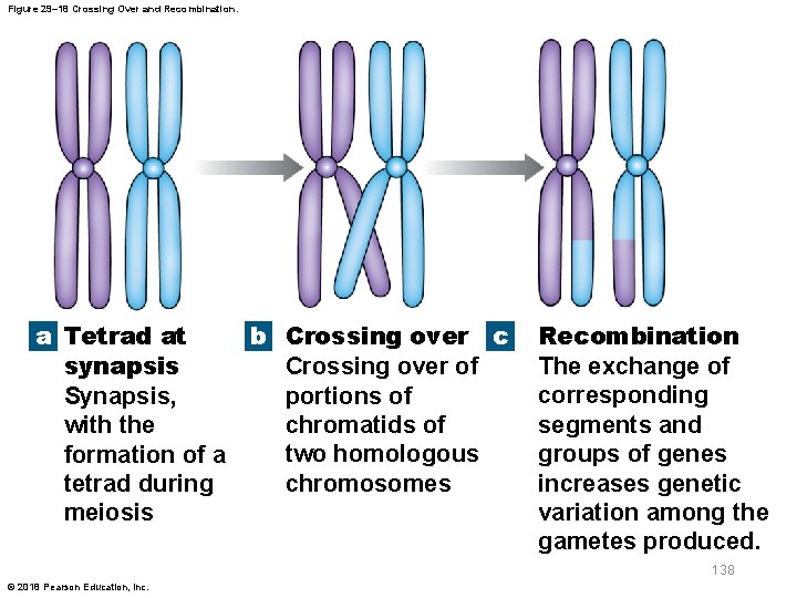
29 -9 Inheritance § Genetic recombination – Crossing over • Parts of homologous chromosomes become rearranged during synapsis • When tetrads form, chromatids may overlap • Chromatids may break and overlapping segments trade places – Translocation • Genetic recombination between nonhomologous chromosomes 137 © 2018 Pearson Education, Inc.

Figure 29– 18 Crossing Over and Recombination. a Tetrad at b Crossing over c synapsis Crossing over of portions of Synapsis, chromatids of with the two homologous formation of a chromosomes tetrad during meiosis Recombination The exchange of corresponding segments and groups of genes increases genetic variation among the gametes produced. 138 © 2018 Pearson Education, Inc.

29 -9 Inheritance § Chromosomal abnormalities – Damaged, broken, missing, or extra copies of chromosomes – Present in 10 percent of zygotes • Most of which do not survive – Present in 0. 5 percent of newborns – Produce a variety of serious clinical conditions 139 © 2018 Pearson Education, Inc.

29 -9 Inheritance § Genetic recombination – Genomic imprinting • Does not change DNA • Results in specific, usually reversible, chemical modifications of DNA and its proteins • Changes dictate whether gene is expressed or not – Epigenetics • Study of inherited traits that are not due to changes in genotype or DNA sequences • Acts by activating or inactivating specific genes 140 © 2018 Pearson Education, Inc.

29 -9 Inheritance § Mutations – Changes in nucleotide sequence of an allele – Spontaneous mutations • Result from random errors in DNA replication • Relatively common, but in most cases error is detected and repaired by enzymes • Errors that go undetected and unrepaired have potential to change phenotype • Can produce gametes that contain abnormal alleles 141 © 2018 Pearson Education, Inc.

29 -9 Inheritance § Carriers – Individuals who are heterozygous for abnormal allele but do not show effects of mutation 142 © 2018 Pearson Education, Inc.

29 -9 Inheritance § Penetrance and expressivity – Penetrance • Percentage of individuals with particular genotype that show “expected” phenotype – Expressivity • Extent to which particular allele is expressed when it is present – Teratogens • Factors that result in abnormal development 143 © 2018 Pearson Education, Inc.

29 -9 Inheritance § Human genome – Full set of genetic material (DNA) in our chromosomes – 3. 2 billion base pairs – 20, 000– 25, 000 protein-coding genes – About 10, 000 different single-gene disorders have been described § Gene therapy – Inserting corrective genes into a patient’s cells – Used to treat genetic disorders or diseases 144 © 2018 Pearson Education, Inc.

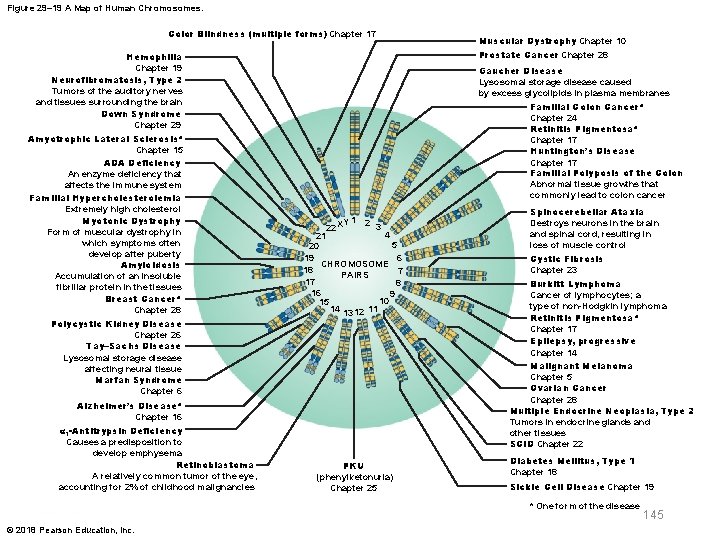
Figure 29– 19 A Map of Human Chromosomes. Color Blindness (multiple forms) Chapter 17 Prostate Cancer Chapter 28 Hemophilia Chapter 19 Neurofibromatosis, Type 2 Tumors of the auditory nerves and tissues surrounding the brain Down Syndrome Chapter 29 Amyotrophic Lateral Sclerosis* Chapter 15 ADA Deficiency An enzyme deficiency that affects the immune system Familial Hypercholesterolemia Extremely high cholesterol Myotonic Dystrophy Form of muscular dystrophy in which symptoms often develop after puberty Amyloidosis Accumulation of an insoluble fibrillar protein in the tissues Breast Cancer* Chapter 28 Polycystic Kidney Disease Chapter 26 Tay–Sachs Disease Lysosomal storage disease affecting neural tissue Marfan Syndrome Chapter 6 Gaucher Disease Lysosomal storage disease caused by excess glycolipids in plasma membranes Familial Colon Cancer* Chapter 24 Retinitis Pigmentosa* Chapter 17 Huntington’s Disease Chapter 17 Familial Polyposis of the Colon Abnormal tissue growths that commonly lead to colon cancer Y 1 2 3 22 X 4 21 5 20 19 6 CHROMOSOME 18 7 PAIRS 17 8 16 9 10 15 14 13 12 11 Alzheimer’s Disease* Chapter 16 α 1 -Antitrypsin Deficiency Causes a predisposition to develop emphysema Retinoblastoma A relatively common tumor of the eye, accounting for 2% of childhood malignancies Muscular Dystrophy Chapter 10 PKU (phenylketonuria) Chapter 25 Spinocerebellar Ataxia Destroys neurons in the brain and spinal cord, resulting in loss of muscle control Cystic Fibrosis Chapter 23 Burkitt Lymphoma Cancer of lymphocytes; a type of non-Hodgkin lymphoma Retinitis Pigmentosa* Chapter 17 Epilepsy, progressive Chapter 14 Malignant Melanoma Chapter 5 Ovarian Cancer Chapter 28 Multiple Endocrine Neoplasia, Type 2 Tumors in endocrine glands and other tissues SCID Chapter 22 Diabetes Mellitus, Type 1 Chapter 18 Sickle Cell Disease Chapter 19 * One form of the disease © 2018 Pearson Education, Inc. 145
 Eleventh edition management
 Management stephen p robbins 11th edition
 Management eleventh edition
 Management eleventh edition
 3 layers of muscle
 Human anatomy & physiology edition 9
 Paratubular cyst
 Chapter 14 anatomy and physiology
 Chapter 1 introduction to human anatomy and physiology
 Anatomy and physiology chapter 8 special senses
 Chapter 13 anatomy and physiology of pregnancy
 Anatomy and physiology chapter 2
 Heat and cold
 Art labeling activity: figure 14.1 (3 of 3)
 Chapter 10 blood anatomy and physiology
 Anatomy and physiology chapter 15
 Anatomy and physiology chapter 1
 Holes anatomy and physiology chapter 1
 Gi tract histology
 Anterior posterior distal proximal
 Chapter 2 human reproductive anatomy and physiology
 Male vs female skeleton pelvis
 Chapter 6 general anatomy and physiology
 Anterior posterior ventral dorsal
 Bhore committee
 Eleventh 5 year plan
 Thfive
 For his eleventh birthday elvis presley
 Physiology of sport and exercise 5th edition
 Upper and lower airway
 Tattoo anatomy and physiology
 Anatomy science olympiad
 Imperfect flowers examples
 Anatomy and physiology of bone
 Triple therapy for peptic ulcer disease
 Sheep liver lobes
 Epigastric region
 Wpigastric region
 Anatomy and physiology of rbc
 Http://anatomy and physiology
 Anatomy and physiology of appendicitis
 Aohs foundations of anatomy and physiology 1
 Aohs foundations of anatomy and physiology 1
 Anatomical planes
 Agriscience unit 26 self evaluation answers
 Science olympiad anatomy and physiology 2020 cheat sheet
 Gastric emptying ppt
 Pancreas anatomy and physiology
 Aohs foundations of anatomy and physiology 1
 Aohs foundations of anatomy and physiology 1
 What produces bile
 Cornell notes for anatomy and physiology
 Holes essential of human anatomy and physiology
 Anatomy and physiology unit 7 cardiovascular system
 Anatomy and physiology
 Aohs foundations of anatomy and physiology 1
 Aohs foundations of anatomy and physiology 1
 Anatomy and physiology exam 1
 Welcome to anatomy and physiology
 Anatomy and physiology of the foot
 Skin cancer
 Physiology vs anatomy
 Pancreas anatomy and physiology
 Anatomy and physiology vocabulary
 Anatomy and physiology
 Biceps muscle names
 Anatomy and physiology
 Anatomy and physiology
 Anatomy and physiology
 Anatomy and physiology
 Anatomy and physiology
 Dorsifelxion
 Anatomy and physiology
 Anatomy and physiology
 Anatomy and physiology
 Anatomy and physiology
 Dorsal root ganglion labeled
 Anatomy and physiology of the eye
 Oblongata
 Irn.org anatomy and physiology
 Anatomy and physiology body parts
 Unit 26 animal anatomy physiology and nutrition
 Figure 14-2 digestive system
 Anatomy and physiology of the retina
 Mucous connective tissue
 Anatomy and physiology of meningitis ppt
 Jeopardy anatomy and physiology game
 What is homeostasis
 Anatomy and physiology
 Respiratory system in humans
 Physiology trivia
 Cycloid scales
 Transverse fissure
 Anatomy and physiology
 Horizontal anatomical plane
 2012 pearson education inc anatomy and physiology
 Fundus
 Fish anatomy and physiology
 Cengage anatomy and physiology
 Fundamentals of information systems 9th edition
 Fundamentals of information systems 9th edition
 No slip condition
 Digital fundamentals by floyd 10th edition
 Machining fundamentals 10th edition
 Fundamentals of organizational communication 9th edition
 Fundamentals of organizational communication 9th edition
 Fundamentals of corporate finance 3rd canadian edition
 Digital fundamentals 10th edition
 Digital fundamentals by floyd
 Dc/ac fundamentals 1st edition
 Computer security fundamentals 4th edition
 Management fundamentals 8th edition
 Fundamentals of information systems
 Fundamentals of corporate finance canadian edition
 Fundamentals of corporate finance fifth edition
 Fundamentals of corporate finance 6th edition
 Abnormal psychology ronald j comer 9th edition
 Fundamentals of information systems 9th edition
 Thermal resistance formula
 The fundamentals of political science research 2nd edition
 Using mis (10th edition) 10th edition
 Zulily case study
 Brunnstrom's clinical kinesiology 7th edition
 Clinical kinesiology and anatomy 6th edition
 Human anatomy fifth edition
 Human anatomy fifth edition
 Fundamentals chapter 1
 Fundamentals of electric circuits chapter 4 solutions
 Fundamentals of corporate finance chapter 6 solutions
 Digital fundamentals chapter 4
 Chapter 24 magnetism magnetic fundamentals answers
 Tire wheel and wheel bearing fundamentals
 Understanding your health and wellness chapter 1
 The circuit chapter 9
 Fundamentals of electric circuits chapter 7 solutions
 Fundamentals of building construction chapter summaries
 Computer fundamentals chapter 1
 Fundamentals of corporate finance chapter 1
 Fundamentals of thermal-fluidsciences chapter 1 problem 25p
 White-collar workers คือ
 Fundamentals of information systems
 Chapter 17 fundamentals of nursing
 Forensic science fundamentals and investigations chapter 6
 Fundamentals in lodging operations chapter 2
 Chapter 2 lab java fundamentals
 Fundamentals of nursing chapter 15 critical thinking


