Fundamentals of Anatomy Physiology Eleventh Edition Chapter 8


- Slides: 87

Fundamentals of Anatomy & Physiology Eleventh Edition Chapter 8 The Appendicular Skeleton Lecture Presentation by Deborah A. Hutchinson Seattle University © 2018 Pearson Education, Inc.

Learning Outcomes 8 -1 Identify the bones that form the pectoral girdles, their functions, and their surface features. 8 -2 Identify the bones of the upper limbs, their functions, and their surface features. 8 -3 Identify the bones that form the pelvic girdle, their functions, and their surface features. 8 -4 Identify the bones of the lower limbs, their functions, and their surface features. 8 -5 Summarize sex differences and age-related changes in the human skeleton. 2 © 2018 Pearson Education, Inc.

Introduction to the Appendicular Skeleton § Appendicular skeleton – Includes 60 percent of bones in the body – Allows us to move and manipulate objects – Includes • Bones of limbs • Supporting bone girdles 3 © 2018 Pearson Education, Inc.

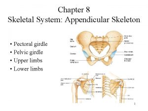
Figure 8– 1 An Anterior View of the Appendicular Skeleton (Part 1 of 2). SKELETAL SYSTEM 206 APPENDICULAR SKELETON Pectoral girdles Upper limbs AXIAL SKELETON 80 126 Clavicle 2 Scapula 2 Humerus 2 Radius 2 Ulna 2 Carpal bones 16 4 60 Metacarpals 10 Phalanges 28 Pelvic girdle 2 Hip bone 2 4 © 2018 Pearson Education, Inc.

Figure 8– 1 An Anterior View of the Appendicular Skeleton (Part 2 of 2). Lower limbs 60 Femur 2 Patella 2 Tibia 2 Fibula 2 Tarsal bones 14 Metatarsals 10 Phalanges 28 5 © 2018 Pearson Education, Inc.

8 -1 The Pectoral Girdles § Pectoral girdle (shoulder girdle) – Connects each arm to the body – Movements position the shoulder joint • And provide a basis for arm movement – Each girdle consists of • One clavicle • One scapula – Connects with the axial skeleton only at the manubrium 6 © 2018 Pearson Education, Inc.

8 -1 The Pectoral Girdles § Clavicles (collarbones) – S-shaped bones – Originate at the manubrium (sternal end) – Articulate with the scapulae (acromial end) 7 © 2018 Pearson Education, Inc.

Figure 8– 2 a The Right Clavicle. Scapula Clavicle Jugular notch a The position of the clavicle within the pectoral girdle, anterior view. © 2018 Pearson Education, Inc. 8

Figure 8– 2 b The Right Clavicle. Acromial end LATERAL Facet for articulation with acromion Sternal end MEDIAL b Superior view of the right clavicle. 9 © 2018 Pearson Education, Inc.

Figure 8– 2 c The Right Clavicle. Sternal end Acromial end MEDIAL LATERAL Conoid tubercle Costal tuberosity Sternal facet c Inferior view of the right clavicle. 10 © 2018 Pearson Education, Inc.

8 -1 The Pectoral Girdles § Scapulae (shoulder blades) – Broad, flat triangles – Articulate with humerus and clavicle – Anterior surface depression is subscapular fossa – Three sides of each scapula • Superior border • Medial border (vertebral border) • Lateral border (axillary border) 11 © 2018 Pearson Education, Inc.

8 -1 The Pectoral Girdles § Corners of scapulae – Superior angle – Inferior angle – Lateral angle § Lateral angle supports the glenoid cavity – Articulates with humerus – To form shoulder joint (glenohumeral joint) 12 © 2018 Pearson Education, Inc.

8 -1 The Pectoral Girdles § Scapular processes – Coracoid process • Small and anterior – Acromion • Large and posterior • Articulates with clavicle at acromioclavicular joint – Spine • Ridge across posterior surface • Divides supraspinous fossa and infraspinous fossa 13 © 2018 Pearson Education, Inc.

Figure 8– 3 a The Right Scapula. Acromion Coracoid process Superior angle Superior border Lateral angle Subscapular fossa Medial border Lateral border Inferior angle a Anterior view © 2018 Pearson Education, Inc. 14

Figure 8– 3 b The Right Scapula. Supraglenoid tubercle Acromion Spine Coracoid process Glenoid cavity Lateral border Inferior angle b Lateral view © 2018 Pearson Education, Inc. 15

Figure 8– 3 c The Right Scapula. Supraspinous fossa Coracoid Acromion process Superior border Neck Spine Infraspinous fossa Medial border Lateral border Inferior angle c Posterior view © 2018 Pearson Education, Inc. 16

8 -2 The Upper Limbs § Skeleton of upper limbs – Consists of bones of the • Arms (arm = shoulder to elbow) • Forearms • Wrists • Hands § Humerus – The only bone in the arm (brachium) – Extends from scapula to elbow 17 © 2018 Pearson Education, Inc.

8 -2 The Upper Limbs § Humerus – Head • Round, proximal portion that articulates with scapula – Greater tubercle • Rounded projection on lateral surface • Forms lateral contour of shoulder – Lesser tubercle • Anterior, medial projection – Tubercles are separated by intertubercular sulcus (bicipital groove) 18 © 2018 Pearson Education, Inc.

8 -2 The Upper Limbs § Humerus – Anatomical neck • Marks extent of joint capsule – Surgical neck • Corresponds to metaphysis of growing bone 19 © 2018 Pearson Education, Inc.

8 -2 The Upper Limbs § Shaft of humerus – Deltoid tuberosity • Large, rough elevation on lateral surface • Attaches deltoid muscle – Radial groove • On posterior surface • For radial nerve 20 © 2018 Pearson Education, Inc.

8 -2 The Upper Limbs § Medial and lateral epicondyles – Distal expansions for muscle attachment § Condyle of the humerus – Articulates with ulna and radius – Trochlea • Extends from coronoid fossa to olecranon fossa – Capitulum forms lateral surface • Radial fossa is superior to capitulum 21 © 2018 Pearson Education, Inc.

Figure 8– 4 a The Right Humerus and Elbow Joint (Part 1 of 1). Greater tubercle Head Lesser tubercle Anatomical neck Intertubercular sulcus Surgical neck Deltoid tuberosity Shaft Radial fossa Coronoid fossa Lateral epicondyle Medial epicondyle Capitulum Trochlea Condyle a Anterior surface © 2018 Pearson Education, Inc. 22

Figure 8– 4 b The Right Humerus and Elbow Joint. Head Greater tubercle Anatomical neck Surgical neck Deltoid tuberosity Radial groove Olecranon fossa Lateral epicondyle Medial epicondyle Trochlea b Posterior surface © 2018 Pearson Education, Inc. 23

Figure 8– 4 c The Right Humerus and Elbow Joint. Humerus Radial fossa Capitulum Trochlea Medial epicondyle Head of radius Coronoid process of ulna Radial notch of ulna c Elbow joint, anterior view 24 © 2018 Pearson Education, Inc.

Figure 8– 4 d The Right Humerus and Elbow Joint. Trochlea of humerus Humerus Olecranon fossa Medial epicondyle Olecranon Head of radius Ulna d Elbow joint, posterior view 25 © 2018 Pearson Education, Inc.

8 -2 The Upper Limbs § Forearm (antebrachium) – Consists of two long bones • Ulna (medial) • Radius (lateral) – Interosseous membrane • Fibrous sheet • Connects lateral margin of ulna to radius 26 © 2018 Pearson Education, Inc.

8 -2 The Upper Limbs § Ulna – Olecranon is proximal end • Point of elbow • Forms superior lip of trochlear notch – Trochlear notch articulates with trochlea of humerus – Coronoid process • Forms inferior lip of trochlear notch 27 © 2018 Pearson Education, Inc.

8 -2 The Upper Limbs § Articulations between ulna and humerus – At limit of extension • Olecranon swings into olecranon fossa – At limit of flexion • Coronoid process projects into coronoid fossa 28 © 2018 Pearson Education, Inc.

8 -2 The Upper Limbs § Other articulations of ulna – Radial notch • Articulates with head of radius • To form proximal radio-ulnar joint – Head of ulna (ulnar head) • Has ulnar styloid process – Attaches to articular disc between forearm and wrist • Articulates with radius to form distal radio-ulnar joint 29 © 2018 Pearson Education, Inc.

8 -2 The Upper Limbs § Radius – Lateral bone of forearm – Disc-shaped head of radius above the neck – Radial tuberosity below the neck • Attaches biceps brachii – Ulnar notch at distal end • Articulates with head of ulna – Radial styloid process on lateral surface • Stabilizes wrist joint 30 © 2018 Pearson Education, Inc.

Figure 8– 5 a The Right Radius and Ulna. Olecranon Proximal radio-ulnar joint Head of radius Neck of radius Ulna Radius Interosseous membrane Head of ulna Ulnar styloid process Ulnar notch of radius Radial styloid process a Posterior view. © 2018 Pearson Education, Inc. 31

Figure 8– 5 b The Right Radius and Ulna. Head of radius Neck of radius Radial tuberosity Radius Trochlear notch Coronoid process Radial notch Ulnar tuberosity Ulna Interosseous membrane Distal radio-ulnar joint Head of ulna Radial styloid process b Anterior view © 2018 Pearson Education, Inc. 32

Figure 8– 5 c The Right Radius and Ulna. Olecranon Trochlear notch Coronoid process Radial notch Ulnar tuberosity Ulna c Lateral view of ulna, showing trochlear notch 33 © 2018 Pearson Education, Inc.

8 -2 The Upper Limbs § Bones of wrist and hand – Eight carpal bones • Four proximal carpal bones • Four distal carpal bones • Allow wrist to bend and twist 34 © 2018 Pearson Education, Inc.

8 -2 The Upper Limbs § Proximal carpal bones – Scaphoid • Near styloid process of radius – Lunate • Medial to scaphoid – Triquetrum • Medial to lunate – Pisiform • Anterior to triquetrum 35 © 2018 Pearson Education, Inc.

8 -2 The Upper Limbs § Distal carpal bones – Trapezium • Lateral – Trapezoid • Medial to trapezium – Capitate • Largest – Hamate • Medial 36 © 2018 Pearson Education, Inc.

Figure 8– 6 Bones of the Right Wrist and Hand. Proximal Carpal Bones Radius Ulna Scaphoid Distal Carpal Bones Proximal Carpal Bones Ulna Distal Carpal Bones Radius Scaphoid Lunate Trapezium Triquetrum Trapezoid Capitate Pisiform Hamate I Pollex V II IV I V Metacarpals IV III II Metacarpals Proximal phalanx Distal phalanx Phalanges Proximal Middle Distal a Anterior view b Posterior view 37 © 2018 Pearson Education, Inc.

8 -2 The Upper Limbs § Metacarpals – The five long bones of the hand – Numbered I–V from lateral to medial – Articulate with proximal phalanges § Phalanges (finger bones) – Pollex (thumb) • Has two phalanges (proximal, distal) – Each of the other four fingers • Has three phalanges (proximal, middle, distal) 38 © 2018 Pearson Education, Inc.

Figure 8– 6 a Bones of the Right Wrist and Hand. Proximal Carpal Bones Radius Ulna Scaphoid Distal Carpal Bones Lunate Trapezium Triquetrum Trapezoid Capitate Hamate Pisiform I Pollex V II IV Metacarpals Proximal phalanx Distal phalanx Phalanges Proximal Middle Distal a Anterior view © 2018 Pearson Education, Inc. 39

Figure 8– 6 b Bones of the Right Wrist and Hand. Proximal Carpal Bones Ulna Distal Carpal Bones Radius Scaphoid Lunate Trapezium Triquetrum Trapezoid Capitate Hamate Pisiform I V IV III II Metacarpals Phalanges Proximal Middle Distal b Posterior view © 2018 Pearson Education, Inc. 40

8 -3 The Pelvic Girdle § Pelvic girdle – Consists of two hip bones • Also called coxal bones or pelvic bones – Attaches to lower limbs – Strong to bear body weight and provide mobility – Part of the pelvis – Each hip bone consists of three fused bones • Ilium • Ischium • Pubis 41 © 2018 Pearson Education, Inc.

8 -3 The Pelvic Girdle § Acetabulum – Socket on lateral surface of each hip bone – Lunate surface articulates with head of femur – Meeting point of ilium, ischium, and pubis § Acetabular notch – A gap in the ridge that forms margins of acetabulum 42 © 2018 Pearson Education, Inc.

Figure 8– 7 a The Right Hip Bone. POSTERIOR Ischium ANTERIOR Pubis Gluteal Lines Iliac crest Anterior superior iliac spine Inferior Posterior Anterior inferior iliac spine Posterior superior iliac spine Acetabulum Lunate surface Acetabulum Posterior inferior iliac spine Acetabular notch Greater sciatic notch Pubis Ischial spine Lesser sciatic notch Superior pubic ramus Obturator foramen Pubic tubercle Ischial tuberosity Inferior pubic ramus Ischial ramus a Right hip bone, lateral views © 2018 Pearson Education, Inc. 43

8 -3 The Pelvic Girdle § Ilium – Greater sciatic notch • For sciatic nerve – Arcuate line • Continuous with pectineal line – Iliac crest • Upper brim – Iliac fossa • Depression between iliac crest and arcuate line 44 © 2018 Pearson Education, Inc.

8 -3 The Pelvic Girdle § Ischium – Ischial spine • Superior to lesser sciatic notch – Ischial tuberosity • Posterior projection you sit on – Ischial ramus • Meets inferior pubic ramus 45 © 2018 Pearson Education, Inc.

8 -3 The Pelvic Girdle § Pubis – Inferior pubic ramus – Superior pubic ramus • Pectineal line is a ridge on anterior, superior surface – Pubic symphysis • Where pubic bones attach to a pad of fibrocartilage § Obturator foramen – Space encircled by ischial and pubic rami – Closed by collagen fibers that attach hip muscles 46 © 2018 Pearson Education, Inc.

Figure 8– 7 a The Right Hip Bone. POSTERIOR Ischium ANTERIOR Pubis Gluteal Lines Iliac crest Anterior superior iliac spine Inferior Posterior Anterior inferior iliac spine Posterior superior iliac spine Acetabulum Lunate surface Acetabulum Posterior inferior iliac spine Acetabular notch Greater sciatic notch Pubis Ischial spine Lesser sciatic notch Superior pubic ramus Obturator foramen Pubic tubercle Ischial tuberosity Inferior pubic ramus Ischial ramus a Right hip bone, lateral views © 2018 Pearson Education, Inc. 47

Figure 8– 7 b The Right Hip Bone. Ilium ANTERIOR POSTERIOR Pubis Ischium Iliac crest Anterior superior iliac spine Auricular surface for articulation with sacrum Iliac fossa Iliac tuberosity Anterior inferior iliac spine Posterior superior iliac spine Posterior inferior iliac spine Greater sciatic notch Pubis Superior pubic ramus Pubic tubercle Inferior pubic ramus Arcuate line Ischial spine Lesser sciatic notch Pectineal line Ischial tuberosity Ischial ramus Location of pubic symphysis b Right hip bone, medial views © 2018 Pearson Education, Inc. 48

8 -3 The Pelvic Girdle § Pelvis – Consists of two hip bones, sacrum, and coccyx – Stabilized by ligaments of pelvic girdle, sacrum, and lumbar vertebrae § Sacro-iliac joints – Where auricular surfaces of ilia articulate with sacrum – Stabilized by ligaments arising at iliac tuberosity 49 © 2018 Pearson Education, Inc.

Figure 8– 8 a The Pelvis of an Adult Male. Sacrum Coccyx Ilium Pubis Hip bone Ischium Lumbar disc Iliac crest L 5 Iliac fossa Sacro-iliac joint Arcuate line Ilium Sacrum Pubis Acetabulum Pubic symphysis Pubic tubercle Obturator foramen © 2018 Pearson Education, Inc. Ischium a Anterior view 50

Figure 8– 8 b The Pelvis of an Adult Male. Sacrum Coccyx Ilium Hip bone Pubis Ischium Iliac crest L 5 Sacral foramina Posterior superior iliac spine Posterior inferior iliac spine Sacrum Greater sciatic notch Ischial spine Ischial tuberosity Coccyx b Posterior view © 2018 Pearson Education, Inc. 51

8 -3 The Pelvic Girdle § Bones of the pelvis enclose the pelvic cavity § True pelvis – Inferior to pelvic brim – Encloses pelvic inlet § False pelvis – Superior to pelvic brim 52 © 2018 Pearson Education, Inc.

8 -3 The Pelvic Girdle § Pelvic outlet – The opening bounded by coccyx, ischial tuberosities, and inferior border of pubic symphysis – Fetus passes through during childbirth § Perineum – Surface region bordered by inferior edges of pelvis – Perineal muscles • Form floor of pelvic cavity • Support organs in true pelvis 53 © 2018 Pearson Education, Inc.

Figure 8– 9 a Divisions of the Pelvis. False pelvis Pelvic outlet Pelvic brim Pelvic inlet a Superior view. The pelvic brim, pelvic inlet, and pelvic outlet. 54 © 2018 Pearson Education, Inc.

Figure 8– 9 b Divisions of the Pelvis. False pelvis Pelvic inlet Pelvic brim True pelvis Pelvic outlet b Lateral view. The boundaries of the true (lesser) pelvis (shown in purple) and the (false) greater pelvis. 55 © 2018 Pearson Education, Inc.

Figure 8– 9 c Divisions of the Pelvis. Pelvic outlet Ischial spine c Inferior view. The limits of the pelvic outlet. 56 © 2018 Pearson Education, Inc.

8 -3 The Pelvic Girdle § Comparing the male and female pelvis – Female pelvis • Broader, smoother, and lighter • Less-prominent markings • Shallower iliac fossa • Wider pelvic outlet • Triangular obturator foramen 57 © 2018 Pearson Education, Inc.

Figure 8– 14 Sex Differences in the Human Skeleton (Part 1 of 4). MALE FEMALE SKULL Heavier, rougher General Appearance Lighter, smoother About 10% larger Cranium About 10% smaller More sloping Forehead More vertical Larger Sinuses Smaller Larger Teeth Smaller Larger, squarer, more robust Mandible Smaller, narrower, less robust 58 © 2018 Pearson Education, Inc.

Figure 8– 14 Sex Differences in the Human Skeleton (Part 2 of 4). PELVIS Narrower, rougher, more robust General appearance More vertical; extends further superior to sacro-iliac joint Long, narrow triangle with pronounced sacral curvature Ilium Sacrum Broader, smoother, less robust Less vertical; less extension superior to sacral articulation Broad, short triangle with less sacral curvature Iliac fossa Shallower Narrower, Heart shaped Pelvic inlet Open, circular shaped Narrow Pelvic outlet Deeper Points anteriorly Coccyx Directed laterally Acetabulum Oval Less than 90º Obturator foramen Pubic angle Wide Points inferiorly Faces slightly anteriorly Triangular 100º or more OTHER Greater More prominent Bone mass and density Bone markings Lesser Less prominent 59 © 2018 Pearson Education, Inc.

8 -4 The Lower Limbs § Functions of the lower limbs – Weight bearing – Movement 60 © 2018 Pearson Education, Inc.

8 -4 The Lower Limbs § Bones of the lower limbs – Femur (in thigh) – Patella (kneecap) – Tibia and fibula (in leg) – Tarsal bones – Metatarsals – Phalanges § Leg = distal portion of limb (from knee to ankle) 61 © 2018 Pearson Education, Inc.

8 -4 The Lower Limbs § Femur – Longest, heaviest bone in body – Head (epiphysis) • Articulates with hip bone at acetabulum • Attaches with ligament at fovea capitis – Neck • Joins shaft at an angle of about 125 degrees 62 © 2018 Pearson Education, Inc.

8 -4 The Lower Limbs § Femur – Greater and lesser trochanters • Large, rough projections at junction of neck and shaft • For tendon attachments – Intertrochanteric line • On anterior surface • Marks edge of articular capsule – Intertrochanteric crest • On posterior surface 63 © 2018 Pearson Education, Inc.

8 -4 The Lower Limbs § Femur – Linea aspera • Ridge along center of posterior surface of shaft • Attaches hip muscles • Divides into two ridges that continue to epicondyles 64 © 2018 Pearson Education, Inc.

8 -4 The Lower Limbs § Femur – Medial and lateral epicondyles • Projections above knee joint – Medial and lateral condyles • Rounded surfaces that form part of knee joint • Separated by intercondylar fossa posteriorly • Separated by patellar surface anteriorly and inferiorly 65 © 2018 Pearson Education, Inc.

Figure 8– 10 Bone Markings on the Right Femur. Neck Greater trochanter Fovea capitis Head Intertrochanteric crest Gluteal tuberosity Intertrochanteric line Lesser trochanter Pectineal line Linea aspera Shaft Lateral supracondylar ridge Medial supracondylar ridge Patellar surface Adductor tubercle Medial epicondyle Lateral condyle Medial condyle a Anterior surface © 2018 Pearson Education, Inc. Intercondylar fossa Lateral epicondyle Lateral condyle b Posterior surface 66

8 -4 The Lower Limbs § Patella (kneecap) – Large sesamoid bone – Forms within tendon of quadriceps femoris – Quadriceps tendon attaches near base – Patellar ligament attaches apex to tibia 67 © 2018 Pearson Education, Inc.

Figure 8– 11 a The Right Patella and Patella with Femur. Base of patella Attachment area for quadriceps tendon Attachment area for patellar ligament Apex of patella a Anterior view 68 © 2018 Pearson Education, Inc.

Figure 8– 11 b The Right Patella and Patella with Femur. Lateral facet, for lateral condyle of femur Medial facet, for medial condyle of femur Articular surface of patella b Posterior view 69 © 2018 Pearson Education, Inc.

Figure 8– 11 c The Right Patella and Patella with Femur. Patella Lateral facet, for lateral condyle of femur Medial facet, for medial condyle of femur Lateral condyle of femur Medial condyle of femur c Inferior view of right femur and patella 70 © 2018 Pearson Education, Inc.

8 -4 The Lower Limbs § Bones of the leg – Tibia and fibula – Bound together by interosseous membrane 71 © 2018 Pearson Education, Inc.

8 -4 The Lower Limbs § Tibia (shinbone) – Large, medial, weight-bearing bone of leg – Medial and lateral tibial condyles • Articulate with medial and lateral condyles of femur • Separated by intercondylar eminence – Tibial tuberosity • On anterior surface • Attaches patellar ligament 72 © 2018 Pearson Education, Inc.

8 -4 The Lower Limbs § Tibia – Anterior margin • Ridge that extends distally from tibial tuberosity – Medial malleolus • Medial projection at ankle 73 © 2018 Pearson Education, Inc.

8 -4 The Lower Limbs § Fibula – Small, lateral bone of leg – Head articulates with tibia • At lateral tibial condyle – No articulation with femur – Attaches muscles of feet and toes – Lateral malleolus • Lateral projection at ankle 74 © 2018 Pearson Education, Inc.

Figure 8– 12 a The Right Tibia and Fibula. Lateral tibial condyle Head of fibula Superior tibiofibular joint Medial tibial condyle Tibial tuberosity Interosseous membrane Anterior margin Tibia Fibula Lateral malleolus (fibula) Medial malleolus (tibia) Inferior articular surface a Anterior view © 2018 Pearson Education, Inc. 75

Figure 8– 12 b The Right Tibia and Fibula. Intercondylar eminence Articular surface of medial tibial condyle Medial tibial condyle Articular surface of lateral tibial condyle Lateral tibial condyle Head of fibula Interosseous membrane Tibia Medial malleolus (tibia) b Posterior view © 2018 Pearson Education, Inc. Fibula Inferior tibiofibular joint Lateral malleolus (fibula) 76

Figure 8– 12 c The Right Tibia and Fibula. Anterior margin Tibia Fibula Interosseous membrane c Transverse section of tibia and fibula, inferior view 77 © 2018 Pearson Education, Inc.

8 -4 The Lower Limbs § Ankle (tarsus) consists of seven tarsal bones – Talus • Transfers weight from tibia across trochlea – Calcaneus (heel bone) • Largest tarsal bone • Transfers weight from talus to ground • Attaches calcaneal tendon (Achilles tendon) – Cuboid • Articulates with calcaneus 78 © 2018 Pearson Education, Inc.

8 -4 The Lower Limbs § Seven tarsal bones – Navicular • Articulates with talus and three cuneiform bones – Medial cuneiform – Intermediate cuneiform – Lateral cuneiform § Cuboid and cuneiform bones articulate with metatarsals 79 © 2018 Pearson Education, Inc.

Figure 8– 13 a Bones of the Ankle and Foot. Tarsal bones Calcaneus Trochlea of talus Navicular Cuboid Cuneiform bones Lateral Intermediate Medial V IV III II I Metatarsals Phalanges Hallux Proximal phalanx Middle Distal phalanx Distal a Superior view, right foot © 2018 Pearson Education, Inc. 80

8 -4 The Lower Limbs § Metatarsals – Five long bones of foot – Numbered I–V, medial to lateral – Articulate with proximal phalanges 81 © 2018 Pearson Education, Inc.

8 -4 The Lower Limbs § Phalanges – 14 bones of the toes – Hallux (great toe) • Has two phalanges (proximal, distal) – Each of the other four toes • Has three phalanges (proximal, middle, distal) 82 © 2018 Pearson Education, Inc.

Figure 8– 13 a Bones of the Ankle and Foot. Tarsal bones Calcaneus Trochlea of talus Navicular Cuboid Cuneiform bones Lateral Intermediate Medial V IV III II I Metatarsals Phalanges Hallux Proximal phalanx Middle Distal phalanx Distal a Superior view, right foot © 2018 Pearson Education, Inc. 83

Figure 8– 13 b Bones of the Ankle and Foot. Talus Cuboid Navicular Cuneiform bones Metatarsals Phalanges Calcaneus b Lateral view, right foot 84 © 2018 Pearson Education, Inc.

8 -4 The Lower Limbs § Arches of the foot – Transfer weight from one part of foot to another – Longitudinal arch • Ties calcaneus to distal portions of metatarsals – Transverse arch • The difference in curvature from one side of foot to the other 85 © 2018 Pearson Education, Inc.

Figure 8– 13 c Bones of the Ankle and Foot. Medial Navicular Talus cuneiform bone Phalanges Metatarsals Calcaneus Medial part of Transverse Lateral part of longitudinal arch c Medial view, right foot 86 © 2018 Pearson Education, Inc.

8 -5 Individual Skeletal Variation § Skeletal features can reveal – Muscle strength and mass (bone ridges, bone mass) – Medical history (condition of teeth, healed fractures) – Sex and age (bone measurements and fusion) – Body size § Modifications in female pelvis for childbearing – Hormone relaxin produced during pregnancy • Loosens pubic symphysis and sacro-iliac ligaments • Increases size of pelvic inlet and outlet 87 © 2018 Pearson Education, Inc.
 Management eleventh edition
 Management stephen p robbins 11th edition
 Management eleventh edition
 Management eleventh edition stephen p robbins
 Endomysium
 Human anatomy & physiology edition 9
 Uterus perimetrium
 The central sulcus divides which two lobes? (figure 14-13)
 Waistline
 Anatomy and physiology chapter 8 special senses
 Chapter 13 anatomy and physiology of pregnancy
 Anatomy and physiology chapter 2
 Heat and cold
 Chapter 14 the digestive system and body metabolism
 Chapter 10 blood anatomy and physiology
 Anatomy and physiology chapter 15
 Anatomy and physiology chapter 1
 Holes anatomy and physiology chapter 1
 Anatomy and physiology chapter 15
 Medial and lateral
 Chapter 2 human reproductive anatomy and physiology
 Anatomy and physiology chapter 8 skeletal system
 Chapter 6 general anatomy and physiology
 Olecranal region
 Chadha committee
 Eleventh 5 year plan
 11th five year plan
 For his eleventh birthday elvis presley
 Physiology of sport and exercise 5th edition
 Pulmonary ventilation
 Tattoo anatomy and physiology
 International anatomy olympiad
 Crown plants examples
 Bone anatomy and physiology
 Peptic ulcer anatomy
 Liver anatomy and physiology
 Podbřišek
 Epigastric region
 Google.com
 Http://anatomy and physiology
 Anatomy and physiology of appendix
 Aohs foundations of anatomy and physiology 1
 Aohs foundations of anatomy and physiology 1
 Anatomical planes
 Unit 26 self evaluation answers
 Science olympiad anatomy and physiology 2020 cheat sheet
 Degluttination
 Physiology
 Aohs foundations of anatomy and physiology 1
 Aohs foundations of anatomy and physiology 1
 Anatomy and physiology
 Cornell notes for anatomy and physiology
 Holes essential of human anatomy and physiology
 Anatomy and physiology unit 7 cardiovascular system
 Anatomy and physiology
 Aohs foundations of anatomy and physiology 1
 Aohs foundations of anatomy and physiology 1
 Anatomy and physiology exam 1
 Welcome to anatomy and physiology
 Physiology of the foot and ankle
 Integumentary system psoriasis
 Pse4u
Pse4u Pancreas anatomy and physiology
 Anatomy and physiology vocabulary
 Anatomy and physiology
 Biceps muscle names
 Anatomy and physiology
 Body landmarks diagram
 Anatomy and physiology
 Anatomy and physiology
 Thyroid anatomy
 Anatomy and physiology
 Figure 11-8 arteries
 Anatomy and physiology
 Anatomy and physiology
 Anatomy and physiology
 Anatomy histology
 Anatomy and physiology of eye
 Telencephalon
 Irn.org anatomy and physiology
 Anatomy and physiology body parts
 Unit 26 animal anatomy physiology and nutrition
 Figure 14-1 digestive system
 Anatomy and physiology of the retina
 Mucous connective tissue
 Anatomy and physiology of meningitis ppt
 Jeopardy anatomy and physiology game
 Short note on homeostasis


