Holes Human Anatomy and Physiology Eleventh Edition Shier


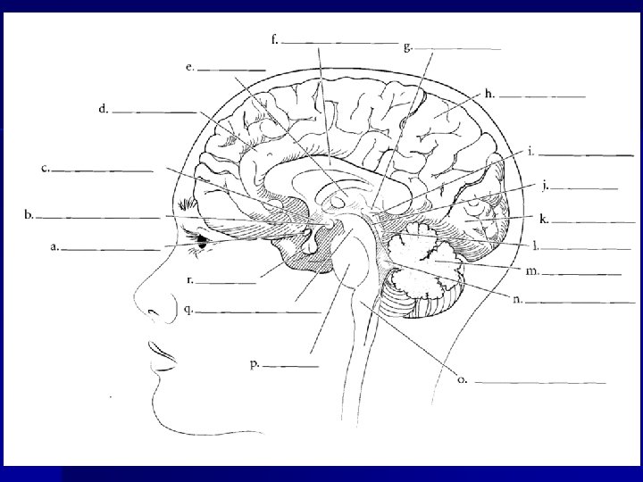
 hypothalamus C. Aquaduct Mammilary")


 Lower portion of Corpus Callosum connects to hippocampus =")
: most basic functions")


 b) c) d) e) f) Limbic System = involved in Emotional")


 n n Inferior part of brain stem, blends into the spinal cord")
 scattered throughout the")
 • 90 min • lasts")


- Slides: 65
Hole’s Human Anatomy and Physiology Eleventh Edition Shier w Butler w Lewis Chapter 11 The Nervous System II Divisions of the Nervous System: The Brain Copyright © The Mc. Graw-Hill Companies, Inc. Permission required for reproduction or display.
Brain Development 1. Embryonic neural tube expands and hollows cranially 2. Three vesicles develop that split and become four adult ventricles 3. The walls of the three vesicles become certain adult brain areas –Forebrain = Cerebrum, basal nuclei, & diencephalons –Midbrain = Midbrain –Hindbrain = Pons, medulla, obloongata, & cerebellum
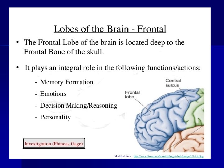
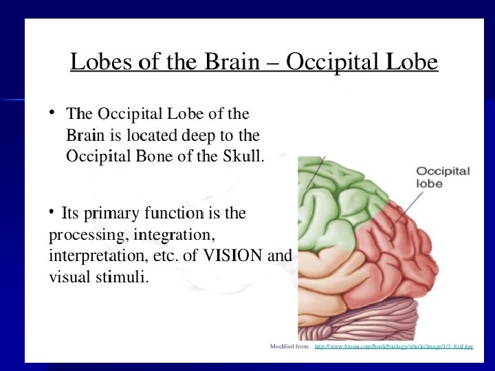
The Brain n n 4 Portions: cerebrum, cerebellum, diencephalon and brain stem Functions: – – – Interprets sensations Determines perception Stores memory Reasoning Makes decisions Coordinates muscular movements – Regulates visceral activities – Determines personality
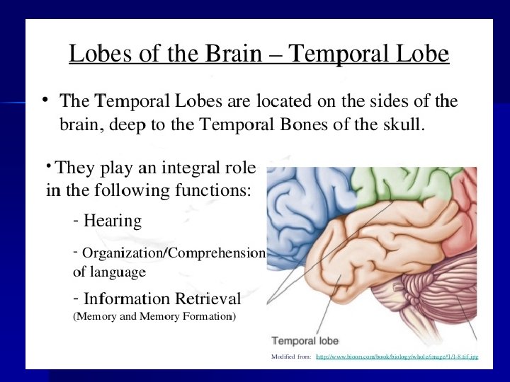
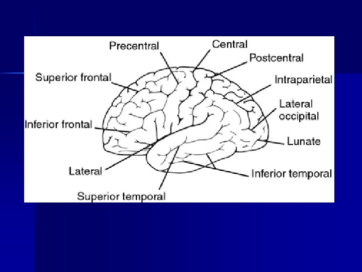
Structure of the Cerebrum n Cerebrum: the largest portion of the brain; two cerebral hemispheres – Corpus callosum: nerve fibers that connect the hemispheres – Convolutions (gyri): surface ridges – Lobes divide the hemispheres: frontal, parietal, temporal and occipital – Tyo types of grooves: n n Sulci: shallow Fissure: deep
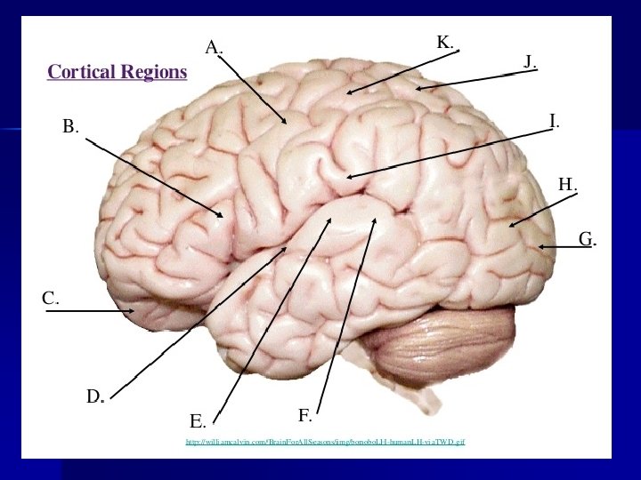
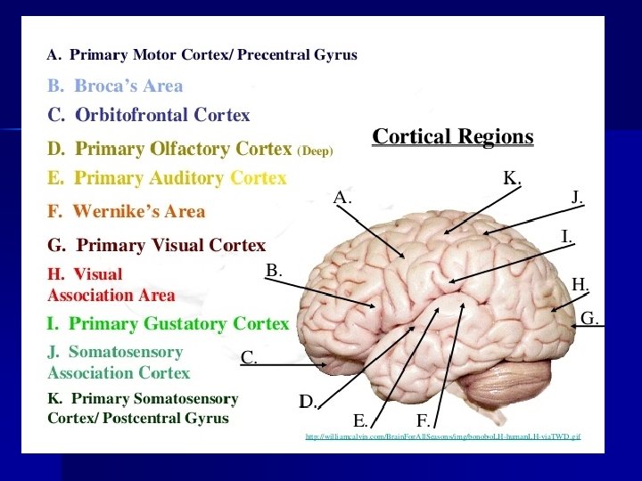
Lobes of Cerebral Hemispheres
*Somato-sensory Cortex “Post-central gyrus”
Functions of the Cerebrum n Functions – – – n Interpreting impulses Initiating voluntary movements Storing information as memory Retrieving stored information Reasoning Seat of intelligence and personality Composition – Bulk white matter (bundles of myelinated nerve fibers, by oligodendrocytes) – Cerebral cortex (outer portion) is gray matter, bundles of neuron cell bodies
Central Sulcus Precentral Gyrus Postcentral gyrus Parietal Lobe Frontal lobe Lateral Fissure Occipital lobe Temporal lobe Cerebellum
Frontal Lobe Longitudinal Fissure Precentral Gyrus Central Sulcus Postcentral Gyrus Parietal Lobe Gyrus Occipital Lobe Sulcus
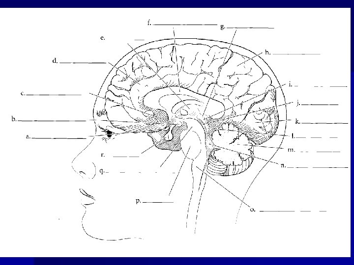
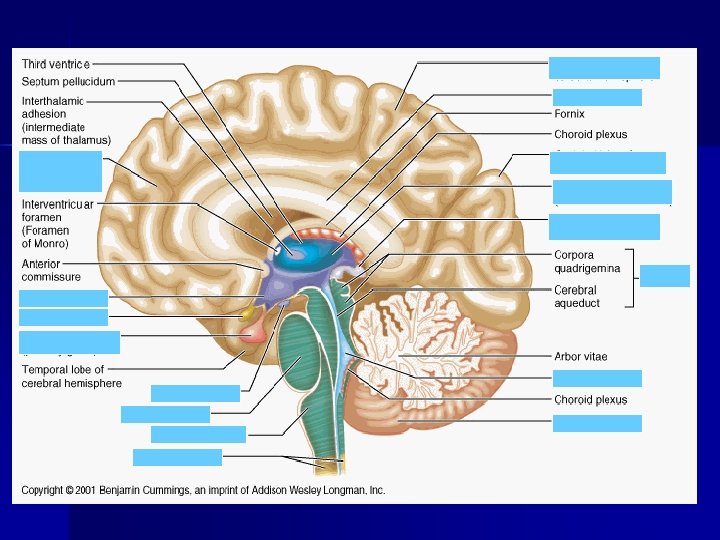
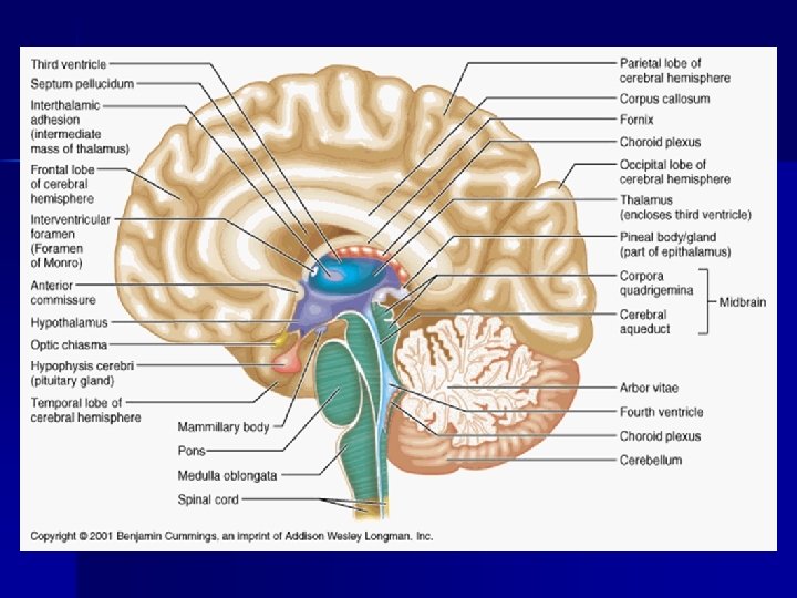
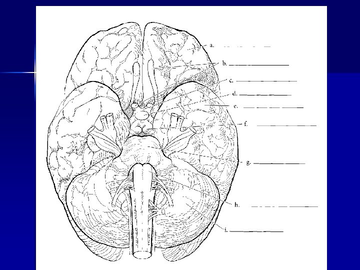
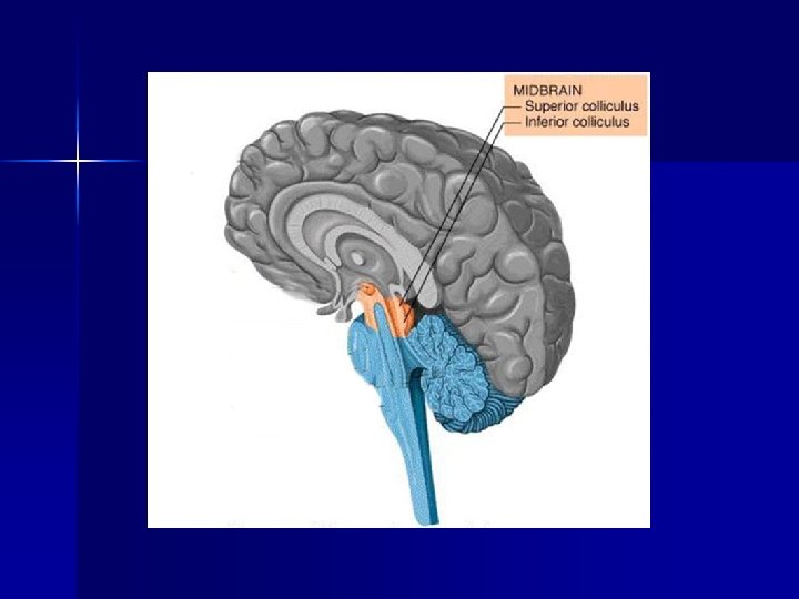
Corpus callosum Thalamus Pineal gland Parietal Frontal Sup. Colliculus (midbrain) hypothalamus C. Aquaduct Mammilary body Occipital Inf. Colliculus (Midbrain) Cerebellum Optic chiasma Pituitary Cerebral Penduncle (midbrain) Pons 4 th ventricle Medulla Oblongata
frontal Cranial nerves Optic chiasma Pituitary gland Temporal lobe Mammillary body pons Medulla oblongata cerebellum
Composition n Bulk of cerebrum is white matter – Bundles of myelinated nerve fibers (by oligodendrocytes) n Cerebral cortex (thin layer, outer portion) is gray matter – Bundles of neuron cell bodies (75% of all neurons in NS) – Recordings of activity or brain waves
Functions of Cerebrum n All conscious behavior via 3 functional areas: sensory, association areas and motor
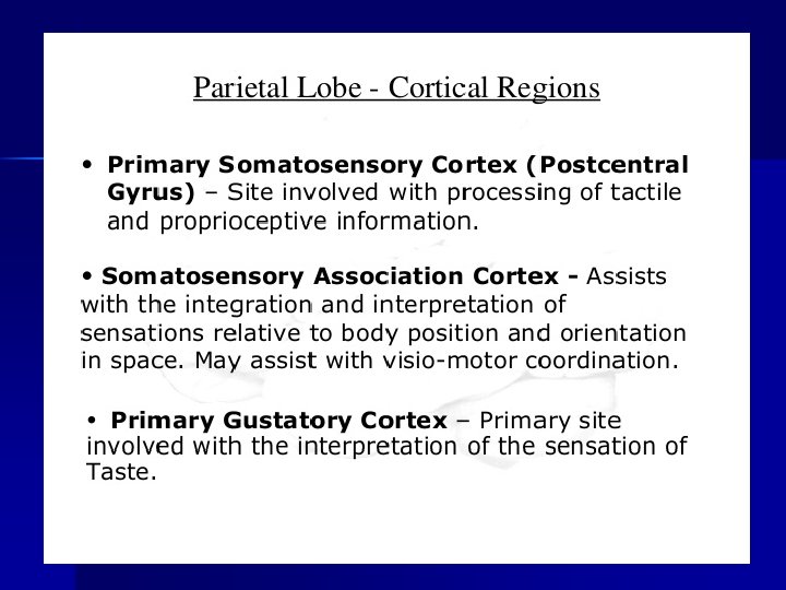
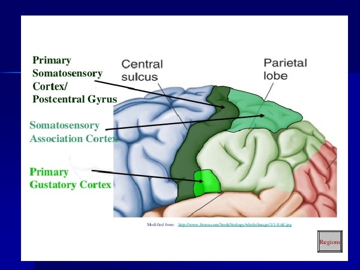
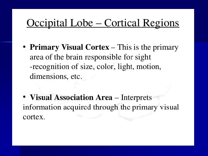
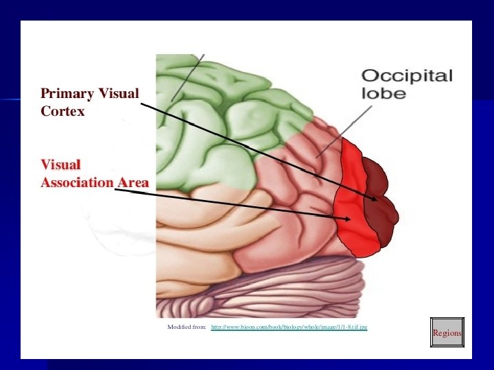
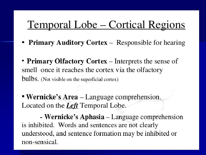
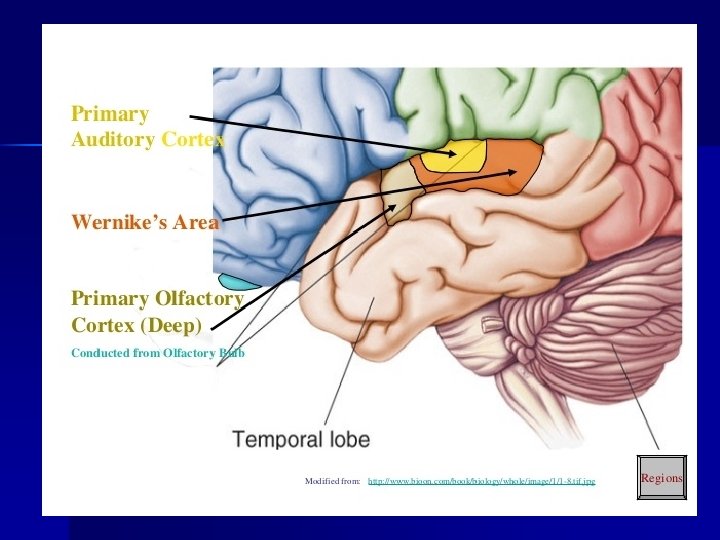
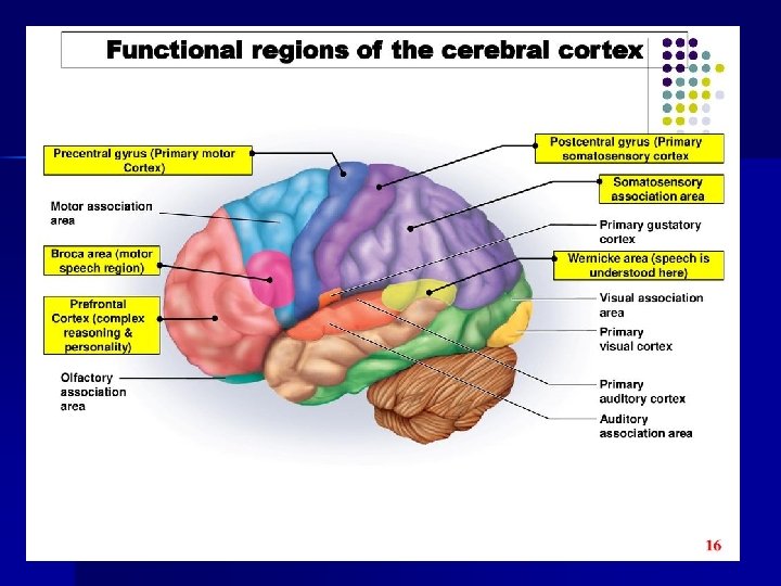
Sensory Areas n Conscious awareness of sensations: – Primary Somatosensory Cortex n n parietal lobe sensations on skin – Visual (Cortex) Area n n occipital lobe interprets vision – Auditory (Cortex) Area n n temporal lobe interprets hearing – Gustatory Cortex n n near bases of the central sulci Taste – Sensory Area for smell n arise from centers deep within the cerebrum
Sensory Areas
Association Areas n General – Areas not directly involved in motor or sensory function – Involved in many traits (analyze, memory, judgement…) – Interconnected – Involve all four lobes – See table 11. 5, page 405
Association Areas Frontal Lobe Association Areas • concentrating • planning • complex problem solving http: //www. philly. com/philly/health/20150201_Te ens__immature_brains_pose_all_sorts_of_danger s. html#pkft 0 p. G 44 AUUWlc. K. 03 Parietal Lobe Association Areas • understanding speech • choosing words to express thought Temporal Lobe Association Areas • interpret complex sensory experiences • store memories of visual scenes, music, and complex patterns Occipital Lobe Association Areas • analyze and combine visual images with other sensory experiences Insular Cortex • empathy and emotion • pain levels
Association Areas
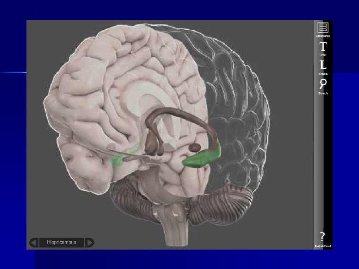
Memory and Emotion (Temporal Lobe) Lower portion of Corpus Callosum connects to hippocampus = memory us la, s ll goe Sme ygda to am halam not t connects to hippocampus = memory Emotional center Memory Mammillary - Fornix – Hippocampus - Amygdala
Functional Regions of Cerebral Cortex n n Hemisphere Dominance (Brain Lateralization): most basic functions are controlled by both left and right hemispheres (communication exists thru corpus callosum) Memory – A consequence of learning whereas learning is the acquisition of new knowledge, memory is the persistence of learning n n n Short Term Long Term A lesion in the cerebral cortex could cause – Motor or sensor deficits – Intellectual impairment – Strange behavior in thought patterns
Hemisphere Dominance • The left hemisphere is dominant is most individuals • Dominant hemisphere controls • speech • writing • reading • verbal skills • analytical skills • computational skills • Nondominant hemisphere controls • nonverbal tasks • motor tasks • understanding and interpreting musical and visual patterns • provides emotional and intuitive thought processes
Memory Short Term • working memory • closed neuronal circuit • circuit is stimulated over and over • when impulse flow ceases, memory does also • unless it enters longterm memory via memory consolidation • 1 st stage Long Term • changes structure or function of neurons • enhances synaptic transmission
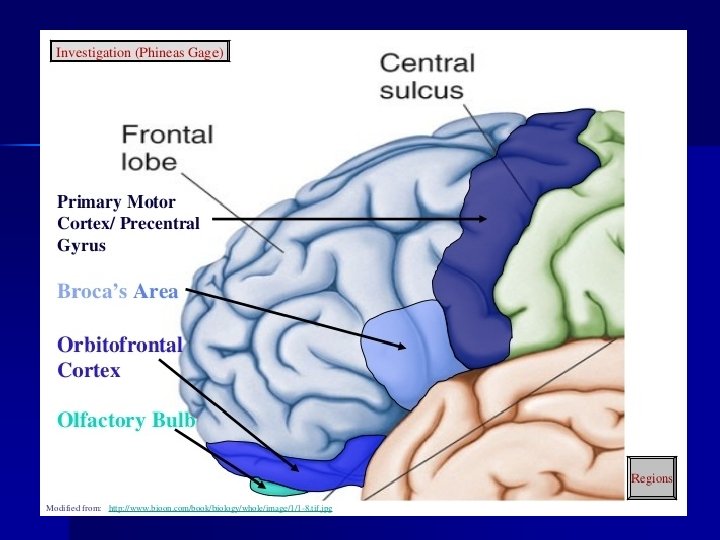
Motor Areas • Primary Motor Areas • frontal lobes • control voluntary muscles • Broca’s Area • anterior to primary motor cortex • usually in left hemisphere • controls muscles needed for speech • Frontal Eye Field • above Broca’s area • controls voluntary movements of eyes and eyelids
Motor Areas
Basal Nuclei n n n Masses of gray matter Deep within cerebral hemispheres “Relay stations, ” for outgoing motor impulses from the brain Produces dopamine Aids in control of motor (muscular) activities – primarily by inhibiting motor functions n Clinical Application pg. 11. 5 Parkinson’s and Huntingdon’s disease result from the degeneration of the basal ganglia Movement disorders – Parkinsons, Huntingtons, Tourettes, OCD
Diencephalon n Between cerebral hemispheres and above the brainstem surrounds third ventricle n Thalamus (gray matter) Hypothalamus (gray matter) Optic tracts Optic chiasma Infundibulum Posterior pituitary Mammillary bodies Pineal gland n n n n
Diencephalon n Thalamus – gateway for sensory impulses heading to cerebral cortex – receives all sensory impulses (except smell) – channels impulses to appropriate part of cerebral cortex for interpretation n Hypothalamus – forms part of the diencephalon – Main visceral control center of the body (i. e. regulates homeostasis) n n n Heart rate & blood pressures Body temperature Water and electrolyte balance Control of hunger, body weight, digestive movements & secretions Regulation of sleep-wake cycles Control of endocrine system functioning
Diencephalon n a) b) c) d) e) f) Limbic System = involved in Emotional response also includes structures in the frontal and temporal cortex, basal nuclei, and deep nuclei controls emotional experience and expression can modify the way a person acts produces feelings of fear, anger, pleasure, and sorrow recognizes life threatening upsets in a person's physical or psychological condition and counters them involved in sense of smell.
Brain Stem Three Parts: 1. Midbrain 2. Pons 3. Medulla Oblongata Serves as a pathway for fiber tracts running to (sensory impulses) and from (motor impulses). The cerebrum and brain stem is the site where many cranial nerve arise (PNS)
Midbrain n n Between diencephalon and pons Corpora quadrigemina = 4 dome-like protrusions on the dorsal midbrain surface Gray matter within white matter Acts in reflex actions (visual and auditory) Contains areas associated with reticular formation.
Pons n n bulging portion of brain stem "bridge" or pathway of conduction tracts location of pneumotaxic area (regulation of breathing rate) of respiratory center also contains areas associated with reticular formation.
Medulla (Oblongata) n n Inferior part of brain stem, blends into the spinal cord at its base Contains an autonomic reflex center involved in maintaining homeostasis of important visceral organs. – – Cardiac center – force and rate of heart contraction Vasomotor center – blood pressure – acts on smooth muscles in arterioles Respiratory center – depth and rhythm of breathing Add’l involuntary activities: vomiting, hiccuping, swallowing, sneezing, coughing.
Reticular Formation n n complex network of nerve fibers (gray matter) scattered throughout the brain stem extends into the diencephalon connects to centers of hypothalamus, basal nuclei, cerebellum, and cerebrum filters incoming sensory information arouses cerebral cortex into state of wakefulness
Types of Sleep Slow Wave Rapid Eye Movement (REM) • 90 min • lasts 15 min • non-REM sleep • paradoxical sleep • person is tired • some areas of brain active • decreasing activity of • heart and respiratory rates reticular system irregular • restful • dreaming occurs • dreamless • reduced blood pressure and respiratory rate • ranges from light to heavy • alternates with REM sleep
Cerebellum • inferior to occipital lobes • posterior to pons and medulla oblongata • two hemispheres • vermis connects hemispheres • cerebellar cortex – gray matter • arbor vitae – white matter • cerebellar peduncles – nerve fiber tracts • dentate nucleus – largest nucleus in cerebellum • integrates sensory information concerning position of body parts • coordinates skeletal muscle activity • maintains posture
Skull bone Meninges brain Cause: Inflammation of the meninges. Can be caused by a virus or bacteria and can be extremely harmful and contagious. Symptoms: stiff neck, sore back, light irritation, nausea, body aches, sleepy, confused, fever
Brain Function Summary Table n Define the Location/Characteristics and Functions for the following parts: – Cerebrum n n n n Primary Motor Cortex Broca’s Area Primary Somatosensory Cortex Visual Cortex Auditory Cortex Gustatory Cortex Association Areas – Basal Nuclei – Diencephalon n n Thalamus Hypothalamus – Brain Stem. Midbrain n n Pons Medulla Oblongata – Cerebellum