Diagnosis management of colorectal cancer Dr Adisai Pattatang





























- Slides: 118
Diagnosis & management of colorectal cancer Dr. Adisai Pattatang
Pathogenesis
Gene mutation ���������� CRC �� 2 5 ����� ���. 1 Tumor suppressor gene ���� adenomatous polyposis coli gene (APC), deleted cancer gene (DCC) ��� P 53 ��������� mutation ��������� loss of heterozygosity (LOH)
. 2 Mismatch repair gene (MMR) ����� Mutation ������� Microsatellite instability (MSI) ��� CRC ������� gene mutation ��� LOH ���������� sporadic colon cancer �����
Adenocarcinomas (98%) comprise most rectal cancers and are the focus of this discussion. Other rare rectal cancers, including carcinod (0. 1%), lymphoma (1. 3%), and sarcoma (0. 3%), are not discussed.
n Squamous cell carcinomas may develop in the transition area from rectum to analverge and are considered analcarcinomas.
n Most (15 -20%) colorectal cancers develop in people with either a positive family history or a personal history of colorectal cancer or polyps.
The remaining cases occur in people with certain genetic predispositions, such as hereditary nonpolyposis colorectal cancer (HNPCC, 4 -7%) or familial adinomatous polyposis (FAP, 1%) or in people with inflammatory bowel disease (IBD, 1%).
Epidemiology
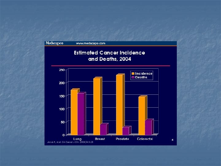
n In USA each year n Approximately 135, 000 new cases and 55, 000 death n Two thirds of these cases occur in colon and one thirds occur in rectum.
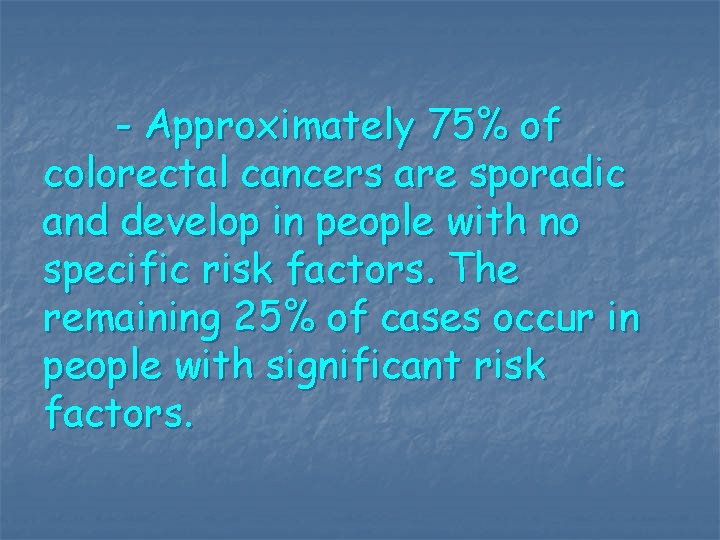
- Approximately 75% of colorectal cancers are sporadic and develop in people with no specific risk factors. The remaining 25% of cases occur in people with significant risk factors.
Race: - Western nations tend to have a higher incidence than Asian and African countries; however, within the United States, little difference in incidence exists among whites, African Ameicans, and Asian Americans.
- Among religious denominations, colorectal cancer occurs more frequently in the Jewish population.
Sex: The incidence of colorectal malignancy is slightly higher in males than in females. Age: Incidence peaks in the seventh decade; however, cases have been reported in young children
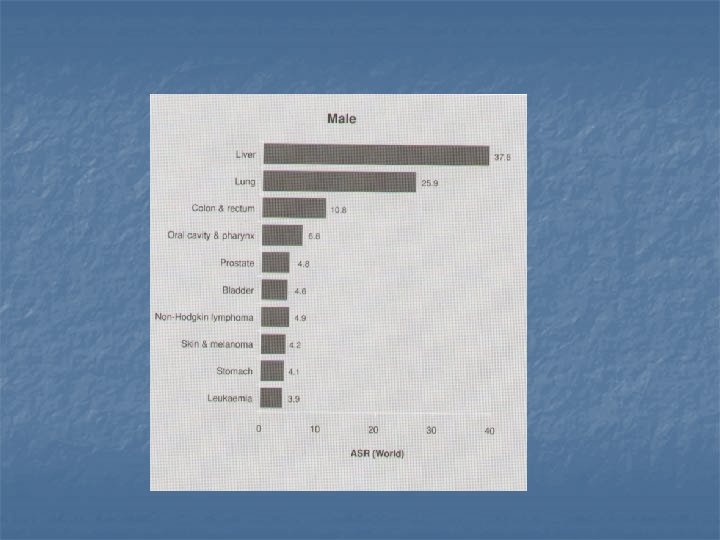
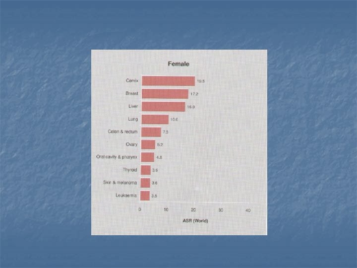
Incidence in Thailand
Clinical manifestation
History: - Bleeding - This is the most common symptom of rectal cancer and occurs in 60% of patients. - Bleeding often is attributed to other causes (eg, hemorrhoids), especially if the patient has a history of other problems.
- Profuse bleeding and anemia are rare. - Bleeding may be accompanie by the passage of mucus, which warrants further investigation.
- Occult bleeding: This is detected on screening fecal occult blood test (FOBT) in 26% of cases
- Change in bowel habits - Present in 43% of patients, this symptom has several different presentations. Often, it occurs in the form of diarrhea, particularly if the tumor has a large villous component.
- These patients may have hypokalemia on laboratory studies. - The capacity of the rectal reservoir may mask the presence of a small lesion. - Some patients experience a change in caliber of the stool.
- Large tumors can cause obstructive symptoms. - Tumors located low in the rectum can cause a feeling of incomplete evacuation and tenesmus.
-Abdominal pain - Partial large bowel obstruction may cause colicky abdominal pain and bloating , present in 20% of cases.
n - Back pain is usually a late sign caused by a tumor invading or compressing nerve trunks. - Urinary symptoms may occur if the tumor is invading or compressing the bladder or prostate.
- Malaise: This nonspecific entity is the presenting symptom in 9% of cases. - Bowel obstruction: Complete obstruction of the large bowel is rare and is the presenting symptom in 9% of cases.
- Pelvic pain: This late symptom usually indicates nerve trunk involvement and is present in 5% of cases. - Other presentations include emergencies such as peritonitis from perforation(3%) or jaundice, which may occur with liver metastases (<1%).
Physical: -Physical examination is performed with specific attention to possible metastatic lesions, including enlarged lymph nodes or hepatomegaly. The remainder of the colon is also examined.
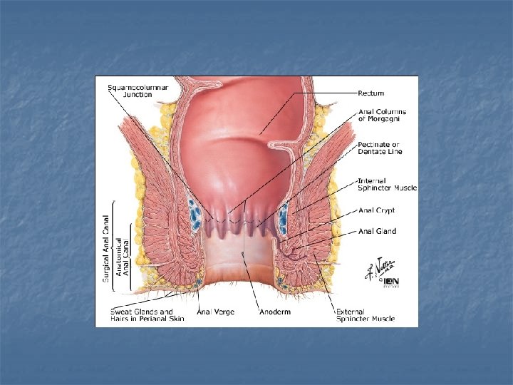
-Digital rectal examination - The easy accessibility of the rectum provides an opportunity to readily detect abnormal lesions via digital rectal examination (DRE). The average finger can reach approximately 8 cm above the dentate line.
- Tumors can be assessed for size, ulceration, and presence of any pararectal lymph nodes. Fixation of the tumor to surrounding strctures (eg. Sphincters, prostate, vagina) also can be assessed.
- DRE also permits a cursory evaluation of the patient’s sphincter function. This information is necessary when determining whether a patient is a candidate for a sphincter-sparing procedure.
IMAGING
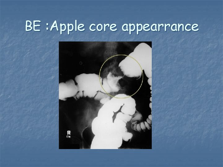
BE : Apple core appearrance
- Chest radiograph: Obtain a chest radiograph to rule out pulmonary metastases and to determine whether the patient has any gross underlying pulmonary disease, including emphysema.
- CT scan : This study is generally used to determine the presence or absence of metastases. - CT scans can identify lesions in the liver, adrenals, ovaries, lymph nodes, and other organs.
n - In 10% of patients, the CT scan misses small liver lesions. - When combined with an angiogram, a CT scan is 95% accurate in identifying liver metastases.
- Some information can be gleaned from a CT scan regarding depth of penetration of the primary rectal tumor. When performed with rectal contrast given as an enema, accurately in 84% of cases.
- CT scan detects lymph nodes larger than 1 cm in 75% of cases - CT scans are helpful in determining whether patients require preoperative chemoradiation therapy.
MRI actually is the most sensitive test for determining the presence of liver metastases and often is used if liver resection is considered.
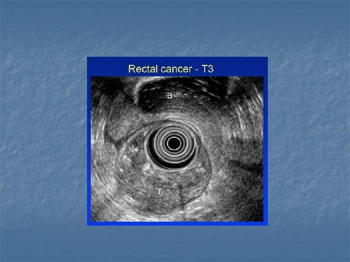
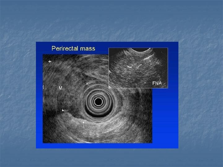
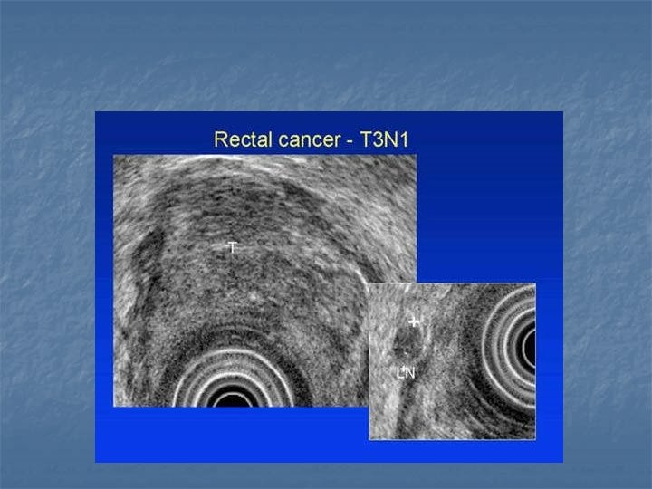
Endorectal ultrasound
- Endorectal ultrasound (ERUS) is an invaluable tool in assessing depth of invasion of rectal cancers; it is 72 -94% accurate.
- The accuracy of detection of lymph node involvement ranges from 7386%. n Most of these nodes are larger than 1 cm. Smaller nodes can be detected, but the accuracy of determining tumor involvement is substantially lower. n
- Positron emission tomography: The major advantage of a positron emission tomography (PET) scan is to differentiate between recurrent tumor and scar tissue by measuring tissue metabolism of an injected glucose-based substance. Scar tissue is inactive, whereas tumor generally is hypermetabolic. This test generally is not used in a routine preoperative metastatic workup.
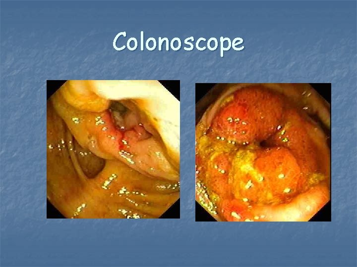
Colonoscope
Lab studies
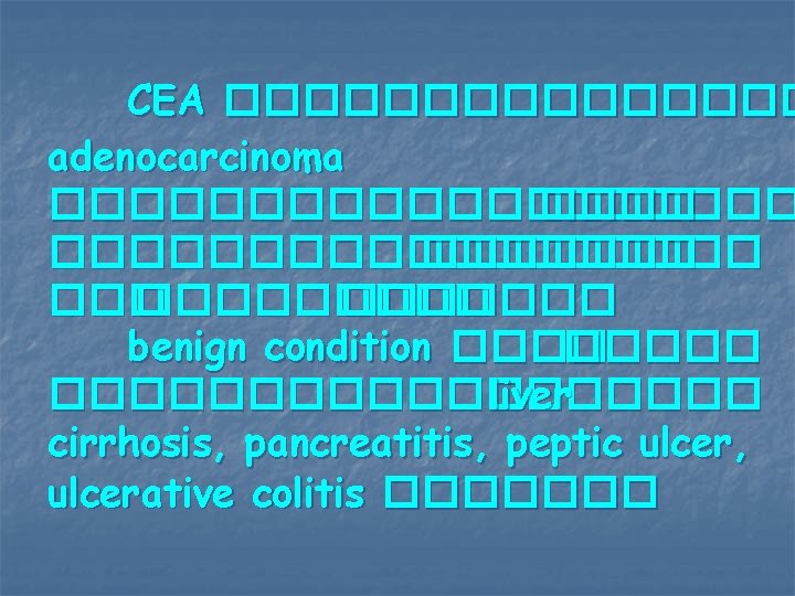
CEA �������� adenocarcinoma ����������� ������� ������� benign condition ��������� liver cirrhosis, pancreatitis, peptic ulcer, ulcerative colitis �������
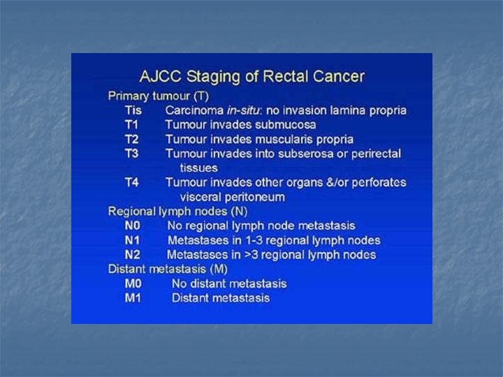
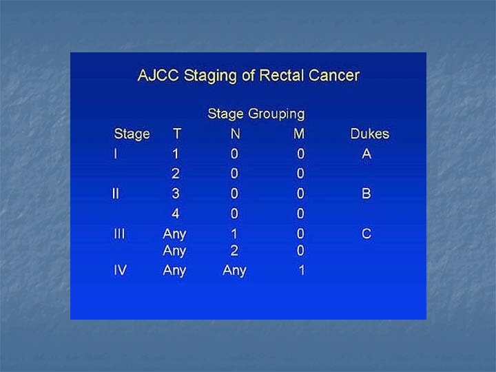
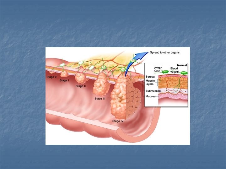
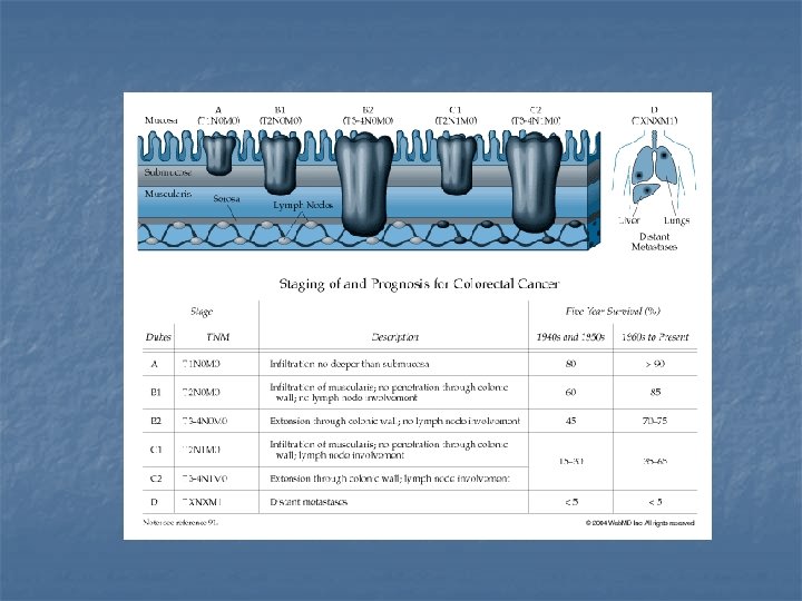
Staging
Treatment ��������� CRC ���� multimoda-lity approach ���������� chemotherapy, radiotherapy ���������
Surgical management of colon cancer
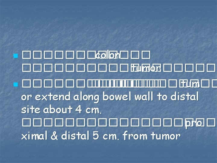
������ colon ��������� tumor n ���������� tum or extend along bowel wall to distal site about 4 cm. ���������� pro ximal & distal 5 cm. from tumor n
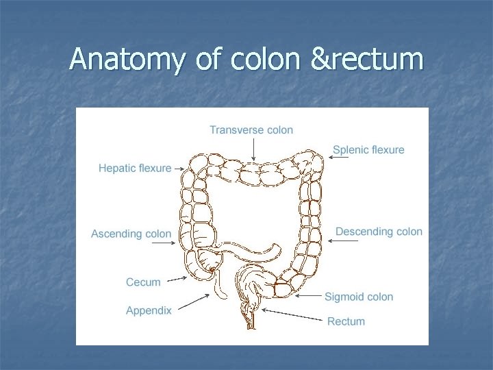
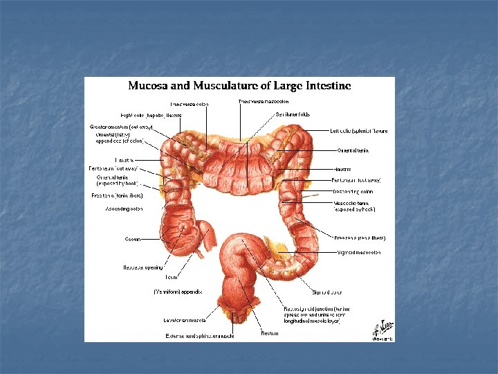
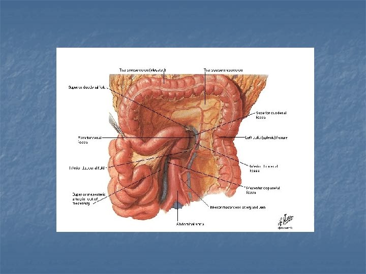
Anatomy of colon &rectum
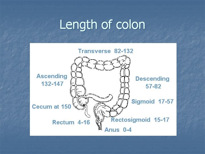
Length of colon
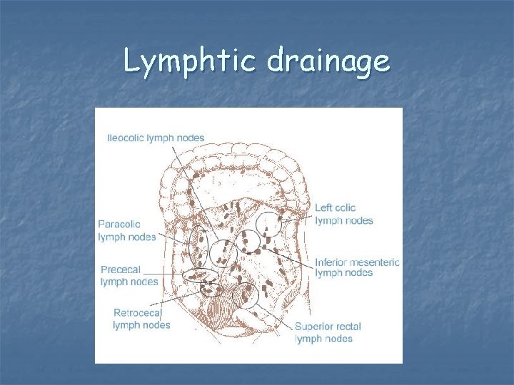
Lymphtic drainage
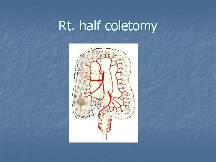
Rt. half coletomy
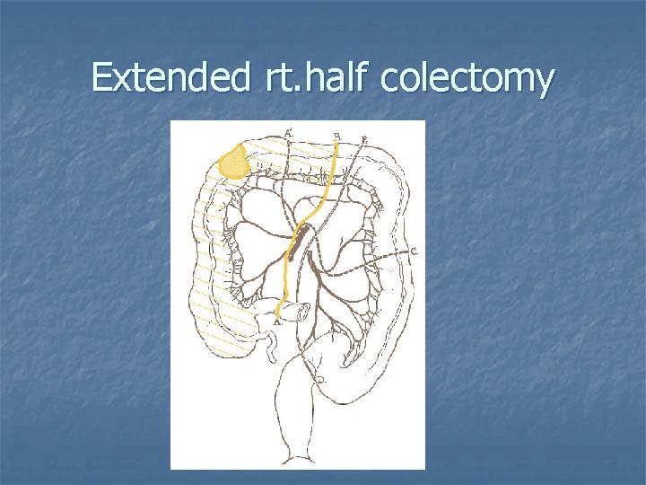
Extended rt. half colectomy
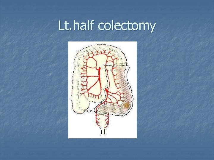
Lt. half colectomy
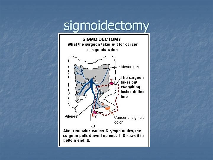
sigmoidectomy
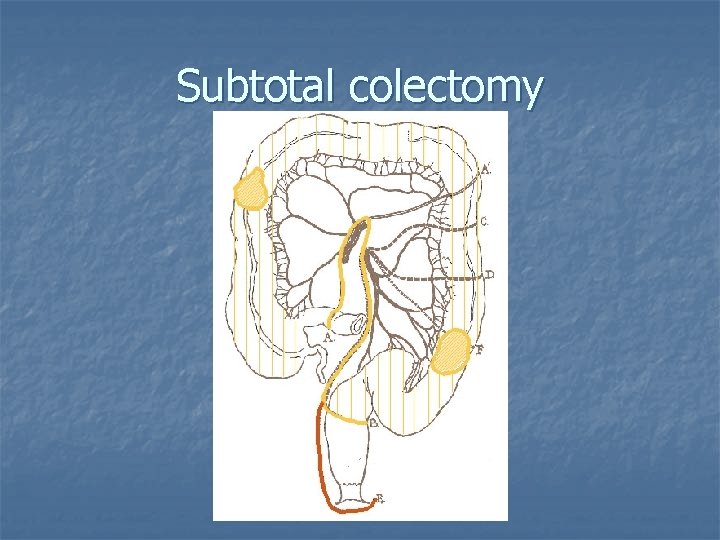
Subtotal colectomy
Surgical management of rectum
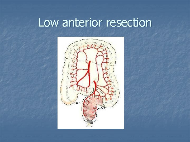
�������� local recurrence ������� 35 -45% ������ low anterior resection ������� lymph node ��������� ���abdominoperineal resection ������ inferior mesenteric artery ��� origin ��� aorta ��� vein ���� DJ junction
Low anterior resection
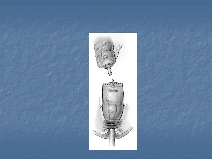
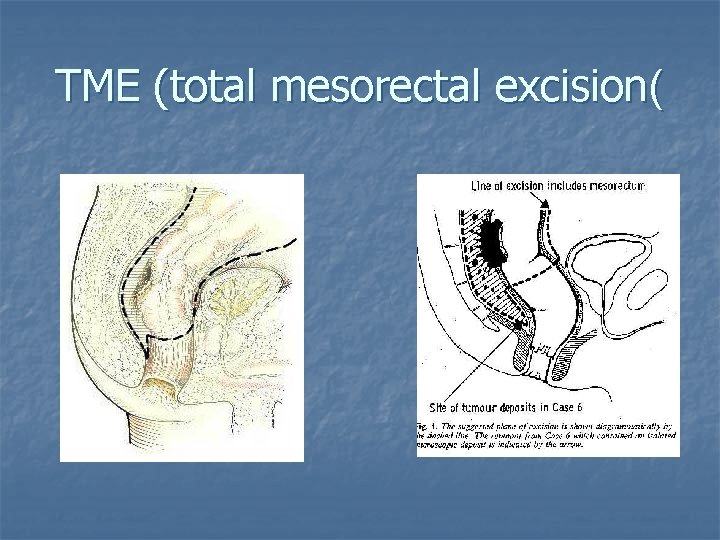
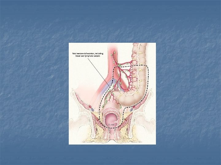
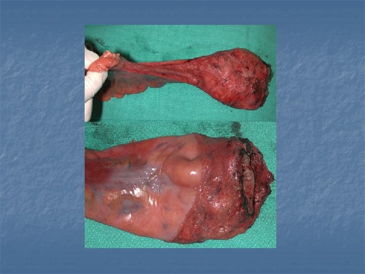
n TME involves sharp dissection in the avascular plane that is created by the envelope that separates the mesorectum from the surrounding structure. This includes the anterior peritoneal reflection and Denovillers fascia anteriorly and preserves the inferior hypogastric plexus posteriorly and laterally.
n TME yields a lower local recurrence rate (4%) than transanal excision (20%) , but it is associated with a higher rate of anastomic leak (11%).
TME (total mesorectal excision(
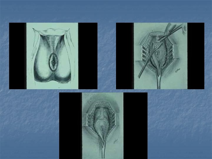
APR abdomino-perineum resection
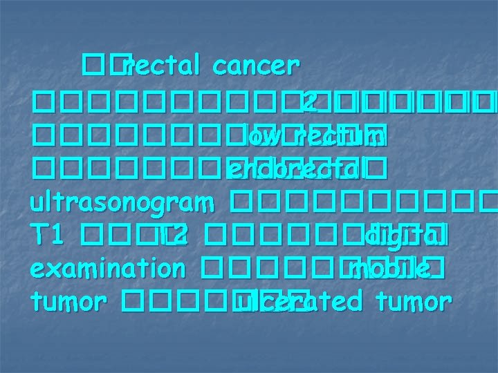
��rectal cancer ��������� 2 ������������� low rectum ������� endorectal ultrasonogram ����� T 1 ���� T 2 ����� digital examination ����� mobile tumor ������� ulcerated tumor
Adjuvant therapy
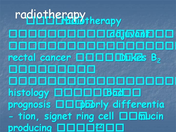
radiotherapy ��������� adjuvant ��������� rectal cancer ������� Dukes B 2 �������������� histology ����� bad prognosis ���� poorly differentia - tion, signet ring cell ��� mucin 22 producing �������
����������������� local recurrence ��� disease-free survival ����� preoperative radiation ��������� postoperative radiation ����� overall survival ��� cancer-specific survival ������������� sphinctorsaving surgery �����
������ radiation �������� standard ������ rectal cancer treatment ����
Chemotherapy �������� adjuvant chemo therapy ����� colon cancer stage lll (Dukes’ C) ������� rectal cancer stage ll B (Dukes’B 2 ) �������������
- ���� 5 FU ������� leucovorin ��regimen ������� Mayo regimen, AIO regimen - ��������� colon cancer stage ll ����� histology ��bad prognosis ���� perineural invasion, venous invasion, lymphatic invasion, poorly differentiation - ������ benefit ������� adjuvant chemotherapy ������ disease free survival ��� overall survival �����
Neoadjuvant chemoradiation for Rectal Carcinoma �������� chemoradiation ��tumor complete response ������ tumor ���������� toxicity �������
������ neoadjuvant chemoradiation ������ tumor ������ down stage ��� tumor ������� sphinctor preserving surgery ������� local recurrence ����� long term survival �����
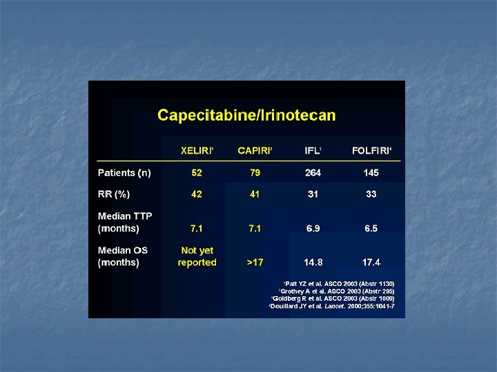
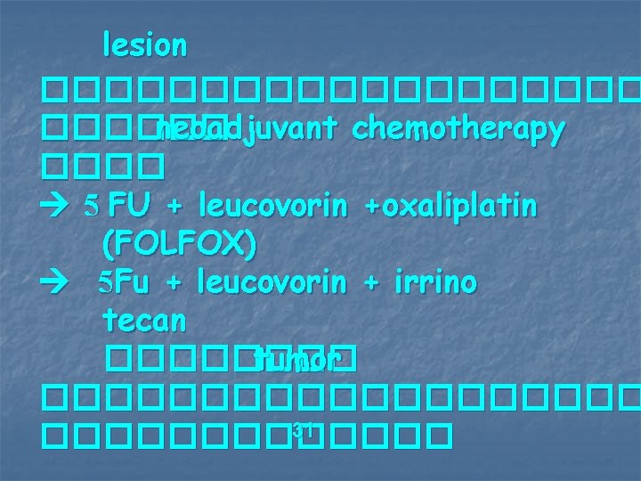
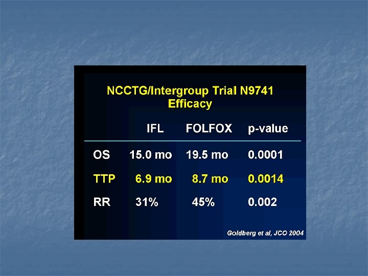
lesion ���������� neoadjuvant chemotherapy ���� 5 FU + leucovorin +oxaliplatin (FOLFOX) 5 Fu + leucovorin + irrino tecan ���� tumor ���������� 31 �������
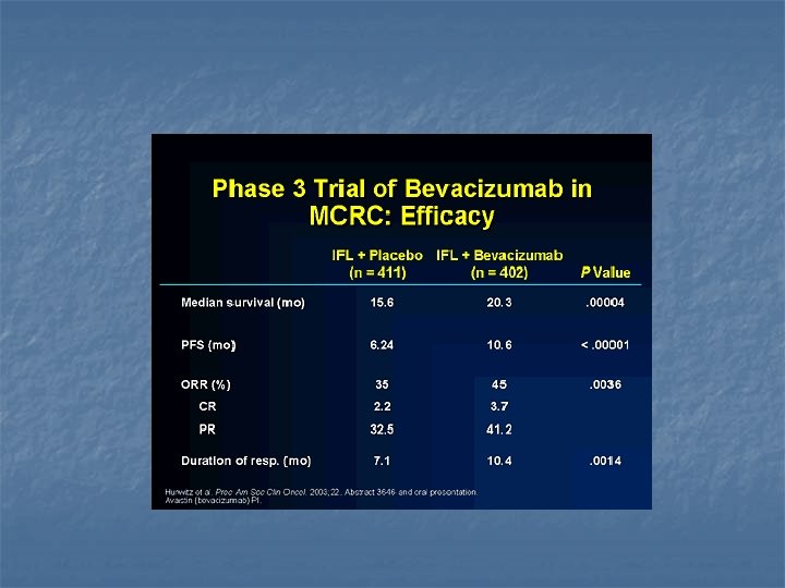
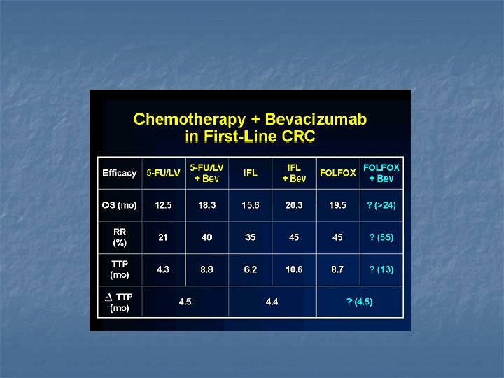
Biological therapy n Bavacizumab (AVASTIN) n Anti vascular endothelial growth factors receptor n IFL +bavacizumab
������� aspirin ��� NSAID ��������� cyclooxygenase enzyme ������ 32 risk ������� CRC �������� cyclooxygenase – 2 enzyme ������ inhibit cell apoptosis ������� angiogenesis
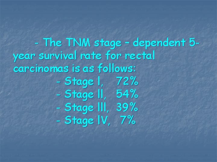
- The TNM stage – dependent 5 year survival rate for rectal carcinomas is as follows: - Stage l, 72% - Stage ll, 54% - Stage lll, 39% - Stage l. V, 7%