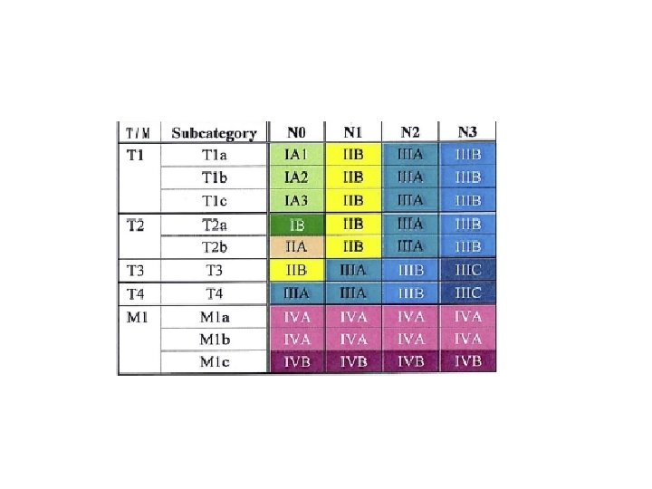
TNM staging system for lung cancer The Eighth

; 151(1): 193")

 Non small cell Adeno Squamous large cell")


")


 survival 24 months 60")


- Slides: 39
TNM staging system for lung cancer
The Eighth Edition Lung Cancer Stage Classification Frank C. Detterbeck CHEST (2017); 151(1): 193 -203
Purpose • Extent of disease • Assist in treatment decision • Prognostic indicator • Compare cohort, measure outcome
Lung cancer -features -treatment -outcome (80 -85%) Non small cell Adeno Squamous large cell (15 -20%) Small cell, Neuroendocrine tumour faster growth, more central/mediastinal, earlier distant mets, shorter survival • Clinical limited • Clinical extensive
Tumor N M • Extent of primary tumour 0 b 1 T 3 T 2 T 1 a (how big is it? ) a c 2 3 T 4 b 4 5 6 7 cm
Tumor N M • Extent of primary tumour b a 0 1 Surrounded by: -lung -visceral pleura -not involving main bronchus T 3 T 2 T 1 a c 2 3 T 4 b 4 5 6 7 cm
Tumor N M • Extent of primary tumour b a 0 1 Surrounded by: -lung -visceral pleura -not involving main bronchus T 3 T 2 T 1 a c 2 3 T 4 b 4 5 6 Involvement of: -main bronchus without carina regardless of distance from carina -atelectasis/post obstructive pneumonitis extending to hilum T 2 Centr -visceral pleura T 2 Visc Pl 7 cm
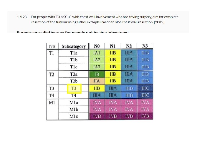
Tumor N M • Extent of primary tumour b a 0 1 Surrounded by: -lung -visceral pleura -not involving main bronchus T 3 T 2 T 1 a c 2 3 T 4 b 4 5 Involvement of: Any size -main bronchus without carina regardless oftumour distanceinvading: from carina -chest wall -atelectasis/post obstructive pneumonitis extending to hilum -pericardium -phrenic nerve -visceral pleura -satellite nodule in the same lobe 6 7 T 2 Centr T 2 Visc Pl T 3 Inv T 3 Satell cm
Tumor N M • Extent of primary tumour b a 0 1 Surrounded by: -lung -visceral pleura -not involving main bronchus T 3 T 2 T 1 a c 2 3 T 4 b 4 5 Involvement of: Any size -main bronchus without carina regardless oftumour distanceinvading: from carina -chest wall Any size tumour invading: -atelectasis/post obstructive pneumonitis extending to hilum -pericardium -mediastinum -phrenic nerve -diaphragm -visceral pleura -heart -great-satellite vessel nodule in the same lobe -recurrent laryngeal nerve -carina -trachea -oesophagus -spine/vertebral bodies 6 cm 7 T 2 Centr T 2 Visc Pl -tumour nodule/seperate tumour in different lobe of ipsilateral lung T 3 Inv T 3 Satell T 4 Inv T 4 Ipsi Nod
T Node M • Lymph node involvement (mets to which nodes? )
T Node M • Lymph node involvement N 0 N 1 N 2 N 3 No regional node metastases ipsilateral pulmonary and/or hilar nodes ipsilateral mediastinal and/or subcarinal nodes contralateral mediastinal, contralateral hilar or any supraclavicular and scalene nodes
IASLC Lymph node map Rusch V, Asamura H, Watanabe H, et al. The IASLC Lung Cancer Staging Project: a proposal for a new international lymph node map in the forthcoming 7 th edition of the TNM classification for lung cancer. J Thorac Oncol. 2009; 4(5): 568 -577
T Node M Tumour right lung
T Node M Tumour left lung
T N Metastastic disease M 0 no distant metastases M 1 distant metastases: • M 1 a malignant pleural/pericardial effusion/nodules contralateral or bilateral pulmonary nodules • • M 1 b M 1 c single extrathoracic metastases multiple extrathoracic metastases either in a single organ or multiple organ
2. 5 cm right lower lobe mass ipsilateral hilar nodes no distant metastases. What stage of disease does this patient have?
2. 5 cm right lower lobe mass ipsilateral hilar nodes no distant metastases. What stage of disease does this patient have? T 1 c N 1 M 0
66 year old female presented to GP with chest pain. She smokes 10 cigarettes daily since her teenage years giving an average of 25 pack years. She denies haemoptysis and tells you that her weight loss is intentional due to a combination of good diet and exercise. She is currently working as a cleaner in a clothing factory. A chest radiograph showed right side opacity and subsequent CT imaging confirmed 2. 5 cm spiculated right lower lobe mass extending to chest wall. There was pathological enlargement of ipsilateral hilar nodes but no distant metastases. CT guided biopsy confirmed lung adenocarcinoma. What stage of disease does this patient have? 1. 2. 3. 4. 5. T 1 c N 1 M 0 T 1 c N 2 M 0 T 2 a N 1 M 0 T 3 N 1 M 0 T 4 N 2 M 0
66 year old female presented to GP with chest pain. She smokes 10 cigarettes daily since her teenage years giving an average of 25 pack years. She denies haemoptysis and tells you that her weight loss is intentional due to a combination of good diet and exercise. She is currently working as a cleaner in a clothing factory. A chest radiograph showed right side opacity and subsequent CT imaging confirmed 2. 5 cm spiculated right lower lobe mass extending to chest wall. There was pathological enlargement of ipsilateral hilar nodes but no distant metastases. CT guided biopsy confirmed lung adenocarcinoma. What stage of disease does this patient have? 1. 2. 3. 4. 5. T 1 c N 1 M 0 T 1 c N 2 M 0 T 2 a N 1 M 0 T 3 N 1 M 0 T 4 N 2 M 0 N 1: ipsilateral hilar, peribronchial and or intrapulmonary node T 2 a: visceral pleura
Lung cancer stage group 1
Lung cancer stage group 1 Stage Median Overall survival (%) survival 24 months 60 months time (m) IA 1 - 97 92 IA 2 - 94 83 IA 3 - 90 77 IB - 87 68 IIA - 79 60 IIB 66 72 53 IIIA 29. 3 55 36 IIIB 19 44 26 IIIC 12. 6 24 13 IVA 11. 5 23 10 IVB 6 10 0 1. Goldstraw. The IASLC Lung Cancer Staging Project: Proposals for Revision of the TNM Stage Groupings in the Forthcoming (8 th) Edition of the TNM Classification for Lung Cancer. J Thorac Oncol 2016; 11: 39
5 cm mass RLL malignant nodule RUL bilateral enlarged hilar lymph node staging? 1. 2. 3. 4. 5. II B III A III B III C IV A
5 cm mass RLL malignant nodule RUL bilateral enlarged hilar lymph node staging? 1. 2. 3. 4. 5. II B III A III B III C IV A • • T 4 -ipsilateral nodule Bil hilar- N 3 No mets =T 4 N 3 M 0
Regardless of cancer stage Curative surgery • Stop smoking intervention ₋ Nicotine replacement ₋ varenicline • ECOG PS • FEV 1, TLCO • Shuttle walk test, CPEX
66 year old female recently diagnosed adenocarcinoma of the lung. Staging is T 2 N 1 M 0 tumour of the right upper lobe confirmed on PET CT. Patient is otherwise fit and well ECOG PS of 1 Which of the following would be the most appropriate initial choice of treatment for this patient? 1. 2. 3. 4. 5. Chemotherapy Lobectomy Pneumonectomy Radiotherapy Wedge resection
66 year old female recently diagnosed adenocarcinoma of the lung. Staging is T 2 N 1 M 0 tumour of the right upper lobe confirmed on PET CT. Patient is otherwise fit and well ECOG PS of 1 Which of the following would be the most appropriate initial choice of treatment for this patient? 1. 2. 3. 4. 5. Chemotherapy Lobectomy Pneumonectomy Radiotherapy Wedge resection • NSCLC • Curative intentlobectomy • Lung sparing (wedge) – Complete resection can be achieved – Small tumours – Borderline fitness levels
• • • EGFR-TK mutation ALK gene rearrangement PDL 1>50% ROS 1 positive No gene mutation or fusion protein or PD-L 1<50% • • Afatinib, erlotinib Ceritinib, alectinib Pemrolizumab, gemcitabine crizotinib
48 year old man attends clinic. He is receiving treatment with crizotinib. What is the mechanism of action for this drug? 1. 2. 3. 4. 5. Checkpoint inhibitor K-RAS inhibitor Monoclonal antibody PDL-1 inhibitor Tyrosine kinase inhibitor
48 year old man attends clinic. He is receiving treatment with crizotinib. What is the mechanism of action for this drug? 1. 2. 3. 4. 5. Checkpoint inhibitor K-RAS inhibitor Monoclonal antibody PDL-1 inhibitor Tyrosine kinase inhibitor