Airway Stenosis Evaluation and Endoscopic Management Murtaza Ghadiali


 n n either cuffed endotracheal or tracheotomy tube")


, secondary (8%),")


, orbit (15 -25%), splenomegaly")


: Investigated histopathologic features of 6 specimens from")

 n Bartels 2002: 108")
: Reported case of complete suprastomal tracheal")
: examined pathophysiology")
/Laryngopharyngeal reflux (LPR) n 1985 – Little")

: Challenged association; largest review of 50")

 Possible hormonal cause n To date, presence of estrogen receptors")


 n Mc. Caffrey (1992) n")


 n Relative reduction in cross sectional area")
 involved")


 n Stenosis induced by resection of anterior")
 n Stents well tolerated, minimal signs of airway irritation, no")


 n Retrospective study of T-tubes in 12 pediatric patients n 10")
 n mean time from insertion to final removal 5. 6 months")
 n 75% success rate of long term stenting comparable to either")

 Strong and Jako (1972) n n")


 Retrospective study of 60 patients: 49 laryngeal (supraglottic,")
 n 39/60 had Silastic stents placed n 1/6")
 n Dilatation employed 8/60 n 0/49 laryngeal n")

: Conclusions n Justified at all levels n Decreased")


 n Retrospective cohort study n Compare efficacy of 3")
 n")


 Pilot Study in Modified Canine Model 8 subjects")


 6 patients with IPSS (single")
 Performed flexible bronchoscopy for")


- Slides: 104
Airway Stenosis: Evaluation and Endoscopic Management Murtaza Ghadiali, M. D. UCLA Division of Head and Neck Surgery November 19 th, 2008
Outline n n Introduction Etiology n Acquired Causes n Role of LPR n IPSS Evaluation n H&P, Grading, DL/B Endoscopic Management n Mitomycin n TGF-β n Lasers/Balloon Dilation n Autoimmune Causes
Introduction Airway Stenosis is both a therapeutic and diagnostic challenge n Presents insidiously with progressive SOB, brassy cough, wheezing/stridor, possible recurrent pneumonitis n Many times misdiagnosed as asthma/bronchitis, COPD, CHF n
Introduction n Common etiology (beginning 1965) n n either cuffed endotracheal or tracheotomy tube Less common: external trauma/compression n high tracheotomy incision n benign tumors n ‘nontraumatic, nonneoplastic’ causes n
Etiology of SGS n I. Congenital SGS n n n Membranous Cartilaginous II. Acquired SGS n n n n Intubation Laryngeal trauma AI (Wegener’s; Sarcoid; Amyloid; Relapsing Polychondritis) Infection IPSS (Idiopatic Subglottic Stenosis) GER/LPR Inflammatory diseases Neoplasms
Nonneoplastic, nontraumatic Subglottic Stenosis Wegener’s Granulomatosis n Amyloidosis n n n Sarcoidosis n n Can present with SG alone Relapsing Polychondritis
SGS – Wegener’s n n n Systemic inflammatory disorder Autoimmune ANCA C + 16 -23% incidence of SG stenosis SGS can be the lone manifestation of WG n Treatment n n Individualized based on degree and acuity of stenosis No major surgery during Wegener’s flare ups
Wegener’s Granulomatosis n Classic triad: necrotizing granulomas of the upper respiratory tract and lungs, focal glomerulitis, disseminating vasculitis n n Treatment: Azathioprine, cyclophosphamide, steroids Laryngeal WG n n Ulcerating lesions induce subglottic stenosis Histopathology: coagulation necrosis from vasculitis, multinucleated giant cells, palisading histiocytes
Amyloidosis n Deposition of extracellular fibrillar proteins in tissues n Primary (56%), secondary (8%), localized (9%), myeloma associated (26%), familial (1%) n Generalized amyloid evaluated by rectal biopsy or FNA anterior abdominal wall fat n Locations n n Tongue > orbit > larynx Laryngeal amyloidosis n n TVC > FVC > subglottic Management n Surgical
Amyloidosis n Diagnosis Congo red staining and green birefringence under polarized light n Fibrillar structure under electron microscopy n Beta-pleated sheet on x-ray crystallography and infrared spectroscopy n n 18 biochemical forms identified n AL (plasma cells), AA (chronic inflammation), Ab (cerebral lesions)
Amyloidosis – Management Step 1 Biopsy the affected organ n Step 2 Rule out generalized amyloidosis n n n Rectal bx, echocardiography, bronchoscopy and PFTs, CT of neck/trachea Step 3 Rule out generalized plasmacytoma n Bone marrow biopsy, bone marrow scintigraphy, serologic and immunologic examininations
Laryngeal Amyloidosis < 1% of benign laryngeal lesions n Most amyloid deposits are AL type n Typically in men in the 5 th decade of life n Sx depends on site (e. g. glottic amyloidosis hoarseness) n
Sarcoidosis n Idiopathic, non-caseating granulomas n Generalized adenopathy (25 -50%), orbit (15 -25%), splenomegaly (10%), neural (4 -6%) n Symptoms: fever, weight loss, arthralgias n Head and neck: cervical adenopathy > larynx n Evaluation: CXR, PPD, skin test for anergy, ACE levels (elevated in 80 -90%) n Treatment n Oral steroids n Laryngeal sarcoidosis n Supraglottic involvement n Typical yellow subcutaneous nodules or polyps n Diffusely enlarged, pale pink, turban-like epiglottis
Relapsing Polychondritis n Inflammation of cartilage and other tissues with high concentration of glycosaminoglycans n Episodic and progressive n Ear > nasal, ocular, respiratory tract n Treatment: symptomatic, steroids n Laryngeal RP n Rare n Inflammation can lead to laryngeal collapse n Treatment usually tracheostomy
Acquired SGS 95% of cases of SGS n Majority due to long-term or prior intubation n Duration of intubation n ETT size n Number of intubations n Traumatic intubations n Movement of the ETT n Infection n
n Poetker DM et al. Association of airway abnormalities and risk factors in 37 subglottic stenosis patients. Otolaryngol Head Neck Surg (2006) 135, 434 -437
Pathogenesis of acquired SGS Initial injury – compression of mucosa by an ETT or cuff n Ischemia n Necrosis n Decreased mucociliary flow n Infection n Three stages of wound healing n n Inflammatory n Proliferative – granulation tissue n Scar formation – contraction and remodeling
Pathogenesis SGS n Mankarious et al (2003): Investigated histopathologic features of 6 specimens from pts that underwent tracheal resection Analyzed levels of hyaline cartilage components: collagen type I and II & aggrecan (secreted by chondrocytes) n Normal tracheal/cricoid: High ratio of type I to II n Specimens: relative decrease in type I and aggrecan n n Regenerative cartilage: greatly increased amounts of type II collagen and aggrecan n Suggests Type I collagen and aggrecan responsibe for cartilage structural integrity n Regenerative fibroblasts do not deposit type I collagen
Acquired SGS and PDT Ciaglia 1985: Percutaneous dilational tracheotomy (PDT) n Bartels 2002: 108 PDT patients; 10 with 6 mo f/u; 1 patient with significant stenosis at f/u n ? Selection Bias n Authors conclude 10% stenosis rate is consistent with open tracheotomy n
Acquired SGS and PDT Klussman et al (2001): Reported case of complete suprastomal tracheal stenosis/atresia after second PDT n ? Initial infection leading to destruction and cartilaginous necrosis/Tracheal ring fracture leading to mucosal tears and cicatricial scarring n Cautioned against use of PDT in secondary tracheotomy n
Acquired SGS and PDT n n n Hotchkiss & Mc. Caffrey (2003): examined pathophysiology of PDT on 6 cadavers 3/6 Trachs were placed incorrectly (range: 3 tracheal rings away to just sub-cricoid) Anterior tracheal wall n n High degree of injury Severe cartilage damage at site of insertion Multiple, comminuted injuries in 2 or more cartilaginous rings Findings suggest acute, severe mechanical injury in PDT
Acquired SGS & LPR n Gastroesophageal reflux (GER)/Laryngopharyngeal reflux (LPR) n 1985 – Little – applied gastric contents/H 2 O to subglottis of dogs n Delayed epithelialization and stenosis formation in lesions treated with gastric contents n 1991 – Koufman – applied acid and pepsin to subglottis of dogs; control was H 2 O n 20 dogs with induced submucosal injury n Increased level of granulation tissue and inflammation n 78% pts with LTS: pharynx reflux abnormal acidic p. H probes; 67%
GER/LPR and SGS 1998 Walner: 74 pediatric patients with SGS had 3 times greater incidence of GER than the general pediatric population n 2001 Maronian: 19 pts with SGS n 9 pts with IPSS; 10 with acquired SGS n 14 pts with p. H testing n n Abnormal (p. H <4): pts 71% IPSS pts and 100% acquired
GER/LPR and SGS n n n Dedo (2001): Challenged association; largest review of 50 pts with IPSS; Only 7/38 patients had reflux symptoms Ashiku (2004): 15/73 IPSS patients had reflux symptoms; No patients had laryngeal signs of reflux Both groups concluded no causal relationship between reflux and stenosis in their groups n Only 2 patients in collective cohorts underwent specific reflux testing
Idiopathic Subglottic Stenosis n n n Rare condition of dense fibrous stenosis of the proximal trachea in absence of inciting event Affects women; primarily involves subglottic larynx and proximal 2 -4 cm of trachea circumferentially May be associated with certain autoimmune states n n Wegener’s Granulomatosis Relapsing Polychondritis Rheumatoid Arthritis SLE Ashiku SK et al. Idiopathic laryngotracheal stenosis. Chest Surg Clin North Am, 2003; 13: 257
IPSS (Idiopathic Subglottic Stenosis) Possible hormonal cause n To date, presence of estrogen receptors in the affected airway has not been conclusively shown in these patients ( Dedo 2001) n ? Possible link between female preponderance and LPR n Progesterone and its impact on LES pressure n Major contributing factor toward heartburn and reflux in pregnancy n Cyclic hormonal variations in normal women found to impact LES pressure leading to possible reflux n
SGS Initial presentation n History of prior intubation and n Progressive SOB and loud breathing
Initial Presentation n History Review intubation records n Pmhx n n Diabetes n Cardiopulmonary disease n Reflux n Systemic steroid use
Initial presentation n Physical exam – Complete H/N exam n Observe n Stridor or labored breathing n Retractions n Breathing characteristics on exertion n Voice quality n Head/Neck n Other abnormalities (congenital anomalies, tumors, infection)
Diagnosis n Differential n Congenital n Laryngeomalacia n Tracheomalcia n VC paralysis n Cysts n Clefts n Vascular compression n Mass
Diagnosis n Differential n Infection/Inflammation n Epiglottitis n GER n Tracheitis n Neoplastic n Malignancy n Recurrent respiratory n Foreign body papillomas; benign lesions
Diagnosis n Radiographs Plain films – inspiratory and expiratory neck and chest n CT n MRI n
Diagnosis n Flexible nasopharyngolaryngoscopy n Nose/Nasopharynx n NP stenosis n Masses, tumor n Supraglottis n Structure abnormalities n Laryngomalacia n Glottis n VC mobility n Webs/masses n Immediate subglottis
Diagnosis n Gold standard for diagnosis of SGS n Rigid endoscopy n Properly equipped OR n Experienced anesthesiologist n Preop discussion about possible need for trach
Operative Evaluation n Endoscopy n n n Perform Rigid DL, B, and E n n n Fiberoptic endoscopic assisted intubation vs. evaluation LMA Spontaneous ventilation, NO PARALYSIS ! Consider awake tracheotomy Closely evaluate the interarytenoid area for stenosis/stricture Evaluate position of cords Determine size, extent, and location of the stenotic lesion n Use an ETT/bronchoscope to measure the lumen Measure from undersurface of the cord to the lesion R/o other stenotic areas
Grading Systems for SGS Cotton-Myer (1994) n Mc. Caffrey (1992) n
Cotton-Myer Grading System
n Cotton-Myer grading system for subglottic stenosis
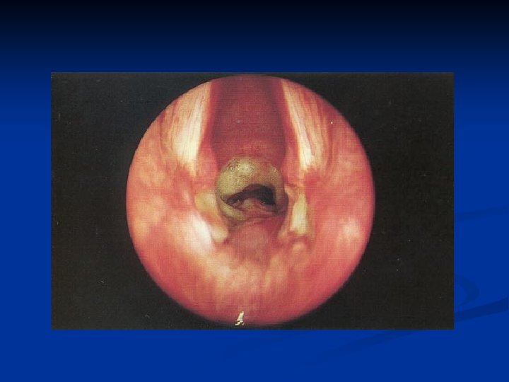
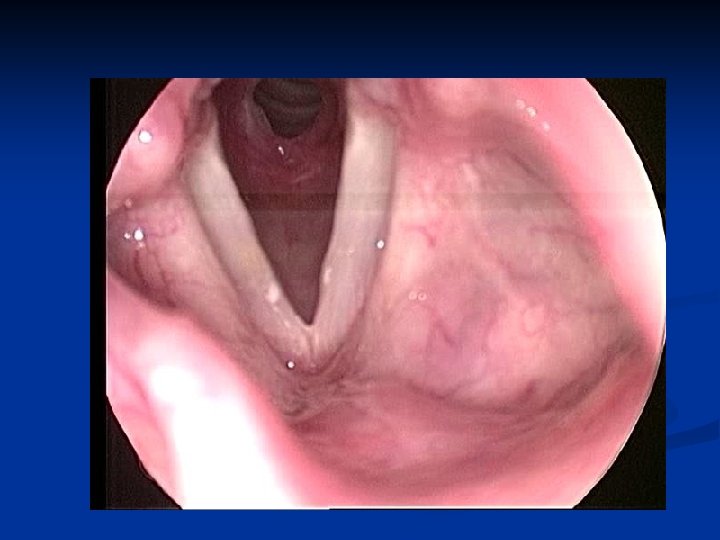
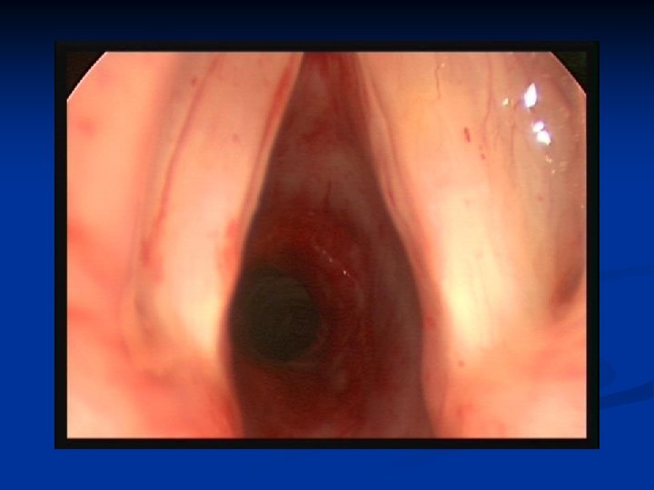
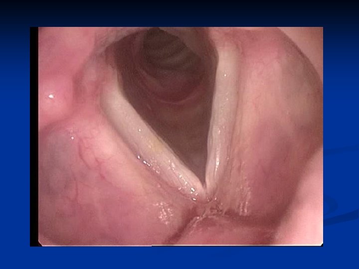
Grade II SGS
Grade III SGS
Grade IV SGS
Myer/Cotton Grading System Multiple revision of original system proposed by Cotton in 1984 n First systems criticized for being based on subjective interpretation, although statistically proven to relate grade with prognosis in children n Myer 1994: used serial ETT measurement to derive Cotton grade n
Grading Systems for SGS n Cotton-Myer Based on relative reduction of subglottic crosssectional area n Good for mature, firm, circumferential lesions n Does not take into account extension to other subsites or length of stenosis n Gold-Standard Staging in pediatric patients n
Mc. Caffrey Grading System Mc. Caffrey (1991) n Relative reduction in cross sectional area not consistently reliable predictor of decannulation in adults n Reviewed 73 cases of LTS in adults finding location of stenosis to be the most significant factor in predicting decannulation
Grading Systems for SGS n Mc. Caffrey Based on subsites (trachea, subglottis, glottis) involved and length of stenosis n Does not include lumen diameter n
Mc. Caffrey Clinical Staging n n Stage I: confined to subglottis/trachea Stage II: SGS, >1 cm, confined to cricoid Stage III: SGS and involving trachea Stage IV: involve glottis with fixation TVC
Grading Systems for SGS n Mc. Caffrey
Mc. Caffrey Conclusions Site: glottic, tracheal, subglottic: major factor in type of surgery thin (<1 cm) subglottic or tracheal lesions-Endoscopic n thick(>1 cm) any site or glottic lesions--Open n Stage: prognostic predictor 90% of Stage I and II successfully treated n 70% of Stage III, 40% of Stage IV n
Management of SGS Medical n Observation n Tracheotomy n Endoscopic Treatment n CO 2 laser (with Mitomycin C/Steroid) n Rigid vs. Balloon Dilation (with Mitomycin) n n Open Airway expansion procedure
Management of SGS n Medical Diagnosis and treatment of GER n Pediatric – consultation with primary physician and specialists (pulmonary, GI, cardiology etc. ) n Adult n n Assess general medical status n Consultation with PCP and specialists n Optimize cardiac and pulmonary function n Control diabetes n Discontinue steroid use if possible before LTR
Management of SGS n Observation n Reasonable in mild cases, esp. congenital SGS (Cotton-Myer grade I and mild grade II) n If no retractions, feeding difficulties, or episodes of croup requiring hospitalization n Follow growth curves n Repeat endoscopy q 3 -6 mo n Adults – depends on symptoms
Surgery for SGS n I. Endoscopic n Dilation +/- stenting n Rigid vs. balloon dilation n n Laser +/- stenting II. Open procedure n Expansion procedure (with trach and stent or SSLTR) n Laryngotracheoplasty (Trough technique with mucosal grafting +/- cartilage grafting) n Laryngotracheal reconstruction n Tracheal Resection with primary anastamosis
Management of SGS n How do you decide which procedure to perform n Status of the patient n Any contraindications n Absolute n Tracheotomy dependent (aspiration, severe BPD) n Severe GER refractive to surgical and medical therapy n Relative n Diabetes n Steroid use n Cardiac, renal or pulmonary disease
Management of SGS n Endoscopic n Dilation n Practiced frequently before advent of open LTP procedures n Often requires multiple repeat procedures n Potentially lower success rate but an option for patients who cannot undergo open procedures
Treatment Options Goals 1. Maintain patent airway 2. Maintain glottic competence to protect against aspiration 3. Maintain acceptable voice
Surgical Management Approaches n Endoscopic: cryotherapy, microcauterization, laser incision or excision of scar tissue, dilatation, stenting n Open surgical: tracheal resection and reanastomosis, external tracheoplasty with/without grafting and possible stenting
Stents n Indwelling expandable stents n n Used in many organ systems: arteries, the urethra, and biliary tree Tracheobronchial system: Lower airways for either tumors, or bronchial stenosis after lung transplantation n Upper airways (Montgomery T-tube, silicone, mesh stents): used alone or with other modalities n
Stents n Stenting Ensure adequate airway during wound maturation n While waiting for pt’s condition to improve prior to definitive surgical resection/treatment n Silastic T-Tubes most commonly used n n Permit better hygiene n Not prone to obstructing granulation n Stent removal possible after 1 -2 years with good results
Expandable Stent Hanna 1997 n Canine model(6) n Stenosis induced by resection of anterior cricoid arch/tracheal wall to reduce airway diameter by 50% n 8 week stenosis maturation period n Tracheostomy performed, followed by introduction of titanium mesh stent (Group A), +/- silicone covering (Group B) n Euthanasia performed at 4 weeks with gross/histologic exam
Expandable Stent Hanna (1997) n Stents well tolerated, minimal signs of airway irritation, no infections n Group A unable to be decannulated due to granulation n Group B all tolerated decannulation without complication
Expandable Stent
Silastic T-Tubes
T-Tubes
Stents Froehlich (1993) n Retrospective study of T-tubes in 12 pediatric patients n 10 acquired after intubation, 2 congenital, (4 extensive tracheomalacia) n 10 with prior tracheotomy n 5 Cotton grade 2, 7 Cotton grade 3 (6 required anterior split to fit T-tube)
Stents Froehlich (1993) n mean time from insertion to final removal 5. 6 months n 9/12 successful tx (mean time from dx to end of tx 15. 3 months) n Complications: tube migration, accidental tube removal, tube occlusion
Stents Froehlich (1993) n 75% success rate of long term stenting comparable to either cricoid split or LTR procedures n stenting takes longer, increased complications n T-tube stenting better reserved for cases not amenable to surgery, i. e. tracheomalacia
Endoscopic Approach Benefits patients due to less morbidity n Shorter hospital stay n Earlier return to work n Tolerance of repeated procedures, if necessary n
“Lasers” n n First medical use (December 1961) Strong and Jako (1972) n n First described CO 2 laser for LTS management Types: n n n CO 2 KTP Nd-YAG
Lasers n Used as both definitive and as an adjunct to open repair n Hall (1971) delayed collagen synthesis in laser incisions n Used in conjuncture with other epithelial preserving techniques
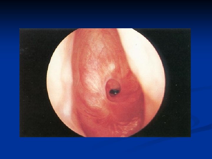
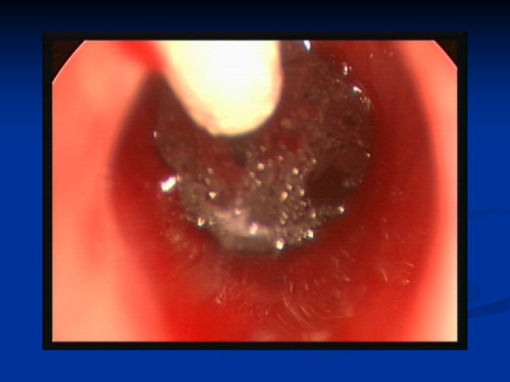
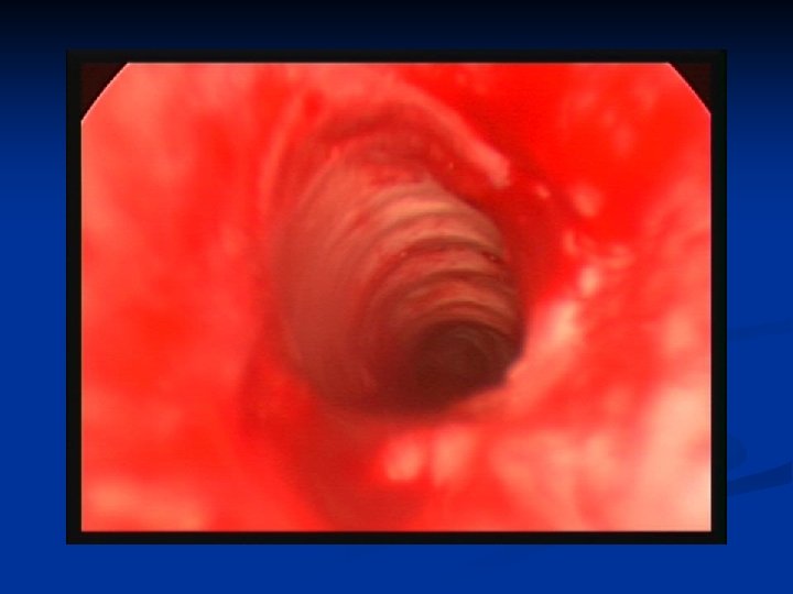
Laser excision of subglottic stenosis
Laser excision of subglottic stenosis
Endoscopic Approach Simpson, et al (1982) Retrospective study of 60 patients: 49 laryngeal (supraglottic, subglottic), 6 tracheal, 5 combined stenosis n Follow up: 1 -8 years n Age: 2 months-72 years old n CO 2 laser used to vaporize scar tissue, divide fibrotic bands, or excise redundant tissue n +/- Silastic stenting, dilatation n
Endoscopic Approach Simpson, et al (1982) n 39/60 had Silastic stents placed n 1/6 supraglottic n 2/12 glottic n 27/31 subglottic stenosis n 4/6 tracheal n 4/5 combined
Endoscopic Approach Simpson, et al (1982) n Dilatation employed 8/60 n 0/49 laryngeal n 4/6 tracheal n 4/5 combined
Endoscopic Approach
Endoscopic Approach Simpson, et al (1982): Conclusions n Justified at all levels n Decreased success with ‘severe’, combined, extensive (>1 cm) or circumferential stenosis; loss of cartilage, and preceding bacterial infection associated with tracheostomy n Age not associated with failure rate
Management of SGS n Endoscopic n Laser n 66 -80% success rate for Cotton-Myer grade I and II stenoses (pediatric cases) n Closer to 50% success rate in appropriately chosen adults n Factors associated with failure n Previous attempts n Circumferential scarring n Loss of cartilage support n Exposure of cartilage n Arytenoid fixation n Combined laryngotracheal stenosis with vertical length >1 cm
Scar Inhibitors n Mitomycin C Antimetabolite of Streptomyces caespitosus n Possesses antineoplastic and antiproliferative properties n Inhibits fibroblast proliferation in vivo and in vitro n Mechanism may involve triggering of fibroblast apoptosis n n 5 -FU & B-aminopropionitrile n n Inhibit collagen cross-linking and scar formation in animal models TGF-β
SGS Comparison Study Shapshay (2004) n Retrospective cohort study n Compare efficacy of 3 endoscopic techniques n CO 2 laser with rigid dilation n CO 2 laser, rigid dilation, steroid injection n CO 2 laser, rigid dilation, topical Mitomycin C application n
SGS Comparison Study n Endoscopic treatment n CO 2 laser radial incision (Shapshay) n n CO 2 laser with steroid injection n 15% success 40 Kenalog in 3 quadrants 18% success CO 2 laser with mitomycin-C topical application n n 0. 4 mg/ml Mitomycin-C topically applied 4 minutes 75% success
Mitomycin C Metanalysis Note: Lone human dissenting study was highest quality randomized clinical trial Warner and Brietzke (2008)
TGF-β: GF secreted by fibroblasts, macrophages and platelets n Implicated in scarring in many different organ systems and in animal models n Biopsy specimens of IPSS and intubation related stenosis patients show high levels of TGF-β-2 n IV and local injection of an antibody available n Used to treat fibrosis in skin, ureters, kidney and eye n Recent study showed inhibition of scarring in rat trachea with continuous infusion of anti-TGFβ n
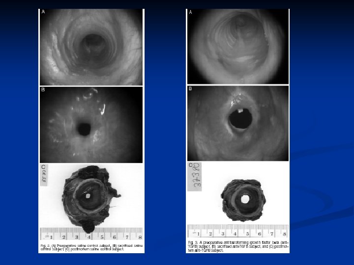
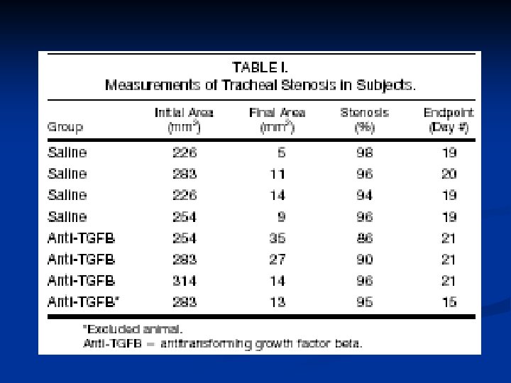
TGF-β Simpson CB et al (2008) Pilot Study in Modified Canine Model 8 subjects underwent cautery injury to subglottis 4 treated with saline injection into injury site 4 treated with combination of IV and local injection of anti. TGFβ at day 0 and day 5
TGF-β n Conclusions: IV and local TGFβ injection resulted in a reduction in tracheal stenosis (p <. 05) and an increase in survival time (p <. 03) when compared to saline control subjects n Anti-TGFβ appears to be useful adjunct in treatment of LTS n Further study needed to determine optimal dosing, route of administration and timing of delivery n
SGS Balloon Dilation of bronchtracheal stenoses with angioplasty balloons described previously in adults and children +/- stents n Advantage compared to rigid or bougie dilation n Balloons maximize the radial direction and pressure of dilation n Less damaging to tracheal wall mucosa n Found to have good initial results n n Often requires stenting of dilated portion n Repeated procedures necessary in active processes, e. g. Autoimmune States
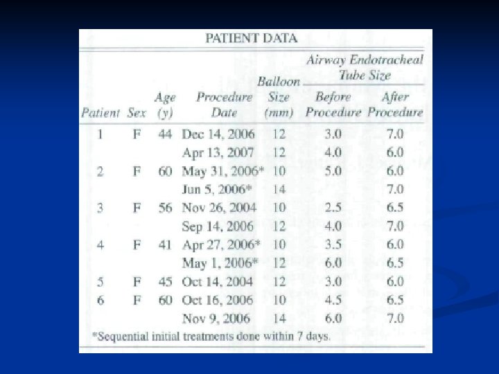
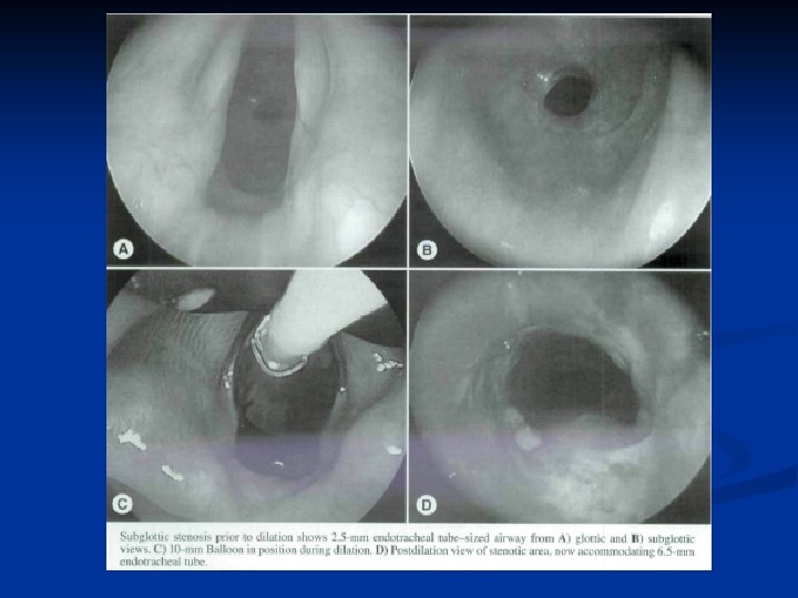
SGS Balloon Dilation n n Lee and Rutter (2008) 6 patients with IPSS (single discrete stenosis) Underwent dilation with 10 to 14 mm balloon in either single or 2 consecutive dilation (in 7 days) F/u between 10 and 30 months in 4 patients n n n No symptoms of recurrent airway stenosis One patient required repeat dilation after 22 mos No adverse effects or complications Recommended burst pressure (8 to 17 atm) 4 cm long catheters, center of balloon positioned at midpoint of stenosis Airway dilated from 2. 0 to 3. 5 ET size larger than initial size
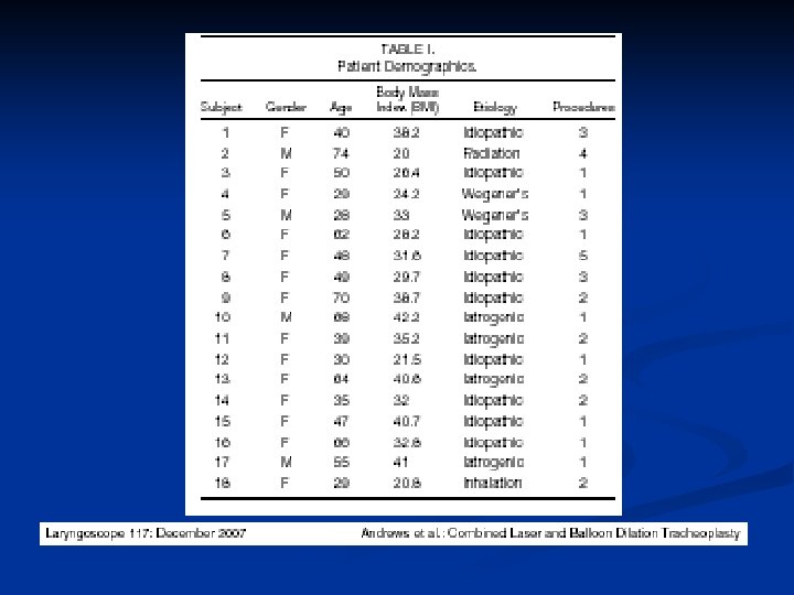
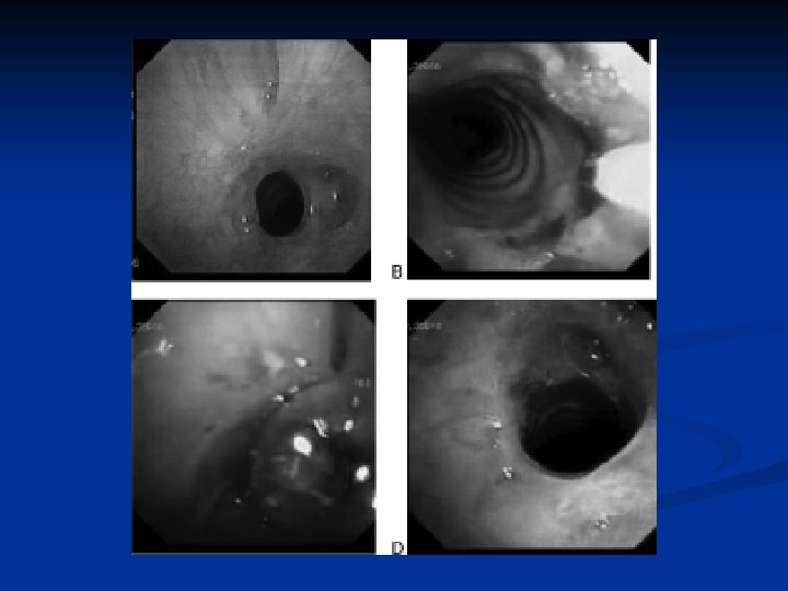
Combined Laser & Balloon Dilation n Andrews et al (2007) Performed flexible bronchoscopy for combined Nd: YAG laser radial incision at site of stenosis and balloon dilation in awake, spontaneously breathing patients Total of 18 patients underwent 36 procedures n n 8 pts required only 1 procedure; 5 pts required 2 procedures (72%) 11/18 patients (60%) were obese or morbidly obese Average f/u 22 mos; avg time b/w procedures 9 mos No complication in study group
Case Example n 58 -year-old female with several month history of hoarseness Also has a history of asthma n Recent PFTs showed no evidence for asthma. n Also had a diagnosis of gastroesophageal reflux disease and feels that her hoarseness has been contributed by the reflux disease n Intermittent dysphagia n
Case Example Laryngo video stroboscopic exam was performed: shows normal vocal fold mobility bilaterally n Presence of mild nodular thickening of the left anterior vocal cord surface n More significantly there is approximately 50% stenoses of her subglottic airway at the level of the cricoid cartilage and erythema of this area n
Endoscopic Balloon Dilation