ENDOSCOPIC THIRD VENTRICULOS TOMY Introduction Endoscopic third ventriculostomy


 Intermediate success rates (50 -75 %) Low success")


")

 patients had improvement")


- Slides: 46
ENDOSCOPIC THIRD VENTRICULOS TOMY
Introduction – Endoscopic third ventriculostomy is a technique to treat non communicating hydrocephalus. – Involves making a hole in the floor of the third ventricle to bypass the obstruction to CSF flow – Involves using an endoscope, (rigid or flexible)
History • L’Espinasse, in 1910, used a cystoscope to perform fulguration of the choroid plexus • Dandy refined this method calling it “ventriculoscopy” • In 1922, he described the use of an endoscope to perform choroid plexectomy. • Mixter in 1923 performed the first ETV in a child with congenital hydrocephalus using a urethroscope and probe.
History • Popularity of ETV waned because of high mortality, large cumbersome instruments, poor magnification and illumination. • In mid 1980’s minimally invasive neurosurgery became popular leading to a renewed interest in ETV and neuroendoscopy. • Advances, which brought together brighter light sources and cameras with improved resolution, were an important part of the rediscovery of neuroendoscopy.
Operative Technique • Tools • Endoscopic Anatomy • Technique
Technique: Tools
Comparison of rigid and flexible scopes Advantages Rigid scopes Flexible scope Better image Steerability Higher resolution Wider view Better colour Better light transmission Disadvantages Less maneuverable Poor image Less true colour Poor light Limited selection of scopes and instruments Narrower view Smaller working channel
Indications - Common • Primary Aqueductal Stenosis – Most common indication • Pineal and Tectal Tumors with HCP – One step procedure to include ETV and biopsy – Risk: Case reports of germinoma recurring along ventriculoscope tract – Wellon et al. , reported 13 patients with tectal plate gliomas and HCP. All underwent ETV with symptoms resolved at 31 months
Indications - Common • Idiopathic Stenosis of foramen of Magendie and Luschka – Several case reports reporting successful management of hydrocephalus • Posterior Fossa Tumors – Saint Rose et al. , performed ETV prior to posterior fossa surgery. They found that this significantly reduced risk of postoperative HCP. • Patients with repeated shunt infections
Indications - Uncommon • Isolated Lateral Ventricle with hydrocephalus – ETV in combination with septal fenestration • Dandy Walker – Case reports of performing ETV and placing a stent from the 3 rd ventricle to the posterior fossa cyst. Successful in two patients, failed in one. • Slit Ventricle Syndrome – Case reports
Indications - Uncommon • Communicating Hydrocephalus – Indications not clearly defined or understood – Some reports of improved symptoms, thought to be due to a functional obstruction – Feng Hailong et al , J Neurosurg 2008, 32 patients with 65. 6 % success
Indications High success rate (>75 %) Intermediate success rates (50 -75 %) Low success rates (<50 %) • Acquired aqueductal stenosis • Tumor obstructing CSF flow • Previously shunted MMC • Congenital aqueductal stenosis • Arachnoid cyst/ Dandy walker • Recurrent shunt infections/malfunction • Post hemorrhagic HCP • Post infectious HCP • MMC
Favourable features for ETV Clinical • Age > 6 months at the time of diagnosis of HCP • Age > 6 m at the time of procedure • No prior radiation therapy • No h/o meningitis/hemorrhage Radiological • Obstructive HCP • Lack of aqueductal flow void • Thinned floor of 3 rd ventricle • Downward bulging floor
Basic Operative Technique • Determine entry point and enter ventricular system • Finding and entering the Foramen of Monro • Inspecting the floor of the third ventricle • Perforating the floor of the third ventricle • Enlarging the perforation (approx 5 mm) • Inspecting the pre-pontine cistern
Targeting: Freehand Method Non Stereotactic Method – Freehand Method • Preoperative MRI evaluation of neurovascular structures below floor • Burr hole 1 cm in front of the palpated coronal suture and at mid-pupillary line, approx 2 -3 cm lateral to midline • Aim endoscope medially toward medial canthus of ipsilateral eye Hellwing et al, Neurosurg Rev 2005
Targeting : Stereotactic Method – Neuronavigation • Allows for optimal burr hole position • Helps to reduce traction on foramina of Monro-related structures • Kelly et al. , described stereotactic third ventriculostomy in 1986 • Target Point: Point in the midline between dorsum sella and the basilar artery • Entry Point: Foramen of Monro (side of entry). Extend line to skin for skin entry point
Technique: Introduce Endoscope • Position – The head is slightly flexed in a sniffing position. • Dura is cut in a cruciate fashion • Pass a 14 French trocar into lateral ventricle • Introduce endoscope through the sheath and navigate through the Foramen of Monro • Identify structures on floor of 3 rd ventricle
Technique: Fenestration • The correct point for the third ventriculostomy is the centre of so-called hypophysiomamillary triangle. • Multiple methods exist for perforating the floor including: – Blunt probe – Electro-coagulation and cutting probes – Laser probe – Ultrasonic contact probe • Dilation of the fenestration is achieved using a Fogarty balloon catheter
Technique • Continuous irrigation with lactated Ringer's solution heated to 37. 5°C temperature • Ventriculomegaly in patients who have undergone ETV decreases at a much slower rate and to a much less extent than in patients with implanted shunts. • Therefore, NCCT is performed at 6 to 8 weeks rather than on postoperative day 1 as is in practice in patients with shunts.
Technique: Fenestration • Must explore the pre-pontine space and open membrane of Liliequist for a successful ETV. • Liliequist membrane extends from the upper border of dorsum sellae to the anterior edge of mamillary bodies.
Complications Postoperative Intra operative Bradycardia – Incidence as high as 41% IVH - Choroid plexus injury Basilar artery injury – incidence 0. 05 % Hypothalamic dysfunction Pneumocephalus Subdural hygromas hematomas Infection CSF leak from wound Memory dysfunction Midbrain injury Delayed Seizures Late failure of ETV – Sudden deaths
Jenkinson et al. Gangemi et al. Drake Hader et al. AGE Adult No of patients 190 110 CSF LEAK (%) 1 1. 8 3. 6 6. 1 MENINGITIS (%) 0 - 2. 8 5. 3 HEMORRHAGE (%) 2 3. 6 1. 4 2. 3 HYPOTHALAMIC INJURY — — SEIZURE (%) — — 3. 75 1 DEATH (%) 0 0 0. 5 — WOUND INFECTION — 1 OVERALL COMPLICATION 5. 8 6. 4 NPH only 0 -20 yrs 368 1. 4 — 13. 6 All 131 6. 1 1. 5 16
Measurable Outcomes in Literature Review • Shunt Independence • Change in ventricular size • Patency of stoma
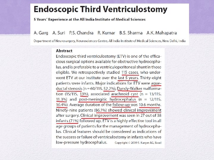
Outcomes Author N Age Mean follow Success up Oumar Sacko et al ( France) J Neurosurg, 2010 368 35 years (2 days– 88 years) 47% < 18 years 47 months (6– 106 months) 68. 5% Michael D. Jenkinson (UK) J Neurosurg, 2009 129 43 years (18 – 79 years) All > 18 yrs 36 months (1– 96 months) 83% Hailong Feng et al (China) J Neurosurg 2004 58 35 years (5 -67 years) 24 months (3 to 41 months) 84% Anil Garg et al (India) Pediatr Neurosurg 2009 115 38 % < 1 yr 48% < 2 yr 10. 6 months (3– 51 months) 86. 1%
Outcomes • Feng et al. ETV in the management of obstructive hydrocephalus. J Neurosurg, 2004. • Method – Retrospective analysis – 58 patients in one institution between 1999 -2003 – Mean age 35 years ( 5 -67 yrs) – Mean follow-up 24 months
Adult Outcomes • Results – At one month, 45/58 (77. 6%) patients had improvement in clinical outcome • Early postoperative improvement (within 2 weeks after ETV, and a patent stoma on cine phase–contrast magnetic resonance (MR) images were significant prognostic factors for a successful ETV. – No predictive value of reduced ventricle size on MRI within 2 weeks, however, it was significant in predicting good outcome at 3 months and 6 months
Pediatric Outcomes • Drake et al, Endoscopic Third Ventriculostomy in Pediatric Patients: The Canadian Experience, Neurosurgery, 2007 – 15 year period, 368 patients • – Average 6. 5 years, 1 day to 20 years, median age 4. 8 yr • 70 % patients were <10 years – 34% due to aqueductal stenosis, 29% due to tumor – 22% had been previously shunted • The 1 - and 5 -year success rates were 65 and 52%, respectively.
Infant Outcomes Author Age group No of Follow patients up Succes Rate Hideki Ogiwara et al (US) Childs Nerv Syst (2010) <6 m 23 57. 4 m 34. 8% Radim Lipina et al (Czech) < 6 m Childs Nerv Syst (2008) 14 21 m 57% Baldauf et al. , (Germany) Child Nervous System, 2007 <15 m 21 26. 2 m 43% Dorothee Koch et al, (Germany) Childs Nerv Syst (2006) <1 yr 28 25 m 46% Anil Garg et al (India) Pediatr Neurosurg 2009 All 38 (< 1 yr) 10. 6 m 71%
Long Term Outcomes • Kadrian et al. Long Term Reliability of ETV. Neurosurgery, 2005. – Retrospective analysis – 181 patients (203 procedures) from one institution over 22 years, from 1979 to 2001 – Age range 2 days to 78 years – 54/118 (54%) patients less than 2 years old – Surgical success: probability of performing an successful ETV was 89%
Long Term Outcomes • Reliability of ETV after 1 year was stratified according to age. – 0 -1 month: 31 % success – 1 -6 months: 50% success – 6 -24 months: 71% success – Greater than 24 months: 84% • 16 patients had ETV revisions with a success rate of 65% at four year follow-up.
ETV following VPS malfunction • Woodworth et al, Neurol Res, 2007 • 11 patients underwent ETV at VPS failure • Compared to patients who underwent ETV as primary procedure (16 patients) • 71% of patients experienced immediate improvement, but only 25% remained recurrence free at 2 years. • ETV after shunt failure was 2. 5 fold more likely to fail
Evaluating Patency of ETV • This has been a challenging problem especially in cases where the clinical course is unclear – Cine phase contrast MRI is able to detect flow across ETV Minor flow appears to be an early sign of closure – Fast spin-echo T 2 (FSE)
ETV Patency on Imaging • Bargallo et al. , AJNR, 2005 • Goal: To determine the utility of cine phase contrast MR imaging • 25 patients reviewed at one year • Results – Restoration of pulsatile motion of CSF circulation observed post operatively. – The stroke volume registered at ventriculostomy was maintained with time. – There was no relationship between ventricular size changes and clinical outcome.
ETV failure Follow-up for 5 -6 years • Maximum incidence of failure: first 1 -2 months Early : within 4 wks • Non-absorption from arachnoid granulation despite good MRI flow Delayed : after 4 wks • Stoma closure gliosis (6 -15%) Jones et al Min. Inv. Tech NS 1993 Siamin et al Childs Nervous System 2001 • ` Cinalli et al Min. JNS 1999
Repeat ETV for Failures 20 patients underwent repeat ETV • Age ranged from 8 months to 53 years • Intraoperative finding at repeat ETV – 1. Occlusion by scar (10 cases) – 2. Virginal floor of the third ventricle (5 cases) – 3. Pinhole ventriculostomy (3 cases) – 4. Incompletely penetrated membrane (1 case) – 5. Blood clot occluding orifice (1 case) • Repeat ETV successful in 65% of patients Sioman et al. , CNS, 2001
ETV Failures • Failure of ETV in infants has been studied by repeat ETV – 1) Reclosure of the ventriculostoma; – 2) Narrowing of the ventriculostoma; and – 3) patent ventriculostoma with new arachnoid membranes in the basal cisterns below the floor of the third ventricle, not present at the time of the first ETV Wagner et al, J Neurosurg, 2005
• Following surgical factors did not correlate with success of ETV: ü intraoperative haemorrhage during ETV, ü presence of haemosiderin deposits indicating previous haemorrhage ü double floor of third ventricle, ü presence of pre-pontine adhesions, ü Thickness of floor of third ventricle • CSF leak was correlated with previous CSF infection (p=0. 013), indicating that previous CSF infection predisposes towards CSF leak • D. Kombogiorgas et al. , Childs Nerv Syst , 2006
ETV in TBM hydrocephalous • Husain M et al, Neurosurg Rev. 2005 • 28 patients (15 males and 13 females) • Average 23 years • Outcome was satisfactory in 14 (50%), acceptable in five (18%) and unsatisfactory in nine (32%) patients
ETV in TBM hydrocephalous Author No of patients Success rate Figaji et al Childs Nerv Syst (2003) 2 pateints 100 % at 1 month Raj Shekhar et al Surg Neurol. 2005 2 100 % at 3 yrs Mazhar Husain et al Neurosurg Rev (2005) 28 50% at 1 year Figaji et al Childs Nerv Syst (2007) 17 41% at 1 year
ETV in NPH • Michelangelo Gangemi et al , Neurosurgery 2004 • Retrospective study • Of the 25 patients, 18 were men and 7 were women, • Age from 53 to 75 years (mean, 69 yr). • Rate of neurological improvement after ETV in this series was 72% • The improvement of gait disturbance was significantly higher (73%) as compared with that of dementia (16%) and urinary incontinence (31%).
ETV in NPH • For this reason, ETV is advised in patients with a clinical evolution of no more than 1 year before the diagnosis • The indications for ETV in patients with INPH may be based on two main clinical criteria: short preoperative evolution and significant prevalence of gait disturbances with little or no dementia.
ETV with CPC The overall surgical mortality rate was 1. 3%, and the infection rate was less than 1%. BENJAMIN C. WARF, M. D. , J Neurosurg Pediatrics, 2005
ETV with CPC in MMC patients with HCP • 115 patients • 93 completed ETV-CPC with > 1 month of follow-up. • Successful in 71 patients (76%), with a mean and median follow-up of 19. 0 months. • The operative mortality rate was 1. 1%, and there were no infections Benjamin C. Warf, J Neurosurg Pediatrics, 2008
New Concepts • Robot assisted ETV: – Robot “Evolution 1” – Used for robot-assisted navigated ETV in six patients with aqueductal stenosis – ETV initially successful in all patients Zimmerman et al. Acta Neurochir, 2004
Thank you r u o y r o f u o y k Than n o i t n e att