INTESTINAL OBSTRUCTION Dr Manish Kumar Gupta Assistant Professor

INTESTINAL OBSTRUCTION Dr. Manish Kumar Gupta Assistant Professor Paediatric Surgery AIIMS, Rishikesh

• • • IHPS Duodenal atresia Annular pancreas Jejunal and ileal atresia Malrotation Necrotising enterocolitis Meconium ileus Duplication cysts Hirschsprung’s disease Anorectal malformation Colonic atresia

Idiopathic hypertrophic pyloric stenosis • • • A disease of neonatal age group or early infancy. Incidence- 1 to 4 per 1000 live births. Age of presentation: 2 weeks to 8 weeks. (most commonly at 4 weeks) Boys : girls = 4: 1 Usually seen in first born male. Exact cause is not known. Seen in children more common who are formula fed than breast feeds. Exposure to erythromycin. Lack of local deficiency of nitric oxide synthetase.

Gradual hypertrophy of circle muscle of the pylorus ↓ Progressive obstruction of gastric outlet ↓ Progressive increase in frequency of vomiting ↓ Loss of gastric contents ↓ Hyponatraemic, hypokalemic, metabolic alkalosis with paradoxical aciduria

• Clinical presentation: • Progressively worsening non bilious emesis • With time, the emesis becomes more frequent, forceful, and projectile in nature. • Visible peristalsis from left to right. • Hungry and dehydrated baby • Palpation of the pyloric tumor (olive-shaped) in the epigastrium or right upper quadrant just below the liver edge. • If untreated, urine output decreases and acute renal injury due to hypovolaemia, hunger stools, hemetemesis Death

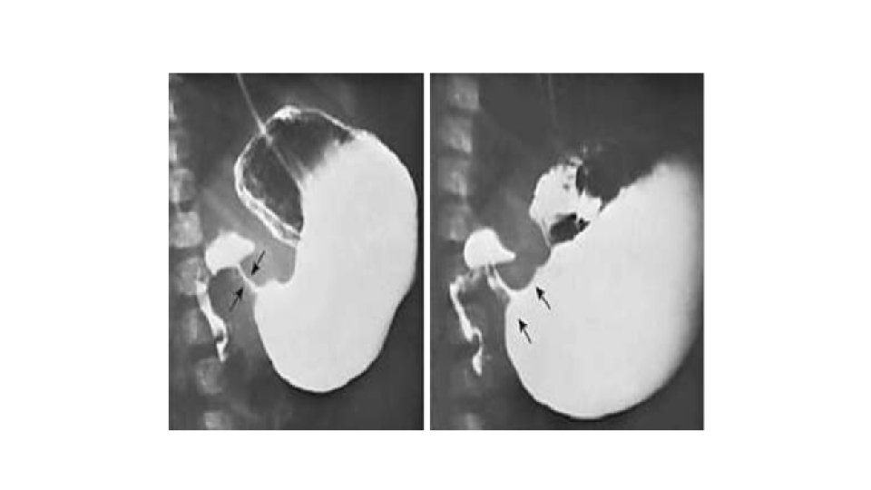
• Diagnosis: • USG: The diagnostic criteria for pyloric stenosis is a muscle thickness greater than or equal to 4 mm and a pyloric channel length greater than or equal to 16 mm. • Upper GI contrast study: String sign

• Treatment: • Ramstedt’s pylomyotomy: incising pyloric musculature without breaching the mucosa • Both open and laparoscopic approach are available.

Incision: Right epigastric incision or periumbilical incision
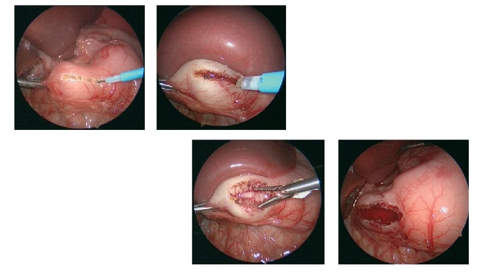

• Postoperatively, infants are usually allowed to resume enteral feedings. • Vomiting after surgery occurs frequently but is generally self-limited. • Potential complications include • incomplete myotomy • mucosal perforation • wound infection

Duodenal Atresia • The duodenum is one of the most common sites of neonatal intestinal obstruction. • Incidence of duodenal atresia is 1 in 6, 000 to 1 in 10, 000 live births. • Commonly associated with Down’s syndrome and annular pancreas.

Associated anomalies

• In the sixth week, the gut epithelium proliferates rapidly, resulting in obliteration of the intestinal lumen. • The intestine is then gradually recanalized over the next several weeks of development. • Errors in recanalization are thought to be the primary cause of duodenal atresia and stenosis.
 : There is")
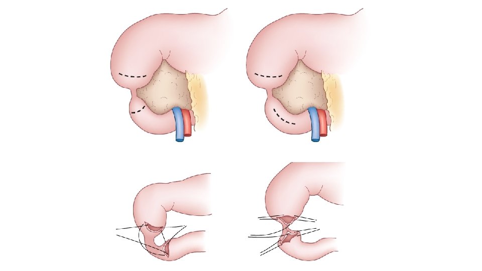
• Types: • Type 1: Type 1 (92% of cases) : There is an obstructing septum(web) formed from mucosa and submucosa with no defect in the muscularis. The mesentery is intact. • A variant of type 1 duodenal atresia, a “windsock deformity, ” can occur if the membrane is thin and elongated. The base of the membrane usually lies in the second portion of the duodenum, but balloons out distally, distending the third and fourth portions. 49 • Type 2 (1% of cases): A short fibrous cord connects the two blind ends of the duodenum. The mesentery is intact • Type 3 (7% of cases): There is no connection between the two blind ends of the duodenum. There is a V-shaped mesenteric defect.

• Most common site of block-near the junction of the first and second portions in 85% of cases. • Site of block is distal to the opening of ampulla of vater.

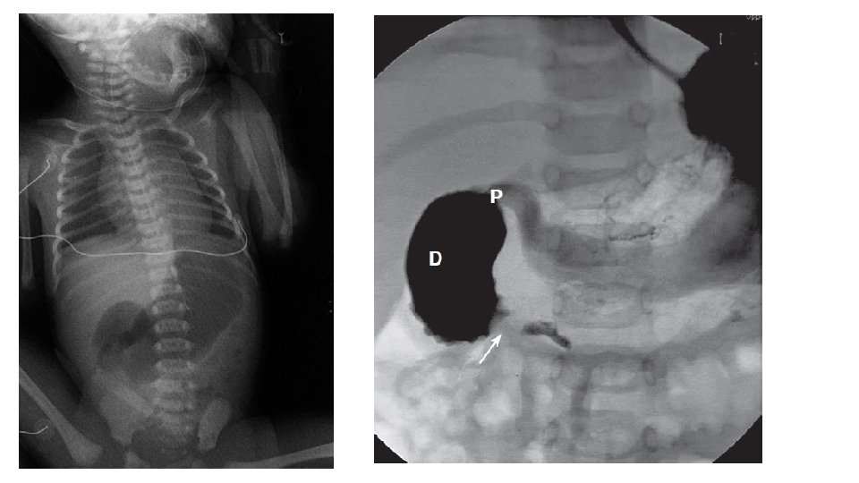
Clinical presentation • Antenatal: Polyhydramnios • Antenatal USG: Double bubble • Prematurity. • Newborn: recurrent bilious vomiting and intolerance to feeds. • Upper abdominal fullness. No gross abdominal distension.

• Treatment: • Duodeno-duodenostomy • Site of abdominal incision : right transverse incision between right costal margin and umbilicus • Other bowel distal to obstruction must be checked after surgical correction to look for any additional blocks or atresia. • Usually a transanastomotic nasojejunal tube is placed during surgery so that feeding may be started early and NJ tube is removed once the aspirates become low.

• Complications: • Leaks • Prolonged dysmotility of duodenum

Annular pancreas
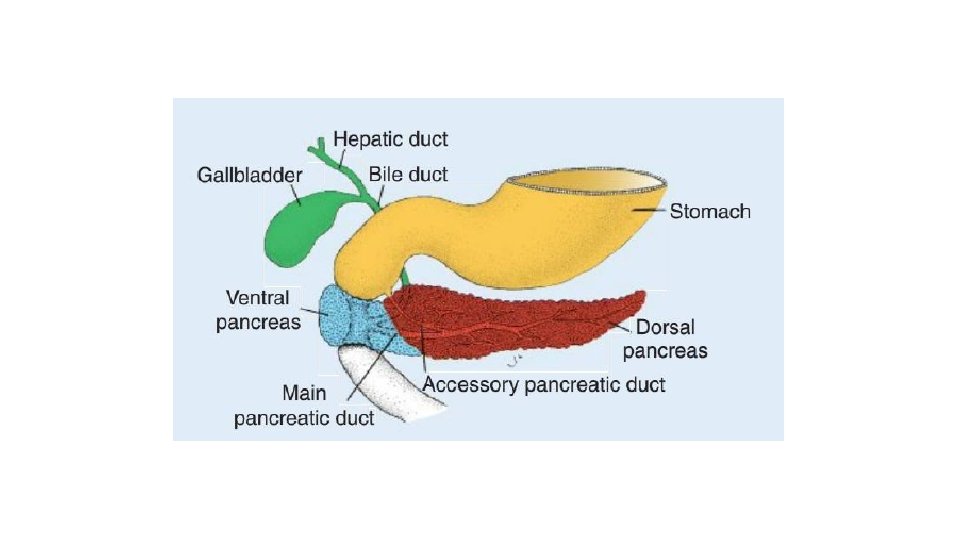

• Clinical Presentation: Similar to Duodenal atresia • Management: Nothing is done to pancreas. • Two ends of duodenum are joined – Duodeno-duodenoplasty

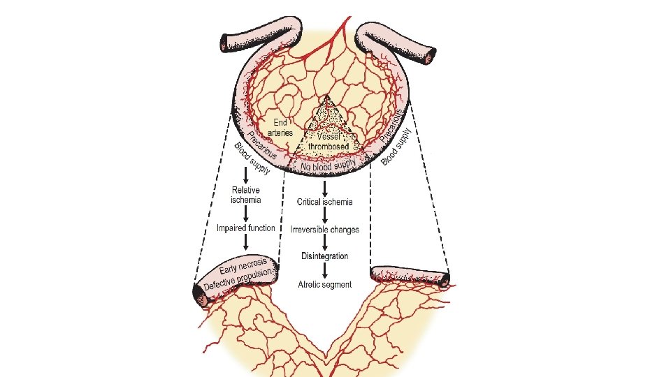
Jejuno-ileal atresias • Jejunoileal atresia is the most common GI atresia. • Incidence is 1 in 2000 live births. • Result of an intrauterine mesenteric vascular occlusion. • Atresias occur slightly more frequently in the jejunum than in the ileum.

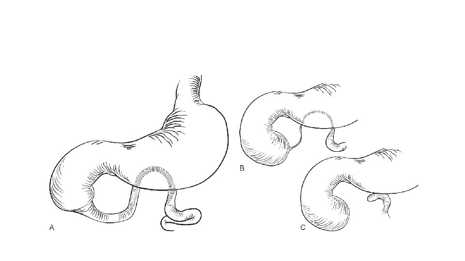
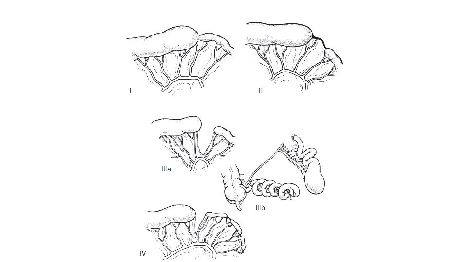
Types • Type I: a mucosal or membranous atresia with intact bowel wall • Type II: an atretic cord between two blind ends of bowel with intact mesentery • Type IIIa: a complete separation of the blind ends of the bowel by a Vshaped mesenteric gap • Type IIIb: an apple peel or Christmas tree deformity with a large mesenteric gap in which the distal bowel receives a retrograde blood supply from the ileocolic or right colic artery. • This tenuous blood supply has implications for reanastomosis and the potential for ischemic necrosis caused by an antenatal volvulus. Thus, many of these infants with this type of atresia are born with reduced intestinal length. • Type IV: there are multiple atresias, with a string of sausage appearance.

Clinical Presentation • Infants present with bilious emesis, abdominal distention, and failure to pass meconium during the early neonatal period. • Overall clinical presentation is dependent on the level of obstruction. • In proximal atresia, abdominal distention is less, but significant bilious emesis is present. • With distal atresias, abdominal distention is present more frequently.

Diagnosis

Management • Initial resuscitation with IV fluids, IV antibiotics and proximal segment decompression with placement of the NG tube and aspiration • Aim: to establish intestinal continuity and save as much bowel as possible. • Early surgery- delayed surgery causes dysfunction of the proximal segment due to pressure ischemia. • End to end or end to side jejunojejunal/ jejuno-ileal/ ileo-ileal anastomosis • Peri-operative TPN

• Problems: • Risk of short bowel syndrome due to loss of significant length of bowel • Bowel lengthening procedures may be needed later in life. • Functional loss of bowel- Poor absorption/ poor peristalsis • Post-op anastomotic leaks and stenosis • Overall survival is 80 -90%.

Intestinal malrotation • The incidence of malrotation has been estimated at 1 in 6000 live births. • It occurs due to abnormal rotation and fixation of the bowel to the retroperitoneum leading to precariously narrow based mesentery. • Presence of abnormal mesenteric bands (Ladd’s bands)-extend from the colon across the anterior duodenum- Predisposing bowel for volvulus and intestinal obstruction. • Up to 75% of patients present during the first month of life, while another 15% will present within the first year

Embryology
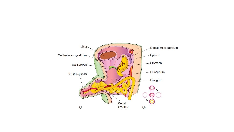
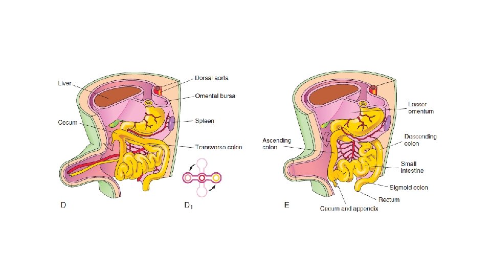

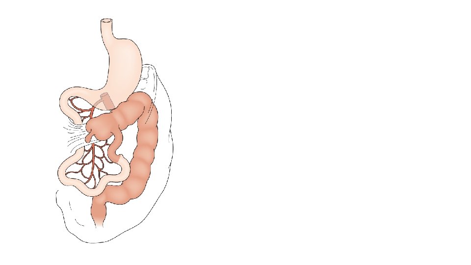
Normal intestinal anatomy results in fixation of the duodenojejunal junction in the left upper quadrant and the cecum in the right lower quadrant. This allows a wide breadth to the mesentery of the small bowel.

Clinical presentation • Recurrent bilious vomiting • Poor growth and failure to thrive • Malabsorption • Chylous ascites • Associated volvulus • • Recurrent pain abdomen Gangrene of the bowel and perforation Peritonitis and shock Hemetemesis

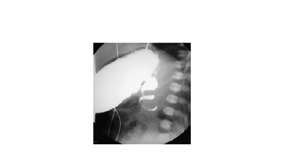
Diagnosis • Upper GI contrast study: • demonstrates an abnormal position of the ligament of Treitz. • Cork screw pattern in case of associated midgut volvulus • USG: • Dilated duodenum • Alteration in the relation of superior mesenteric vessels • Whirl pool sign on colour doppler in case of volvulus

Management • Aggressive resuscitation especially when midgut volvulus is suspected. • Emergency laparotomy. • Laparoscopic approach is also done in many centres. • Name of the surgery: Ladd’s procedure/operation.

Entry into the abdomen through right supraumbilical incision. Delivery of the bowel. Derotation of the bowel. Excision of the ladd’s band. Straightening of the duodenum Broadening of the small intestinal mesentery Appendicectomy Placement of the small bowel along the right lateral gutter and large bowel along the left lateral gutter. • Closure of abdomen. • •

• If gangrenous bowel is present, excision of the gangrenous portion is done primarily or after second look re-exploration. • Aims is to save as much bowel as possible to avoid development of short bowel syndrome. • Post-operative bowel function resumes in 3 -5 days when oral feeds are started. • Complications: Adhesive obstruction later which may require a surgical management.


Necrotising enterocolitis • NEC is the most common GI emergency in the neonatal period. • Usually seen in premature and low birth weight babies. • Usually develops after the starting of feed. • Mortality rate is high. (10 -50%) • Most common cause of short bowel syndrome in children.

Pathology • After feeds if excess glucose comes to ileum and colon due to poor absorption proximally in the intestines or high glucose content of the feeds • Bacterial overgrowth • Local acid production by bacteria • Poor gut development of premature baby • Inflammation and necrosis of bowel due to translocation of bacteria into the bowel wall and mesentery • Bowel gangrene, perforations and severe sepsis

Clinical presentation • Acute abdominal distention, tenderness, and feeding intolerance with gross or occult blood in the stool are hallmark features for NEC. • Other nonspecific clinical signs include irritability, temperature instability, and episodes of apnea or bradycardia. • NEC typically occurs in the first few days of life with the initiation of enteral feedings. • In approximately 80% of cases, it occurs within the first month of life. • As NEC progresses, systemic sepsis develops, with hemodynamic deterioration and coagulopathy.

• The distal ileum and ascending colon are the usual affected areas. • The entire GI tract (NEC totalis) may also be involved.


Diagnosis • The pathognomonic radiographic feature of NEC is pneumatosis intestinalis. • Pneumatosis is composed of hydrogen gas generated by the bacterial fermentation of luminal substrates. • Other radiographic findings may include portal venous gas, ascites, fixed loops of small bowel, and free air.


Medical Management • Initial medical management consists of • • nasogastric tube decompression fluid resuscitation blood and platelet transfusion administration of broad-spectrum antibiotics. • In general, NEC can be successfully treated medically in approximately 50% of cases with a 7 - to 10 -day course of antibiotics.

• The absolute indication for operative management of NEC is the presence of intestinal perforation, as revealed by free air on plain abdominal radiographs. • Other relative indications for surgery include • • • overall clinical deterioration abdominal wall cellulitis worsening acidosis falling white blood cell or platelet count a palpable abdominal mass persistent fixed radiographic bowel loop

Surgical management • The general principles of surgical management of NEC include resection of all nonviable segments of intestine with the creation of a stoma. • All efforts need to be made to preserve as much intestinal length as possible. • It may be necessary to resect multiple intervening necrotic segments of bowel, preserving all viable intestine. • In cases in which the bowel is ischemic but not frankly necrotic, a second-look operation may be performed after 24 hours.

• Once the child is stable and started on feeds with increase in the weight, further surgery may be done to maintain the instestinal continuity.

To be continued… • Meconium ileus • Hirschsprung’s disease • Anorectal malformation • Duplication cysts • Colonic atresia

MECONIUM ILEUS

INTRODUCTION • Meconium ileus is a subset of CF. • It represents a disorder of neonates characterized by intestinal obstruction secondary to the intraluminal accumulation of inspissated and desiccated meconium. • Earliest clinical manifestation of CF.
 • Meconium obstructing the small bowel + pathologic")
history • First described by Landsteiner(1905) • Meconium obstructing the small bowel + pathologic changes in pancreas + pancreatic enzymes deficiency. • Term “cystic fibrosis” of the pancreas by Fanconi(1936) • Association of chronic pulmonary disease of infancy + pancreatic insufficiency

• CF is now recognized as the most common potentially lethal autosomal recessive disease in the Caucasian population.

INCIDENCE • Incidence of 1 per 1000 to 2500 live births. • Rare in blacks (1/17, 000 live births). • Very rare in in Asians (1/90, 000 live births) and Africans. • Meconium ileus is presenting feature of 20. 8% of the CF population in the United States.

GENETICS • Locus for CF- 7 q 31. • codes for the cell membrane protein termed the CF transmembrane regulator(CFTR). • cyclic adenosine monophosphate–induced chloride channel that regulates ion flow across the apical surface of epithelial cells. • More than 400 mutations known for CFTR.

Abnormal electrolyte content along the external apical environment of epithelial membranes Tubular structures lined by affected epithelia Desiccation and reduced clearance of their secretions
 • Biliary-")
• Respiratory- recurrent pneumonia • Gastrointestinal- meconium ileus (10% to 20%) • Biliary- obstructive biliary disease (15% to 20%) • Pancreatic- pancreatic insufficiency(90%) • Reproductive systems- azoospermia(nearly 100%)

• The ΔF 508 mutation- most common CFTR gene locations. • Homozygous individuals nearly always express pancreatic exocrine insufficiency • also present with a higher incidence of meconium ileus.

PATHOGENESIS • Exocrine-eccrine gland dysfunction • mucus-secreting and sweat glands. • Intestinal mucosal gland abnormalities caused production of tenacious material that causes the intraluminal obstruction. • Intestinal glandular disease -dominant role • Pancreatic disease -secondary role

• Defect in electrolyte resorption of sodium and water by ductular cells • Impermeability of these cells to chloride ions Excretory product high in both sodium and chloride. desiccation and reduced clearance of such secretions

• Analysis of the protein content of abnormal meconium • Most of this protein is albumin. • In the absence of degrading enzymes, they impart a highly viscid rubbery character to the involved secretions.

• Thickened meconium accumulates in utero obstruct the intestine lumen complications of meconium ileus (i. e. , twist of a heavy loop with perforation, peritonitis, and cyst or atresia) and the pattern of abdominal distention and obstruction seen in the neonate.

• The proximal ileum dilates, and its wall thickens as it becomes filled with the tenacious and tarry meconium. • The narrowed distal small bowel and the colon contains beaded or “boxcar” concretions of graywhite, putty-like inspissated meconium. • The more distal colon is small or unused, a microcolon.

Clinical Features • Presents in the neonate as: • uncomplicated or simple • Complicated

• Uncomplicated meconium ileus • typically presents immediately at birth with abdominal distention. • In utero genetic diagnosis of CF and an ultrasound suggestive of intestinal obstruction with echogenic bowel. • Bilious vomiting and failure to stool.

• Complicated meconium ileus • will present either in utero or postnatally with evidence of bowel obstruction complicated by evidence of previous intestinal perforation and/or necrosis • Crescents or speckles of intra-abdominal calcification may be present • On clinical assessment there may be evidence of peritonitis including an erythematous or edematous abdominal wall • +/- demonstrable abdominal tenderness.

Meconium peritonitis • Meconium pseudocyst: • meconium accumulating in the peritoneal cavity for weeks to months. • Calcified pseudocyst • fibrous wall forms around an accumulation of meconium • spared bowel loops are peripheral to this cyst

• Adhesive meconium peritonitis: • Meconium contamination of the peritoneal cavity • Dense and vascular adhesions • Scattered calcifications

• Meconium ascitis: • When intestinal perforation occurs only a few days before delivery • an abdomen filled with meconium • calcification is absent • Bacterially infected ascites • colonized intestinal organisms penetrate from the perforated intestine into the peritoneal cavity.

CLINICAL HISTORY • A family history of CF is present in 10% to 33%. • This history + in utero amniocentesis permits the accurate diagnosis of the fetus afflicted with CF. • Above + the results of serial in utero sonography permits the accurate predication of which infants are at risk for the development of the intestinal obstruction of meconium ileus.

• Maternal polyhydramnios may be a feature of in utero meconium ileus due to high-grade intestinal obstruction. • In utero growth retardation is common, prematurity and other associated anomalies are rare.

PHYSICAL EXAMINATION • Abdominal distention • Visible peristaltic waves and palpable, doughy bowel loops are often present. • Finger pressure over a firm loop of bowel may hold the indentationputty sign.

• In the presence of an in-utero perforation with meconium peritonitis and “cyst” formation • a palpable abdominal mass, discoloration of the abdominal wall, and signs of peritoneal irritation are often observed. • Physical evidence of hypovolemia may rapidly develop in infants with peritonitis. • On passage of a nasogastric tube, a quantity of bile-stained gastric fluid usually exceeds 20 m. L.

RADIOLOGIC STUDY • In utero, meconium ileus bowel may be distended but an echogenic bowel wall in the third trimester may be diagnostic as well.

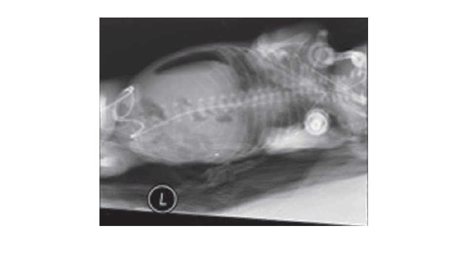
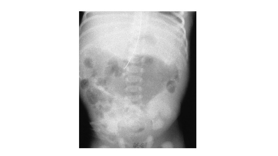
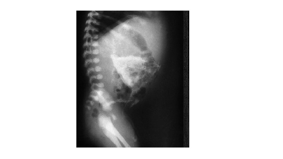
• Uncomplicated meconium ileus is • supine and erect films appearing remarkably similar 1. great disparity in the size of the intestinal loops 2. no or few air-fluid levels on the erect film because swallowed air cannot layer above thickened inspissated meconium 3. a granular, “soap bubble, ” or “ground-glass”
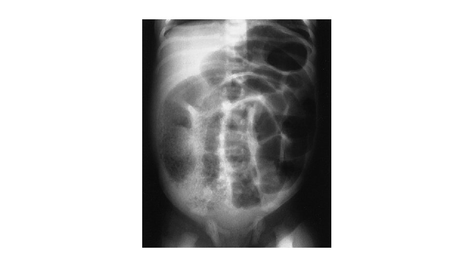

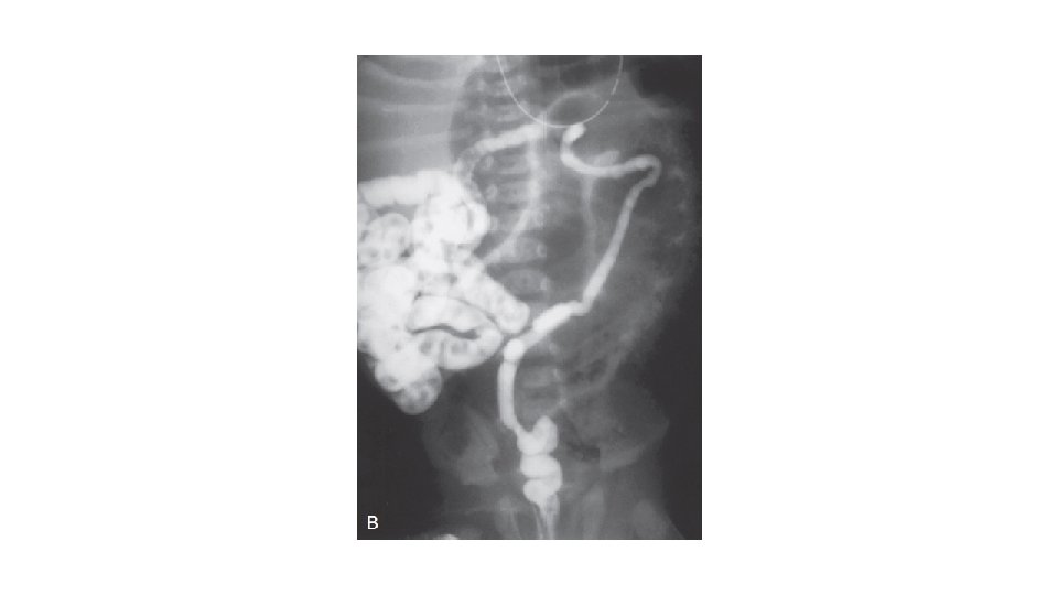
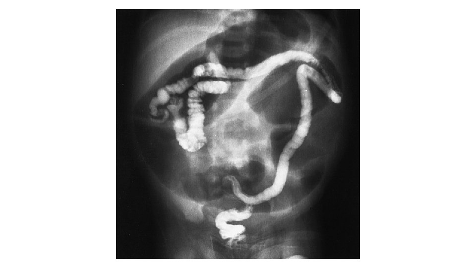
• A contrast enema will outline a normally positioned unused colon • It will be empty or will contain pellets of inspissated meconium. • The colon will be the typical “unused” colon or “microcolon. ” • If reflux of contrast agent into the terminal ileum occurs, it will outline pellets of inspissated meconium.

Laboratory Testing • Sweat test: • The minimum amount of sweat to be collected is 100 mg, and a measured concentration of sweat chloride in excess of 60 m. Eq/L is diagnostic of CF.

• Genetic testing for CF: • can be done by analyzing cellular DNA for CFTR, • If a family has known CFTR mutations, then amniocentesis with fetal DNA analysis may predict a fetal CF diagnosis.

• Increased albumin concentration in meconium may be useful as a diagnostic screening tool for meconium ileus. • Meconium from normal neonates has an albumin concentration of less than 5 mg/g of stool, whereas meconium from neonates with CF has values at times in excess of 80 mg/g.

• Operative tissue specimens that aid in the diagnosis of CF include intestinal (rectal) or appendiceal pathognomonic changes • goblet cell hyperplasia • Accumulation of secretions within the crypts or within the lumen.

Nonoperative Management • Constitutes dissolution of the inspissated intraluminal meconium in an otherwise patent and uncompromised ileocolon.

Prerequisites: • intravenous antibiotics should be administered • the patient should have a full fluid resuscitation with fluids given aggressively (one to three times maintenance) during the procedure • exclude other causes of distal intestinal obstruction • the complications of volvulus, atresia, perforation, or peritonitis must be excluded • The patient should be prepared for imminent operation should complications develop.

• A Gastrografin enema has been the standard of nonoperative treatment. • Meglumine diatrizoate is a hyperosmolar, water-soluble, radiopaque solution containing 0. 1% polysorbate 80 • 40%sodium diatrizoate

• 5 m. L of a 10% N-acetylcysteine solution should be administered every 6 hours through a nasogastric tube to liquefy upper gastrointestinal secretions.

• After successful conservative management: • supplemental pancreatic enzymes must be administered with each feeding when feeding is started.

• If the evacuation of meconium is incomplete and obstruction persists, the enema may be repeated. • If either no evacuation of meconium, then this technique should be abandoned and operative intervention planned. • Similarly, signs of worsening obstruction, clinical distention, greater distention of loops on radiograph, or signs of peritonitis resulting from a possible perforation are also indications for operative intervention.
 • Failed to respond")
Operative management • Indications: • Simple: (1/3 rd to ½) • Failed to respond to conservative management • Complicated(2/3 rd to ½) • • • Intestinal atresia Volvulus Perforation Meconium cyst formation with peritonitis Intestinal gangrene, or combinations of these events.

SIMPLE MECONIUM ILEUS • Goal of operation is the relief of intraluminal ileocolonic obstruction • evacuation of the adherent intraluminal meconium • resection of the portion of bowel filled with inspissated material. • Enterotomies with irrigation coupled with a limited resection.

• Irrigating solutions: • • • warmed saline 50% diatrizoate solution 1% solution of pancreatic enzymes hydrogen peroxide Most commonly, either a 2% or a 4% solution of Nacetylcysteine

• Three options: • The enterotomy and the abdomen may then be closed • enterostomy may be created • the site can be controlled by insertion of a T tube.

• Appendectomy with appendicostomy, with meconium evacuation or irrigation through this route.

• Relieving stoma with or without an associated partial resection. • Mikulicz double-barreled enterostomy
:")
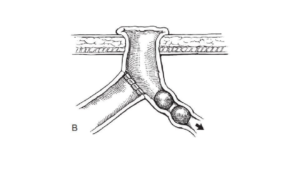
• Primary resection and anastomosis • Distal chimney enterostomy (Bishop-Koop procedure):

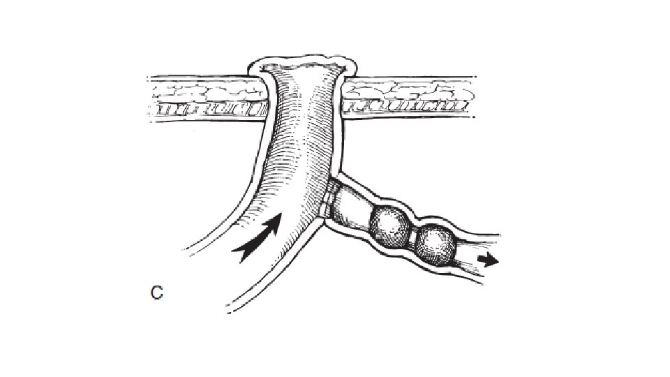
• Santulli proximal chimney enterostomy

COMPLICATED MECONIUM ILEUS • Operative indications include: • Persisting intestinal obstruction • Enlarging abdominal mass • Signs of peritonitis.

Rehbein’s two-stage operation • After initial resuscitation, a proximal dilated loop stoma is created to the obstructive site. • Once the general condition of baby improves, further laparotomy and resection and anastomosis of distal segment is done.

• Meconium cyst • Decortication of the cyst. • Ultrasound guided drainage.
 delivered through a nasogastric")
Postoperative Management • Instillation of 2% or 4% acetylcysteine (Mucomyst) delivered through a nasogastric tube, which will solubilize the residual meconium. • When enteral feedings begin, supplemental pancreatic enzymes must also be started. • If postoperative ileus is prolonged or if short-bowel syndrome →total parenteral nutrition.
- Slides: 117