THE FACIAL NERVE Topics Gross applied anatomy Pathology






- Slides: 77
THE FACIAL NERVE
Topics • Gross applied anatomy • Pathology of nerve injury and regeneration • Principles of the electrophysiological tests • Causes of facial nerve paralysis • Principles of treatment of facial paralysis • Bell’s palsy
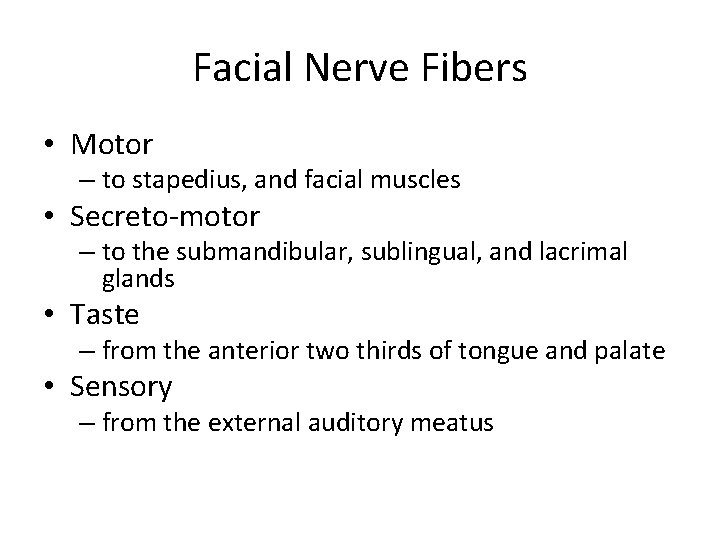
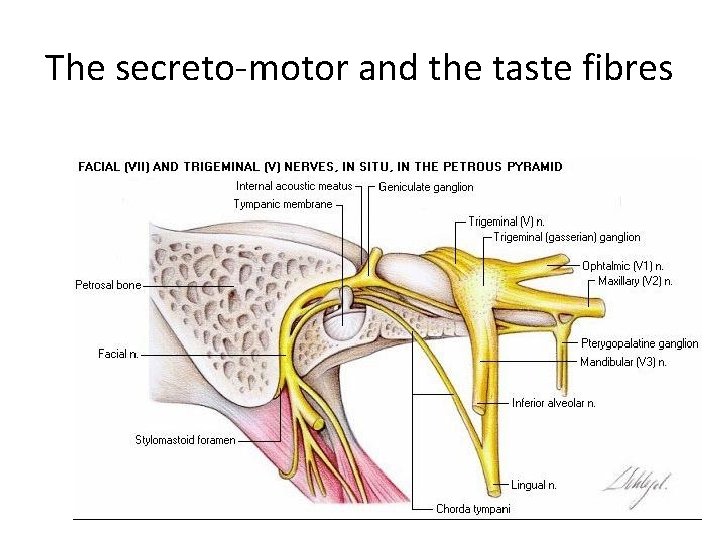
Facial Nerve Fibers • Motor – to the stapedius and facial muscles • Secreto-motor (parasympathetic) – to the lacrimal gland the submandibular and sublingual salivary glands • Taste – from the anterior two thirds of the tongue and palate • Sensory – from the external auditory meatus
Anatomical Divisions • Intracranial • Cranial (intratemporal) • Extracranial (extratemporal)
The Intracranial Part • Include the nuclei and the Cerebello Pontine angle semgents
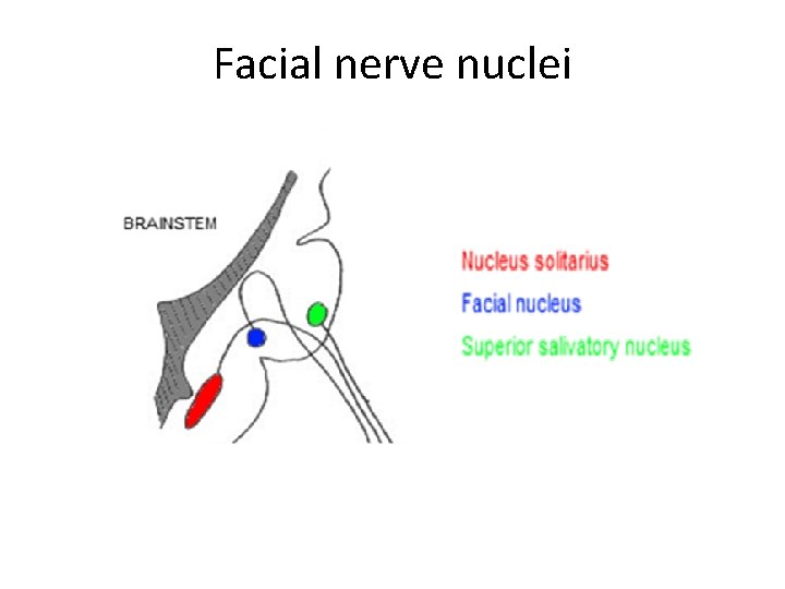
Facial nerve nuclei
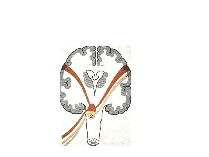
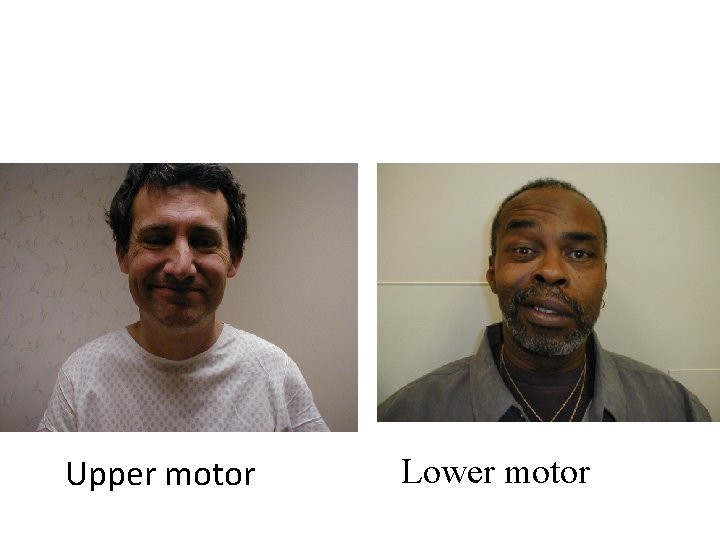
• Lower motor lesions affect all the ipsilateral facial muscles • Upper motor lesions spare the upper facial muscles and affect the contralateral lower face
Upper motor Lower motor
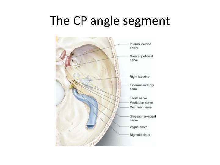
The CP angle segment
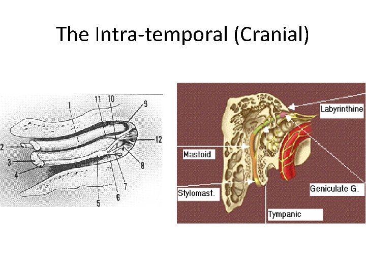
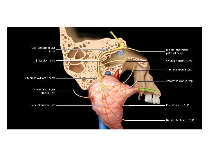
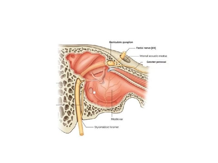
The Intra-temporal (Cranial)
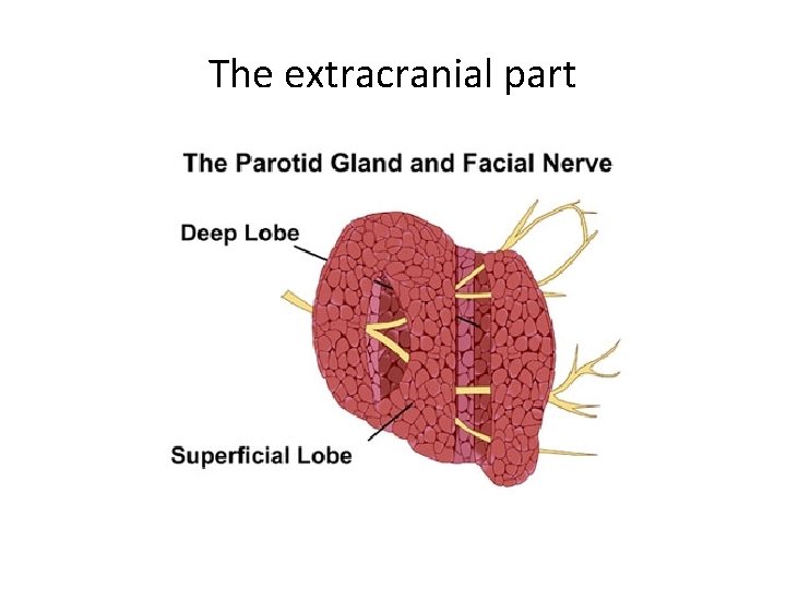
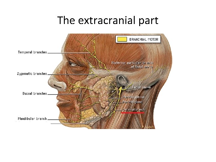
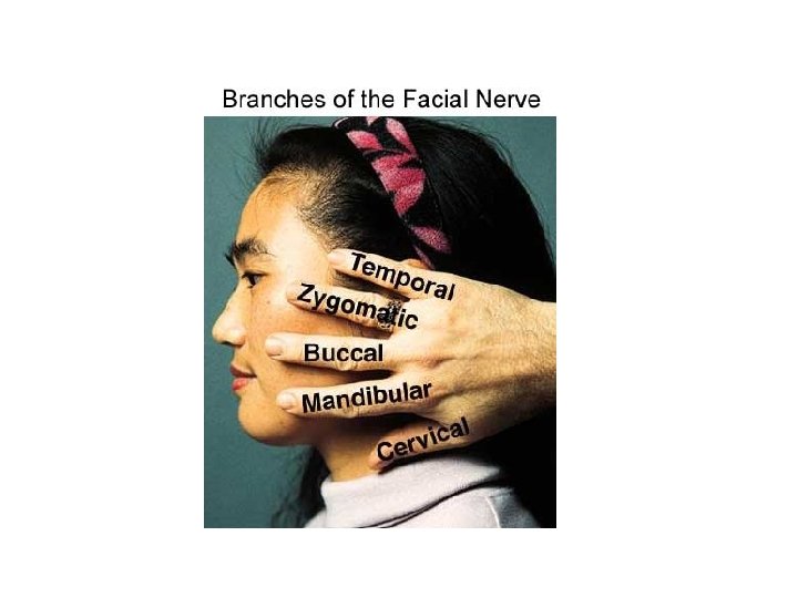
The extracranial part
The extracranial part
Facial Nerve Fibers • Motor – to stapedius, and facial muscles • Secreto-motor – to the submandibular, sublingual, and lacrimal glands • Taste – from the anterior two thirds of tongue and palate • Sensory – from the external auditory meatus
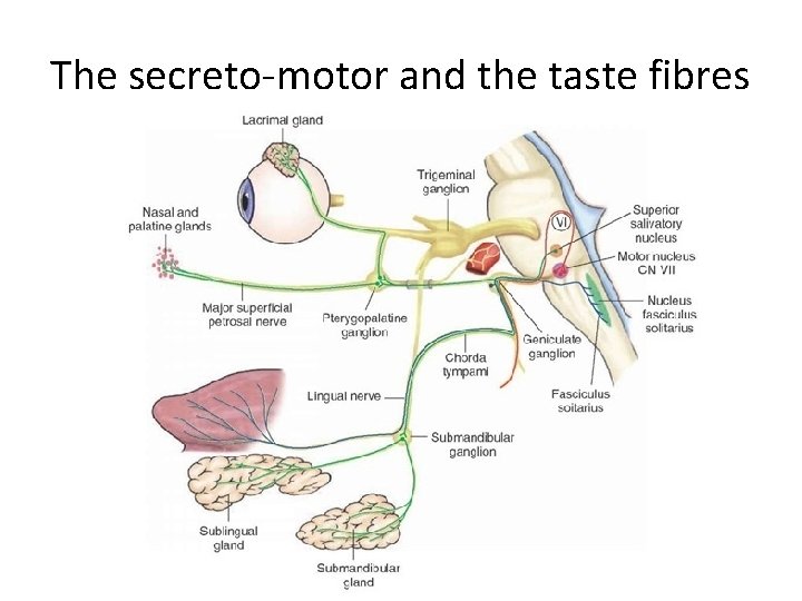
The secreto-motor and the taste fibres
The secreto-motor and the taste fibres
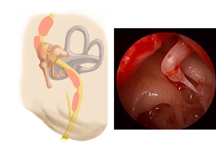
Variations and Anomalies
Facial Nerve Paralysis
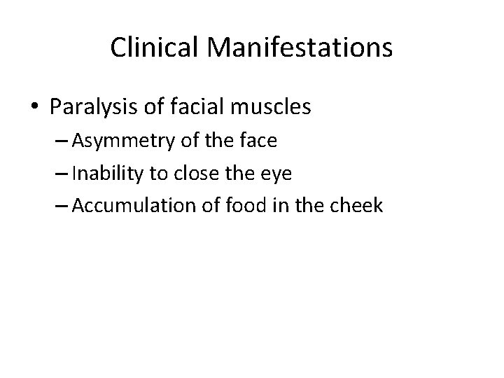
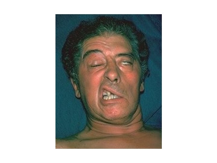
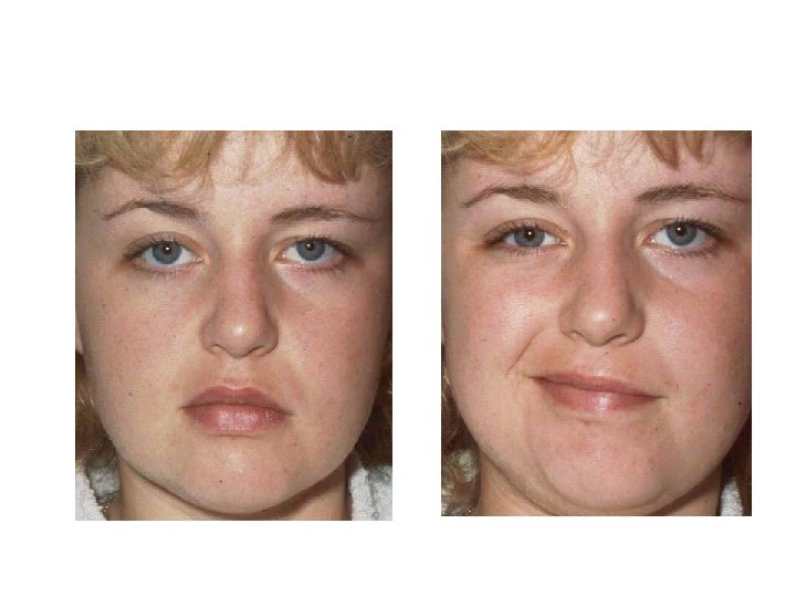
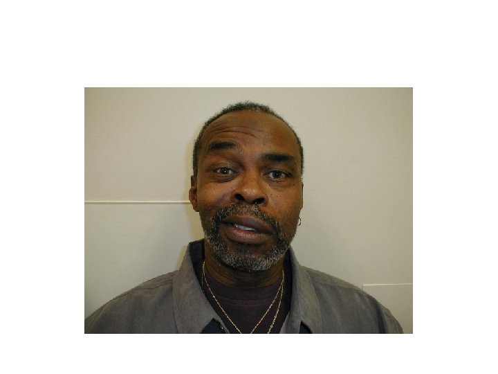
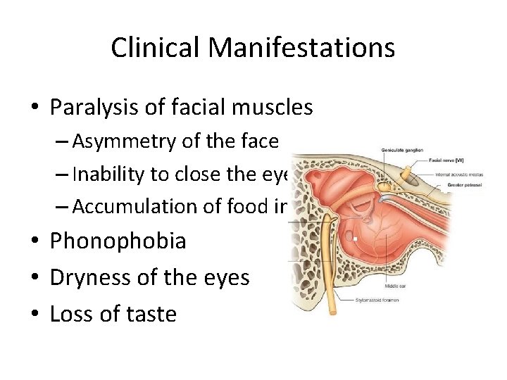
Clinical Manifestations • Paralysis of facial muscles – Asymmetry of the face – Inability to close the eye – Accumulation of food in the cheek
Clinical Manifestations • Paralysis of facial muscles – Asymmetry of the face – Inability to close the eye – Accumulation of food in the cheek • Phonophobia • Dryness of the eyes • Loss of taste
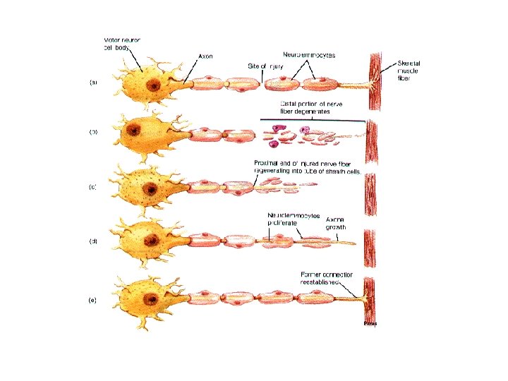
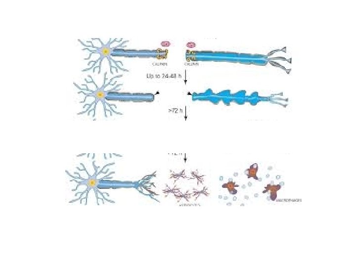
Pathophysiology of Nerve Injury
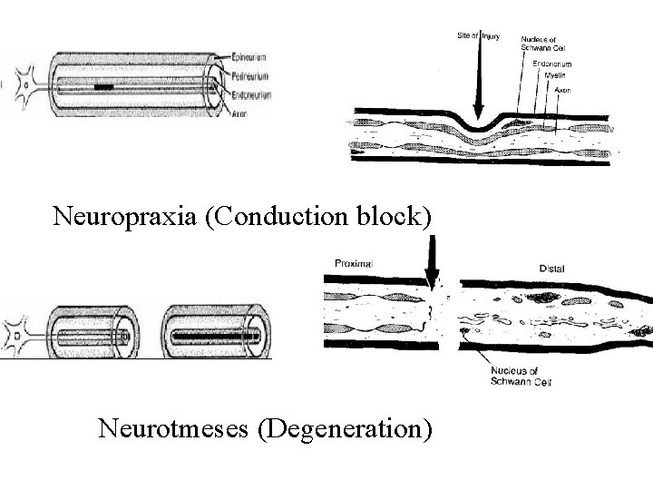
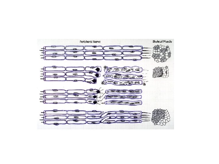
Neuropraxia (Conduction block) Neurotmeses (Degeneration)
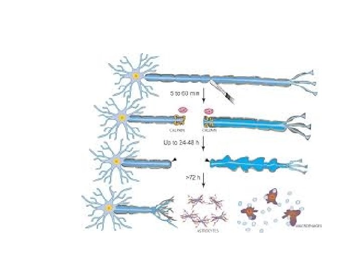
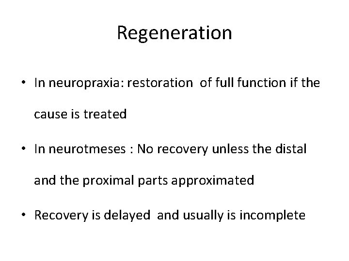
Regeneration • In neuropraxia: restoration of full function if the cause is treated • In neurotmeses : No recovery unless the distal and the proximal parts approximated • Recovery is delayed and usually is incomplete
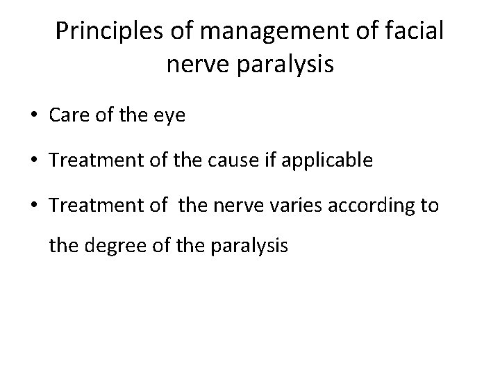
Principles of management of facial nerve paralysis • Care of the eye • Treatment of the cause if applicable • Treatment of the nerve varies according to the degree of the paralysis
Partial facial paralysis • Being partial means that some of the nerve fibres are in continuity and recovery is expected by conservative treatment (e. g. removal of pressure, steroid etc)
Complete facial paralysis • Complete paralysis may be a result of neuropraxia or/and degeneration • If it is due to neuropraxia, recovery is expected by conservative treatment • If it is due to degeneration, surgical treatment is required • To differentiate between degeneration and neuropraxia electrophysiological tests are required
Electrophysiological Tests • It detect degeneration of the nerve fibres • Useful only 48 -72 hours following the onset of the paralysis
Electrophysiological Tests • Nerve Excitability Test (NET) • Electroneurography (ENo. G)
Nerve excitability test (NET) • The current’s thresholds required to elicit justvisible muscle contraction on the normal side of the face are compared with those values required over corresponding sites on the side of the paralysis
Electroneurography (ENo. G) • The amplitude of action potentials in the muscles induced by the maximum current is compared with the normal side ; and used to calculate the percentage of intact axons.
Interpretation of the tests • Not useful in the first 48 – 72 hours • After 48 -72 hours (the time required for degeneration to take place) – Normal results means that there is no degeneration (Neuropraxia) – Abnormal results means degeneration
Topognostic Tests • Indicated in some cases to locate the site of the injury
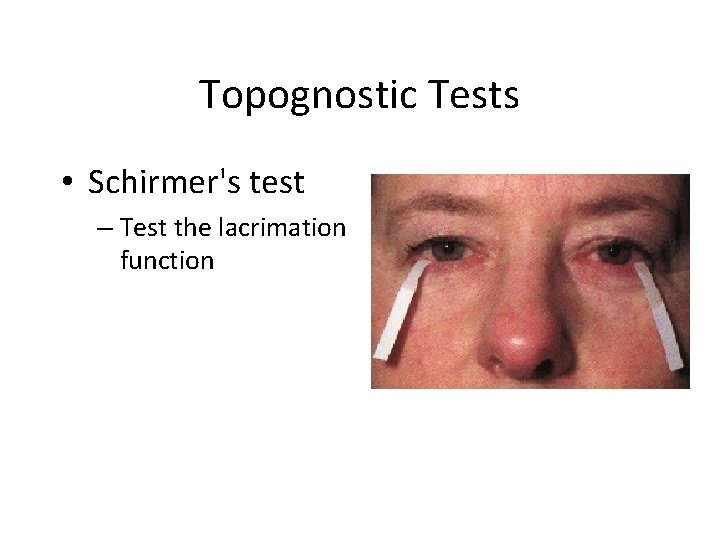
Topognostic Tests • Schirmer's test – Test the lacrimation function
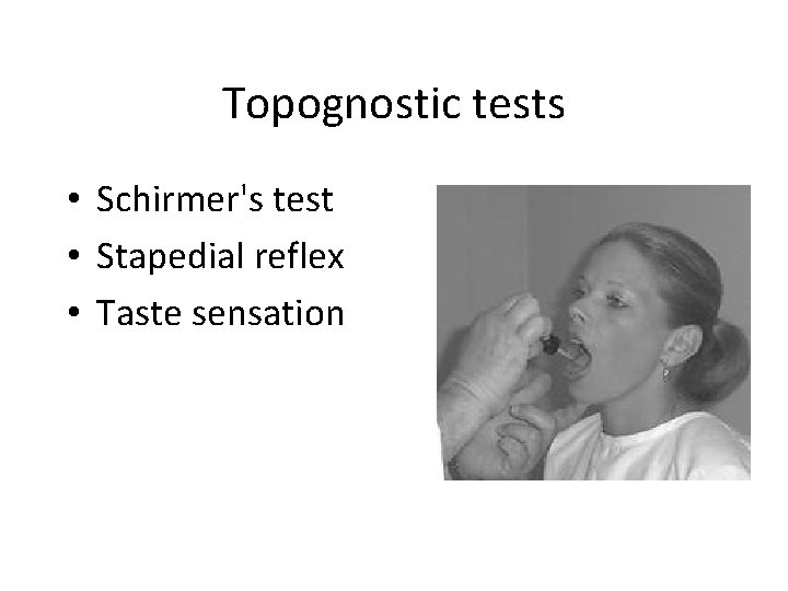
Topognostic tests • Schirmer's test • Stapedial reflex • Taste sensation
Topognostic tests • Schirmer's test • Stapedial reflex • Taste sensation
Causes of facial paralysis • Intracranial causes • Cranial (intratemporal) causes • Extracranial causes
Causes of facial paralysis • Congenital: Birth trauma • Traumatic: Head and neck injuries & surgery • Inflammatory: O. M, Necrotizing O. E. , Herpes • Neoplastic: Meningioma, malignancy ear or parotid • Neurological: Guillain-Barre syndrome, multiple sclerosis • Idiopathic: Bell’s palsy
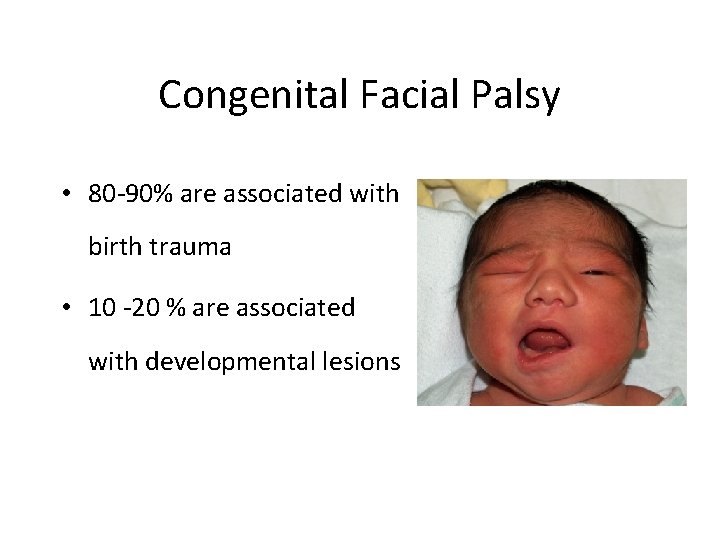
Congenital Facial Palsy • 80 -90% are associated with birth trauma • 10 -20 % are associated with developmental lesions
Inflammatory causes of facial paralysis
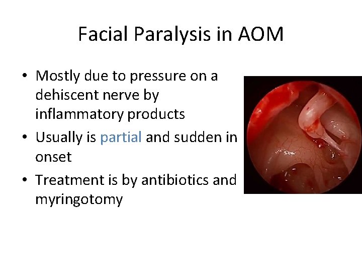
Facial Paralysis in AOM • Mostly due to pressure on a dehiscent nerve by inflammatory products • Usually is partial and sudden in onset • Treatment is by antibiotics and myringotomy
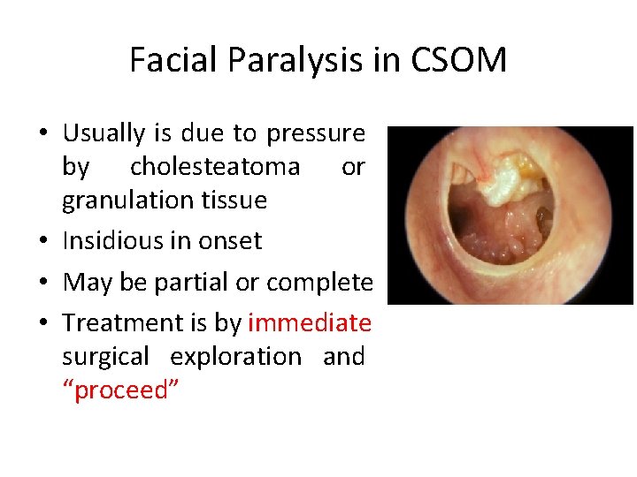
Facial Paralysis in CSOM • Usually is due to pressure by cholesteatoma or granulation tissue • Insidious in onset • May be partial or complete • Treatment is by immediate surgical exploration and “proceed”
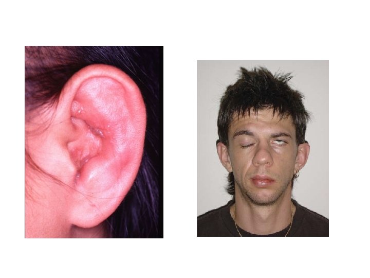
Herpes Zoster Oticus (Ramsay Hunt Syndrome) • Herpes zoster affection of cranial nerves VII, VIII, and cervical nerves • Facial palsy, pain, skin rash, SNHL and vertigo
HERPES ZOSTER OTICUS (RAMSAY HUNT SYNDROME) • Herpes zoster affection of cranial nerves VII, VIII, and other nerves • Facial palsy, pain, skin rash, SNHL and vertigo • Vertigo improves due to compensation • SNHL is usually irreversible • Facial nerve recovers in about 60% • Treatment by: Acyclovir, steroid and symptomatic
Traumatic Facial Injury • Iatrogenic • Temporal bone fracture
Iatrogenic Facial Nerve Injury • Operations at the CP angle, ear and the parotid glands
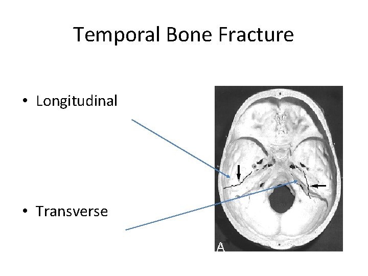
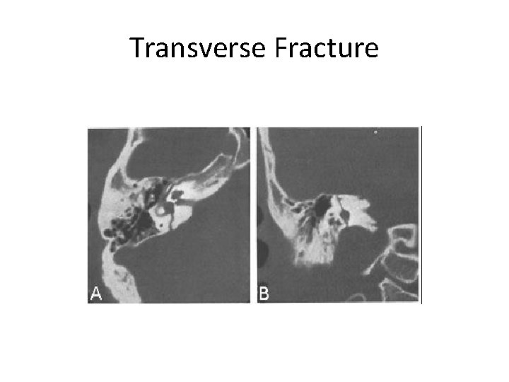
Temporal Bone Fracture • Longitudinal • Transverse
Transverse Fracture
Pathology • Edema • Transection of the nerve
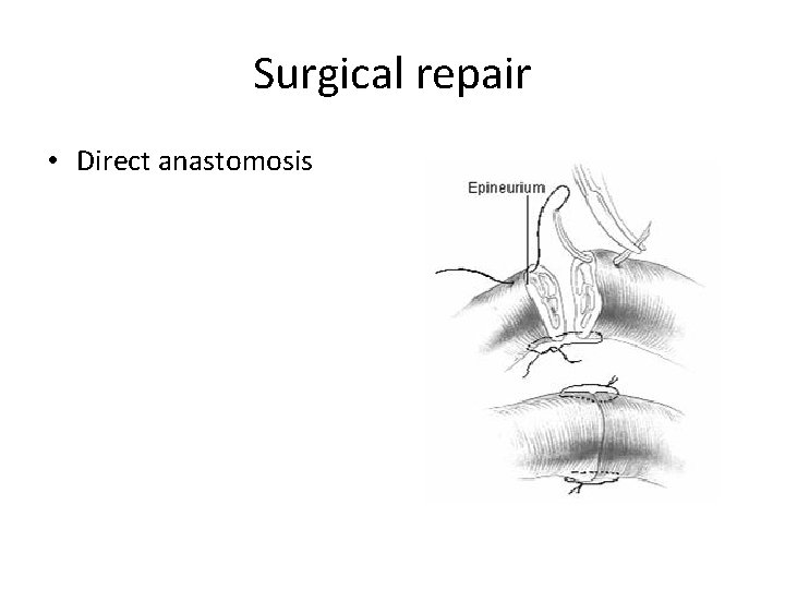
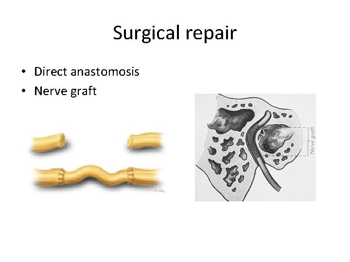
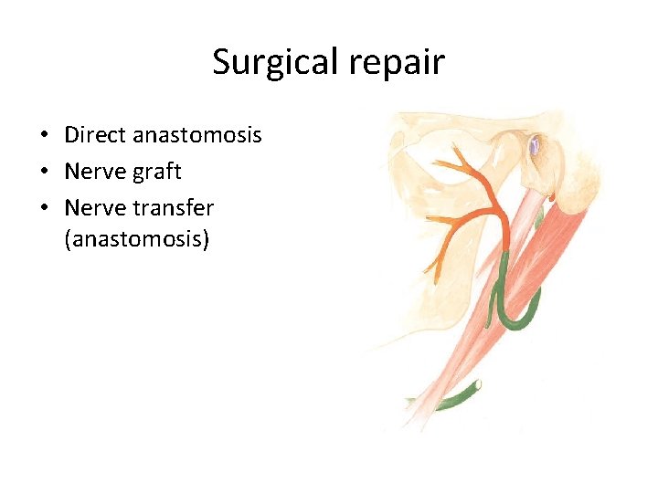
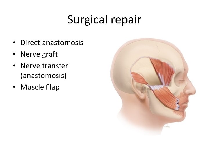
Management of Traumatic Facial Nerve Injury • If it is delayed in onset, it is usually incomplete and is due to edema – Conservative • If of immediate onset, it is usually complete and due to transection of the nerve – Surgical repair
SURGICAL REPAIR
Surgical repair • Direct anastomosis
Surgical repair • Direct anastomosis • Nerve graft
Surgical repair • Direct anastomosis • Nerve graft • Nerve transfer (anastomosis)
Surgical repair • Direct anastomosis • Nerve graft • Nerve transfer (anastomosis) • Muscle Flap
Bell’s Palsy • Most common diagnosis of acute facial paralysis • Diagnosis is by exclusion
Pathology • Edema of the facial nerve sheath along its entire intratemporal course (Fallopian canal)
Etiology • Vascular vs. viral
Clinical Features • Sudden onset unilateral FP • Partial or complete • No other manifestations apart from occasional mild pain • May recur in 6 – 12%
Prognosis • 80% complete recovery • 10% satisfactory recovery • 10% no recovery
Treatment • Reassurance • Eye protection • Physiotherapy • Medications (steroids, antivirals, vasodilators) • Surgical decompression in selected cases
Surgical Management • Debate over years • Patients with 90% degeneration • Within 14 days of onset
OFFICE HOURS Prof. YOUSRY EL-SAYED • Flat 407, Building 5 , King Abdel-Aziz Universty Hospital • Mondays from 11 am to 1 pm • Thursdays 11 am to 1 pm
THANK YOU