Physiology of CNS Somatic Motor System By Dr

Physiology of CNS Somatic Motor System By Dr. Abdel Aziz M. Hussein Assist prof of Physiology

Extrapyramidal Tracts
 and area (4) → descends to")
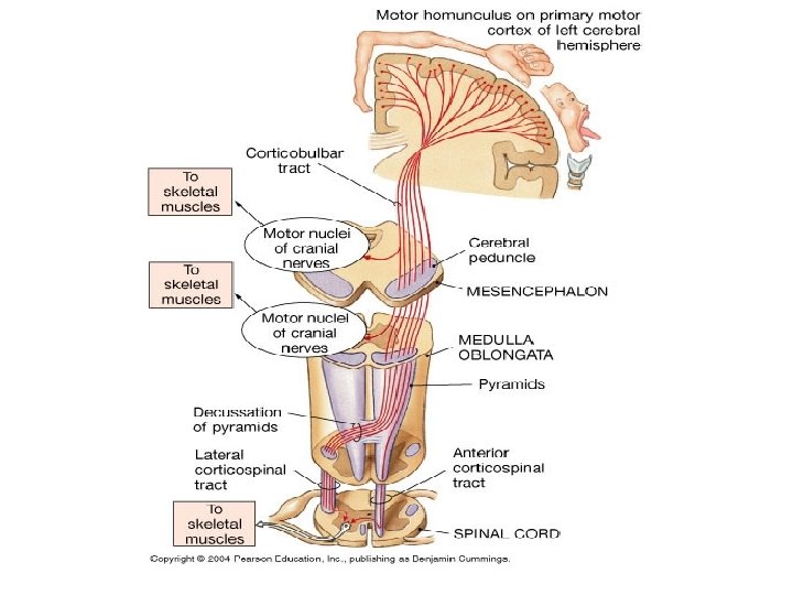
Extrapyramidal Tracts • Origin: • From area (6) and area (4) → descends to corpus striatum → Globus pallidus→ from the globus pallidus fibers pass to 1. Reticular formation 2. Vestibular nuclei 3. Red nucleus 4. Tectum of midbrain. • From these nuclei the following extrapyramidal tracts arise:

Extrapyramidal Tracts Motor areas 4 and 6 Corpus striatum Basal Ganglia Globus pallidus RF Vest. Nuclei Red Nucl. Tectum Ret. Spin T. Vest. Spin. T. Rubrospin. T. Tectospin. T.

Extrapyramidal Tracts

Rubrospinal Tract

Cerebrum Rubrospinal Tract Cerebellum Basal ganglia Red nucleus Rubrospinal T. Lateral AHCs ++

Rubrospinal Tract Functions: 1. Mediates motor signals concerned with the skilled voluntary movements performed by distal limb muscle. 2. Also facilitates & motor neurons of the distal flexors

Reticulospinal Tracts

Reticulospinal Tracts Medullary RF Pontine RF Medial RST Lateral RST. -- ++ -Medial AHCs

Reticulospinal Tracts • Functions: • a. Medial R. S. T. • It mediates facilitatory effect on the motor neurons of the antigravity ms to maintain postural support. • b. Lateral R. S. T. • It inhibits the tone in the antigravity ms under some postural conditions, which provides background to perform other motor activities.

Vestibulospinal Tracts

Vestibulospinal Tracts Medial VN Medial VST. Lateral V. N. Lateral VST ++++ ++ Medial AHCs

Vestibulospinal Tracts • Functions: • a. Lateral V. S. T. • It is facilitatory to & motor neurons of the antigravity ms to maintain body posture & equilibrium in response to impulses from the vestibular apparatus which evoked by charges in head position or exposure to acceleration. • b. Medial V. S. T. • It is facilitatory to & motor neurons of the neck and upper limb muscles that are involved in regulation of the head & upper limb position during exposure to acceleratory movements

Tectospinal Tracts

Tectospinal Tracts Superior colliculus Inferior colliculus Medial T. S. T Lateral TST ++++ Medial AHCs

Tectospinal Tracts • Functions: • This tract mediates reflex turning of the head in response to sudden visual or auditory stimuli.
")
Upper and Lower Motor Neuron Lesion (UMNL and LMNL)
")
Upper Motor Neuron Lesion (UMNL)

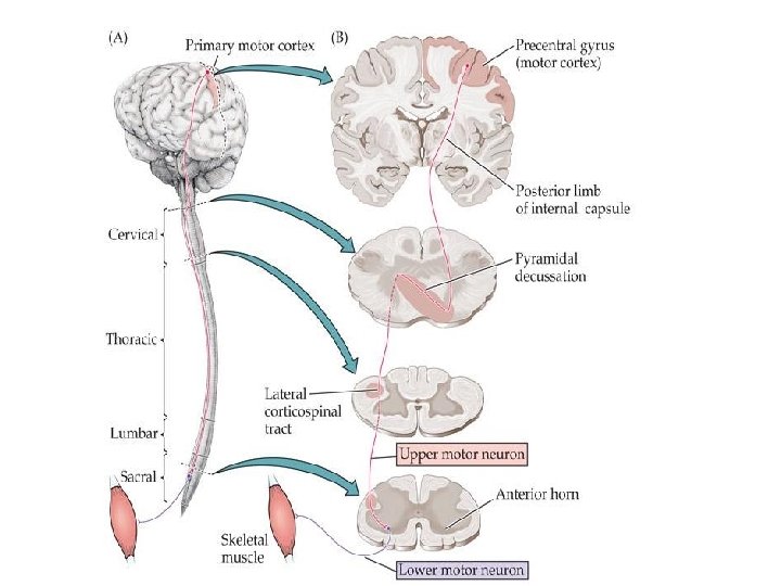
Upper Motor Neuron Lesion MOTOR CORTEX INTERNAL CAPSULE BRAIN STEM SPINAL CORD

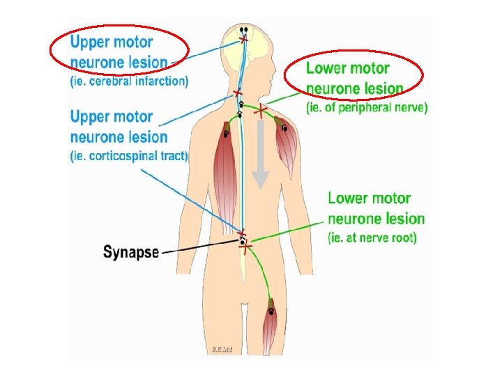
Upper Motor Neuron Lesion Def. , • It is the damage of upper motor neuron in the higher center or the descending motor tract. Causes 1. Trauma 2. Tumour 3. Vascular disorders as thrombosis or hemorrhage. Sites: • Most common site of UMNL is the internal capsule.

Internal Capsule
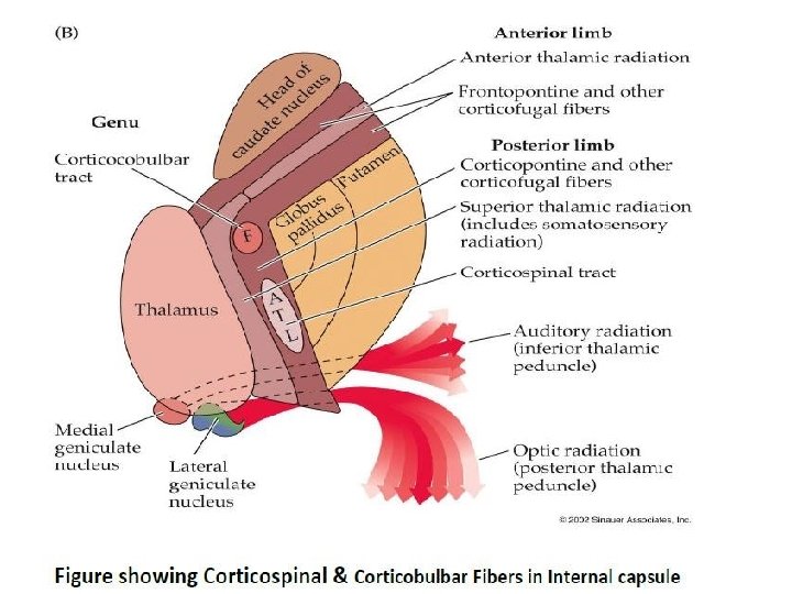

Thrombosis of Lenticulostriate artery

Damage of Posterior Limb of Internal Capsule Internal capsule Motor loss Hemiplegia Sensory loss Loss of somatic sensations, vision and hearing
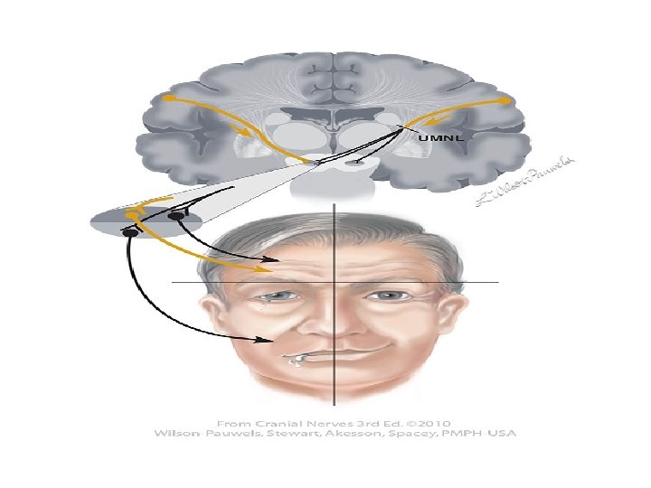
 1. Contralateral paralysis (loss of only voluntary movements) of the distal")
Motor Loss (Hemiplegia) 1. Contralateral paralysis (loss of only voluntary movements) of the distal ms of the limbs, lower facial ms and ms of the tongue. 2. Contralateral paresis (weakness i. e. , the ms retains some movements) of the axial ms and upper facial ms. • Axial ms are supplied by descending motor tracts other than CBS whereas ms of the upper face are ipsilaterally innervated by CBS tract.
")
Motor Loss (Hemiplegia or UMNL)
 3) Spasticity (increased ms tone) of the skeletal ms due to")
Motor Loss (Hemiplegia) 3) Spasticity (increased ms tone) of the skeletal ms due to increased supraspinal facilitation to -motor neurons. • A lesion at the level of internal capsule interrupts the descending inhibitory cortical fibers which feeds the inhibitory reticulospinal tract leaving the facilitatory vestibulospinal and reticulospinal to act. • This spasticity is of the clasp-knife type

Spasticity Facilitatory RF and VST Inhibitory RF
 4) Exaggerated tendon jerk & clonus: due to increased supraspinal facilitation.")
Motor Loss (Hemiplegia) 4) Exaggerated tendon jerk & clonus: due to increased supraspinal facilitation. 5) Positive Babinski's sign
 6) The paralyzed ms show no or minimal atrophy as the")
Motor Loss (Hemiplegia) 6) The paralyzed ms show no or minimal atrophy as the lower motor neuron is intact and the ms contracts reflexly. 7) Normal response of the paralyzed ms to electric stimulation a) Faradic current produces clonic or tetanic contractions b) Galvanic current produces contractions that occur only at closing (make) and opening (break) of the circuits. CCC > AOC > COC
 Contralateral hemianaesthesia i. e. loss of all sensations on the opposite")
Sensory Loss 1) Contralateral hemianaesthesia i. e. loss of all sensations on the opposite side of the body
 Contralateral homonymous hemianopia i. e. , loss of vision in the")
Sensory Loss 2) Contralateral homonymous hemianopia i. e. , loss of vision in the two opposite halves of the field of vision
 Bilateral diminution of hearing acuity. q No complete loss of hearing")
Sensory Loss 3) Bilateral diminution of hearing acuity. q No complete loss of hearing as both ears are bilaterally represented in both cortices.
")
Lower Motor Neuron Lesion (LMNL)

Lower Motor Neuron Lesion Def. , • It is damage of the lower motor neurons (the spinal AHCS and the cranial motor nuclei or their axons) resulting in skeletal ms paralysis Causes 1. Trauma 2. Neuropathy
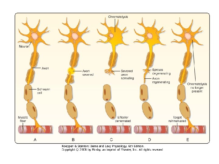
 Structural changes q. In Nerve (degeneration and regeneration q. In")
Effects of LMNL I) Structural changes q. In Nerve (degeneration and regeneration q. In muscle (atrophy and increase Ach receptors II) Functional changes 1. Flaccid paralysis 2. Fasciculation and fibrillation 3. Dennervation supersensitivity 4. Reaction of degeneration

Structural Effects of LMNL Retrograde degeneration Wallerian degeneration Regeneration Atrophy of muscles
 Flaccid paralysis: q Paralysis of denervated ms with loss")
Functional Effects of LMNL A) Flaccid paralysis: q Paralysis of denervated ms with loss of all types of movements; "voluntary, postural and reflex". q All reflexes are lost including stretch reflex resulting in loss of ms tone and tendon jerk (flaccidity). q The extent of paralysis is usually limited to a small group of ms
 Fasiculations and fibrillations: • Appears few days or weeks")
Functional Effects of LMNL B) Fasiculations and fibrillations: • Appears few days or weeks after denervation • Disappear when the motor nerve completely degenerates or successful re-innervation of the ms occurs.
 Fasiculations : • Synchronous visible contraction of the motor")
Functional Effects of LMNL B) Fasiculations : • Synchronous visible contraction of the motor unit (all ms fibers) supplied by the injured axon. • Result from spontaneous generation of action potential (injury potentials) in distal segment of the injured axon

 Fibrillations: • As degeneration of the injured axon continues,")
Functional Effects of LMNL B) Fibrillations: • As degeneration of the injured axon continues, the axon terminals are now separate from the main axon and hence, from each other. • Injury potentials are still generated along the terminals leading to asynchronous contraction of the individual ms fibers attached to terminals. • Invisible to the observer and detected only by electromyogram (EMG).

Fibrillations
 Denervation supersensitivity: • Denervated ms becomes supersensitive to acetylcholine.")
Functional Effects of LMNL C) Denervation supersensitivity: • Denervated ms becomes supersensitive to acetylcholine. • This is due to increase in the number of A. Ch. receptors which cover the entire surface of ms cell membrane.
 Reaction of degeneration: • - This means abnormal response")
Functional Effects of LMNL D) Reaction of degeneration: • - This means abnormal response of the denervated muscle to electric stimulation. • Reaction of degeneration (response of denervated ms): • INCOMPLETE REACTION: • i) Faradic current produces no response. • The denervated muscle has a prolonged chronaxia. • The faradic current is faster than the excitation time needed to produce a response. • ii) Galvanic current produces an abnormal response. • The response in general is weaker than normal and ACC > CCC.
 Reaction of degeneration: COMPLETE REACTION: • When the denervation")
Functional Effects of LMNL D) Reaction of degeneration: COMPLETE REACTION: • When the denervation is prolonged, the ms is atrophied and fibrosed. • Thus, no response occurs to both faradic and galvanic currents.

UMNL and LMNL

UMNL and LMNL

UMNL and LMNL

UMNL and LMNL

THANKS
- Slides: 58