THE CIRCULATORY SYSTEM ANATOMY Introduction The circulatory system


 separate the atrium and ventricle on each side of")


 control the beat of")


 valve into the right ventricle.")


 valve into")


,")


, splenic vein (spleen), pancreatic vein (pancreas), and mesenteric veins (small")

 right lobe, (2) left lobe, (3) caudate lobe, (4)")


- Slides: 93
THE CIRCULATORY SYSTEM ANATOMY
Introduction The circulatory system is comprised of the heart, veins, capillaries, arteries, lymph vessels, and lymph glands, which work together to supply the body tissues with nourishment and collect waste materials.
Functions of the circulatory system: Distribute nutrients, Transport and exchange oxygen and carbon dioxide, Remove waste materials, Distribute secretions of endocrine glands, Prevent excessive bleeding
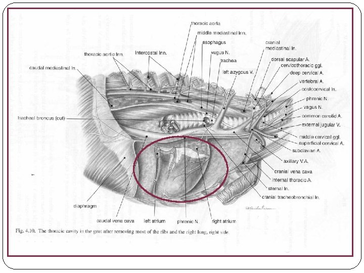
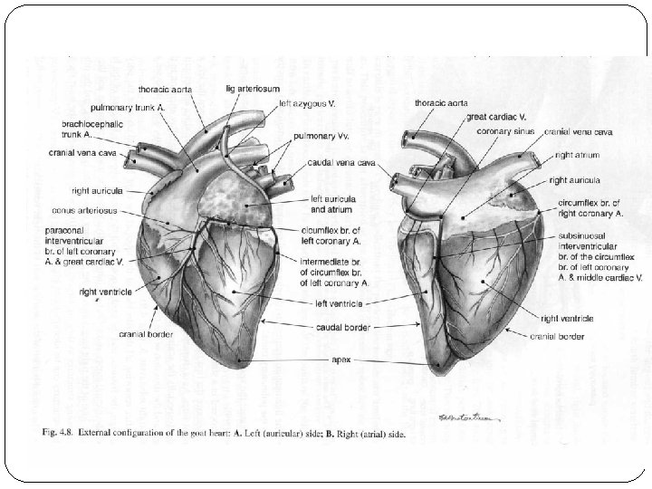
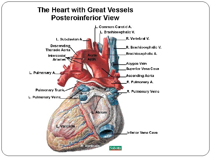
Anatomy of the heart �The heart is a funnel-shaped, hollow, muscular organ that is responsible for pumping blood to all parts of the body. �The heart is located near the center of the thoracic cavity between the lungs and is contained in the pericardial sac
Anatomy of the heart �The pericardial sac supports the heart and contains some fluid for lubrication. �The broad end, or base, of the heart is also supported by large arteries and veins. �The pointed end, or apex, of the heart is directed toward the
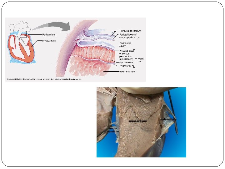
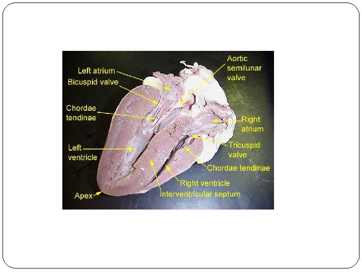
The heart wall is made up of three layers. • Epicardium – outer layer of heart wall, which is also the inner layer of epicardial sac; • Myocardium – middle layer composed of cardiac muscle • Endocardium – inner layer that consists of endothelial cells, which line the heart, covers the heart valves, and lines the blood vessels.
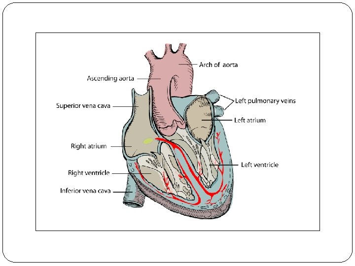
�In mammals and birds, the heart is divided into a right and left side and each side is divided into an atrium and ventricle. �Therefore, the heart is said to have four chambers (right atrium, right ventricle, left atrium, and left ventricle).
�The atrioventricular valves (AV valve) separate the atrium and ventricle on each side of the heart. �The AV valves have flaps of tissues, called leaflets or cusps, which open and close to ensure that the blood flows only in one direction and does not backflow into the atriums.
�The AV valve on the right side of the heart is called the tricuspid valve because it has three leaflets (cusps). �The AV valve on the left side of the heart is called the bicuspid valve (or mitral valve) because it has two leaflets.
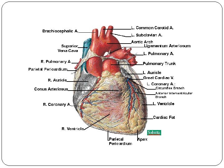
�The pulmonary valve and the aortic valve prevent blood from back-flowing into their respective ventricles. �The pulmonary valve is located between the right ventricle and the pulmonary artery. �The aortic valve is located between the left ventricle and the aortic artery.
�A group of cells called the sinoatrial node (SA node) control the beat of the heart by sending out electrical signals to make the heart pump.
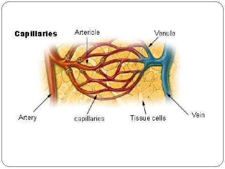
Anatomy of the Vascular System The vascular system is made up of three types of blood vessels: • Arteries, • Capillaries, and • Veins
�Arteries are blood vessels that carry blood, rich in oxygen, from the heart to other parts of the body. �The large arteries have thick walls of elastic-like tissue that enables them to withstand the blood pressure created by the heart’s beating.
�Arterioles branch into smaller vessels called capillaries. �At this junction, the arterioles have an especially thick layer of smooth muscle in their walls that carefully controls the amount of blood each capillary receives.
�Capillaries are tiny, thin-walled blood vessels that connect arteries to veins and are located in all body tissues. �Capillaries are so small in diameter that blood cells pass through in a single file
�The semi-permeable membrane of capillary walls allows nutrients, oxygen, and water to diffuse from the blood to the tissues. �Waste products, like carbon dioxide, diffuse from the tissues into the blood.
�Larger tubular connectors, which also connect arterioles to venules, are located within the capillary beds. �These tubules allow more blood to flow through an area, help warm tissues, and increase the return of blood pressure to the heart.
�Once blood passes through the capillary beds, it begins its return to the heart. �Veins are the blood vessels that return blood to the heart from all parts of the body.
�Capillaries unite to form small veins called venules. �The venules join together to form larger veins, which have thin walls and are collapsible. �For each artery, there is a much larger vein counterpart.
�Veins have valves that aid the return flow of blood and prevent the blood from reversing flow. �These valves allow for muscle contractions and movement of body parts. �The valves also assist the return flow of blood to the heart when blood pressure is low.
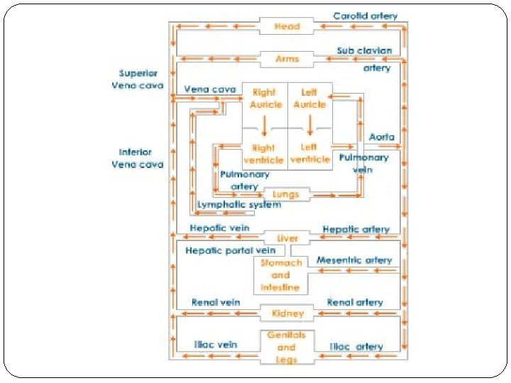
Parts of the Circulatory System �The total circulatory system is divided into two main parts: • Pulmonary circulation, and • Systemic circulation.
Pulmonary Circulation System Red portion of heart and red blood vessels carry oxygen-rich blood. Blue portion of heart and blue blood vessels carry oxygen-poor blood.
�Pulmonary circulation is the part of the circulatory system that takes the blood from the heart to the lungs, where it is oxygenated, and returns it to the heart. �The main parts of the pulmonary circulation system include the heart, pulmonary arteries, capillaries of the lungs, and pulmonary veins.
Circulation �Blood that is low in oxygen returns to the heart through two large veins called the superior (or cranial) vena cava and the inferior (or caudal) vena cava. �The un-oxygenated blood enters the right atrium of the heart.
�The blood then passes through the right atrioventricular (tricuspid) valve into the right ventricle. �The right ventricle pumps the blood through the pulmonary valve into the pulmonary artery.
�The pulmonary artery quickly divides into two branches. �Each branch of the pulmonary artery carries blood to a lung. �In the lungs the pulmonary arteries branch into capillaries that surround the alveoli
�Through diffusion, carbon dioxide moves from the blood into the alveoli and oxygen moves from the alveoli into the blood. �The oxygenated blood then returns to the heart through the pulmonary vein into the left atrium.
�From the left atrium, the blood flows through the left atrioventricular (bicuspid) valve into the left ventricle.
�The thick-walled left ventricle pumps the blood through the aortic valve into the aorta. �The amount of pressure that is required for pulmonary circulation is much less than what is required for systemic circulation. �Therefore, the muscle mass developed in the right ventricle is much less that of the left ventricle.
�Un-oxygenated blood is dark or brownish red, while oxygenated blood is bright red. �In the pulmonary system, un- oxygenated blood is carried by the pulmonary arteries and oxygenated blood is carried by pulmonary veins. �In the systemic system, arteries carry oxygenated blood and veins carry un-oxygenated blood.
System
�The systemic circulation includes the flow of oxygenated blood from the heart to the tissues in all parts of the body and the return of unoxygenated blood back to the heart. �The blood vessels, including the arteries, capillaries, and veins, are the main parts of systemic circulation.
�Through systemic circulation, oxygen and nutrients are delivered to the body tissues via the arteries. �Blood is filtered during systemic circulation by the kidneys (most of the waste) and liver (sugars).
�The systemic circulatory system is complex and its functions vary. �The systemic circulatory system is divided into subsystems for particular regions of the body.
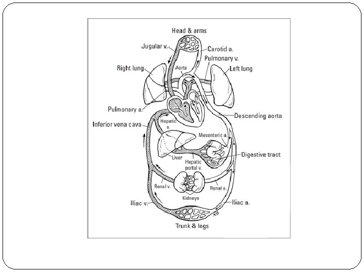
The Flow of Blood Through the Systemic Circulatory System �Oxygenated blood leaves the left ventricle of the heart through the aorta, the largest artery in the body. �The left and right coronary arteries immediately branch from the aorta and carry fresh blood to the heart muscle itself. �The coronary veins quickly return that blood back to the heart.
�The brachiocephalic trunk is the next branch from the aorta. �The carotid arteries branch off the brachiocephalic trunk and carry oxygenated blood to the neck and head region. �Blood from the neck and head region returned by the jugular
�The left and right brachial arteries also branch from the brachiocephalic trunk to supply blood to the shoulders and forelegs. �The thoracic aorta refers to the portion of the aorta that goes from the heart, through the thoracic cavity to the diaphragm. �The portion of the aorta that goes from the diaphragm, through the abdominal region, to the last lumbar vertebrae is called the abdominal aorta.
�Branches from the thoracic aorta supply oxygenated blood to the lungs (via bronchial arteries), esophagus, ribs and diaphragm. �The celiac artery branches from the aorta immediately past the diaphragm and itself branches into the gastric, splenic, and hepatic arteries.
�The gastric artery supplies blood to the stomach. �The splenic artery supplies blood to the spleen. �The hepatic artery supplies blood to the liver.
�The cranial and caudal mesenteric arteries branch from the abdominal aorta and carry blood to the small and large intestines. �The renal arteries are next to branch from the abdominal aorta
�The renal arteries have two important functions: • supply blood to the kidneys, and • carry large volumes of blood to the kidneys for filtration and purification.
�From the renal arteries arise arteries that supply blood to the testicles in males (internal spermatic arteries) and parts of the reproductive system in females (uteroovarian arteries).
�The abdominal aorta ends where it branches into the internal and external iliac arteries. �The internal iliac artery supplies blood to the pelvic and hip region. �The external iliac artery branches into the femoral arteries.
�The femoral arteries and their branches supply oxygenated blood to the hind legs.
�Veins normally accompany arteries and often have similar names. �Veins are always larger than the arteries and are sometimes more visible than arteries because they are closer to the skin surface. �Most veins eventually empty the un- oxygenated blood into the vena cavas.
�The cranial veins return the blood from the head, neck, forelegs, and part of the thoracic cavity to the right atrium of the heart via the superior vena cava. �These cranial veins include the jugular vein, brachial veins, internal thoracic veins, and the vertebral veins.
�The caudal veins return blood from the iliac, lumbar, renal, and adrenal veins to the right atrium of the heart via the inferior vena cava. �Before blood is returned to the heart from the stomach, pancreas, small intestine, and spleen, it goes through the liver for filtration. This portion of the systemic system is known as the hepatic portal system.
�The gastric vein (stomach), splenic vein (spleen), pancreatic vein (pancreas), and mesenteric veins (small intestines) empty into the portal vein that carries the blood to the liver.
�In the liver, the portal vein branches into smaller venules and finally into capillary beds. �In the capillary beds of the liver, nutrients are exchanged for storage and the blood is purified. �The capillaries then join into venules that empty into the hepatic vein, which carries blood to the inferior (caudal) vena cava.
Liver of a sheep: (1) right lobe, (2) left lobe, (3) caudate lobe, (4) quadrate lobe, (5) hepatic artery and portal vein, (6) hepatic lymph nodes, (7) gall
Anatomy of the limphatic system �The lymphatic system is part of the immune system and acts as a secondary (accessory) circulatory system.
system: • remove excess fluids from body tissues, • absorb fatty acid and transport fat to circulatory system, and • produce immune cells (lymphocytes, monocytes, and plasma cells).
�Blood fluid escapes through the thin-walled capillaries into spaces between body tissue cells. �Lymph vessels, which have very thin walls, pick up these fluids called lymph.
Flow of Blood & Lymph Within Tissue
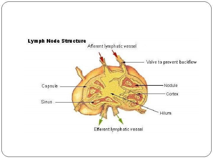
�The lymph vessels join to form larger ducts that pass through lymph nodes (or glands). �Each lymph node has a fibrous outer covering (capsule), a cortex, and a medulla.
�Lymph nodes filter foreign substances, such as bacteria and cancer cells, from the lymph before it is re-entered into the blood system through the larger veins. �Lymph nodes, which are scattered among the lymph vessels, act as the body’s first defense against infection.
�Lymph nodes produce the following cells: • Lymphocytes – a type of white blood cell, • Monocytes – a leukocyte that protects against blood-borne pathogens, and • Plasma cells – produce antibodies.
�Each lymph node has its own blood supply and venous drainage. �The lymph nodes usually have names that are related to their location in the body.
�When a specific location gets infected, the lymph nodes in that area will enlarge to fight the infection. �If the lymph node closest to an infected area is unable to eliminate the infection, other lymph nodes in the system will attempt to fight the infection.
Anatomy of the blood �Blood is an important component of the circulatory system. The amount of blood that a domestic animal has is expressed in terms of percentage of body weight Domestic Animal % of Body Weight Cattle Sheep 7. 7 % 8. 0% Horses 9. 7%
�Plasma, which makes up 50 – 65% of the total volume of blood, is a straw-colored liquid containing water (90%) and solids (10%). �The solids in plasma include inorganic salts and organic substances such as antibodies, hormones, vitamins, enzymes, proteins, and glucose (blood sugar).
The non-plasma, or cellular, portion of blood is composed of red blood cells, white blood cells, and platelets. �From left to right: Red blood cell (erythrocyte); Platelet (thrombocyte); White blood cell (leukocyte).
�Red blood cells, called erythrocytes, are responsible for carrying oxygen from the lungs to various body tissues. �Red blood cells contain hemoglobin, which gives them their characteristic red color and helps them carry the oxygen.
�Red blood cells are biconcave discs, a shape that provides a large area for oxygen exchange.
Red blood cells are produced in the red marrow of bones.
�Most domestic animals have a red blood cell count of seven million cells per cubic millimeter of blood. �Red blood cells will last from 90 to 120 days and are removed from the blood by the spleen, liver, bone marrow, or lymph nodes when they are worn out.
�Blood platelets, or thrombocytes, are oval-shaped discs that are formed in the bone marrow. �Blood platelets help prevent blood loss from injuries to blood vessels by forming clots (white thrombus).
�Platelets may secrete a substance that causes the clot to contract and solidify. �Platelets may also secrete a substance that causes an injured vessel to constrict at the injury.
White blood cells, or leukocytes, are divided into two general categories: • Granulocytes, and • Agranulocytes.
�Granulocytes are the category of leukocytes that contain granules within the cytoplasm. �Granulocytes include: • Neutrophils, • Eosinophils, and • Basophils.
�Neutrophils – produced by bone marrow, neutrophils fight disease by migrating to the point of infection, absorbing bacteria, and destroying them.
�Eosinophils - a type of granulocyte that plays a role in combating infection by parasites, as well as, impacting allergies and asthma.
�Basophils – rare granulocytes that are responsible for the symptoms of allergies, including inflammation.
�Agranulocytes are the category of leukocytes that contain very little, if any, granules. �Agranulocytes are produced by the lymph nodes, spleen, thymus, and other lymphoid tissue.
�There are two types of agranulocytes: • Lymphocytes, and • Monocytes.
�Lymphocytes – agranulocytes that produce and release antibodies at site of infections to fight disease.
�Monocytes are agranulocytes that absorb disease-producing materials, such as bacteria that cause tuberculosis, through phagocytosis.
�In domestic animals, approximately 85% to 90% of the leukocytes in domestic mammals are neutrophils and lymphocytes. �The total number of neutrophils and lymphocytes are about equal, but temporary stress increases the ratio of neutrophils to lymphocytes until that stress is removed.
�When bacterial infections occur, the number of white blood cells normally increases. �When viral infections occur, the number of white blood cells normally decreases. �Therefore, the concentration of white blood cells can help diagnose
Tugas Makalah tentang anatomi unggas : 1 -15 Osteology 16 -30 Digestive system 31 -45 Respiratory system 46 -60 Nervous system