Open Vs Closed Circulatory System n Open ended


 divide into")


 muscle, surrounded by a double")


, located in the")


 PM")
 Atria relax, impulses from")

 ventricle contracts, pressure of blood forced in the aorta")


- Slides: 37
Open Vs Closed Circulatory System
n Open ended vessel which blood leaves Blood flows around cells Enters heart through openings n Closed Blood remains in vessels Materials exchanged by diffusion More efficient then open system, Faster and flow can be increased/decreased
Blood Vessels 3 main types 1. Arteries (A for Away from heart) divide into smaller vessels- Arterioles 2. Veins (to heart) divide into smaller veins- Venules 3. Capillaries tiny vessel (1 cell in thickness), link arteries & veins
Arteries Vs Veins What have they both got in common? What is different?
Arteries Vs Veins Both have the same 3 layers. Main difference is in the middle layer. n 3 layers 1. Outer, is tough, made of protein called collagen which prevents wall from over expanding. 2. Middle, is muscle and elastic, it can alter the size of the vessel 3. Inner, are living cells called endothelium.
Valves n Blood pressure is the force blood applies against the wall of a blood vessel. n Blood pressure is highest in arteries, this causes the arteries to expand, which causes our pulse. n Pressure in veins is very weak. Ordinary muscles help push the blood by contracting and squeezing to return blood to the heart. n Because pressure is low, must prevent backflow n Valves control direction of blood flow
Artery Carries blood away from heart Vein Carries blood to heart Blood under high pressure Blood under low pressure Thick walls Thin walls Small Lumen Large Lumen Blood flows in pulse No pulse Blood rich in oxygen Blood low in oxygen Valves absent Valves present
Capillaries n Tiny, branched vessels n Walls are permeable, allow materials to move across the walls n 100, 000 km
The Heart n Made of cardiac (slow to fatigue) muscle, surrounded by a double membrane called Pericardium. n It contracts 100, 000 times a day n Pumps 5 -20 litres of blood per minute n Why such a difference? 5 -20
Structure of the Heart Divided into 2 side by a wall called Septum. 4 chambers, 2 upper = Atria, 2 lower = Ventricles n Atria & ventricles are separated by valves n These valves are held in place by tendons n These tendons are held in place by Papillary Muscles. n Valve on right has 3 flaps, tricuspid valve n Valve on left has 2 flaps, bicuspid valve n Semilunar valves prevent blood returning to heart n
Pulmonary artery Semilunar valve Superior vena cava Aorta Semilunar valve Pulmonary vein Pacemaker Left atrium Right atrium Bicuspid valve Inferior vena cava Tendon Tricuspid valve Right ventricle Cardiac Muscle Pericardium Papillary Muscle Left ventricle Septum
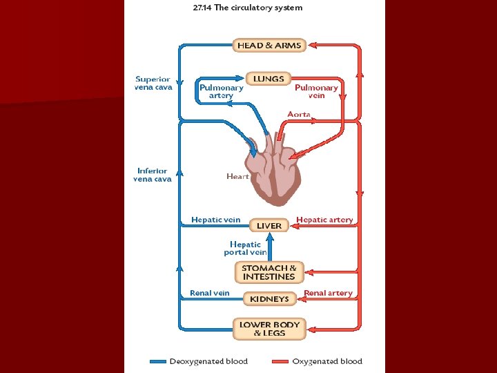
Blood flow in the heart Step 1 n Deoxygenated blood enters the right atrium through 2 venae cavae. Superior - head, arms, chest. Inferior – Lower part of body. n The right atrium contracts forcing blood into right ventricle n The venae cavae close to prevent backflow
Blood flow in the heart Step 2 n When the right ventricle contracts the tricuspid valve is forced closed n Blood is then forced out of the heart and into the lungs through the semilunar valve in the pulmonary artery.
Blood flow in the Heart Stage 3 n Oxygenated blood returns to the heart from the lungs and enters the left atrium through the pulmonary veins (only vein to ever carry oxygen rich blood) n Pumped through bicuspid valve into the left ventricle
Blood flow in the heart Stage 4 n When the left ventricle contracts the bicuspid valve closes. n Blood is pumped out of the heart through the semilunar valve in the aorta n When the ventricles relax these valves close to stop backflow
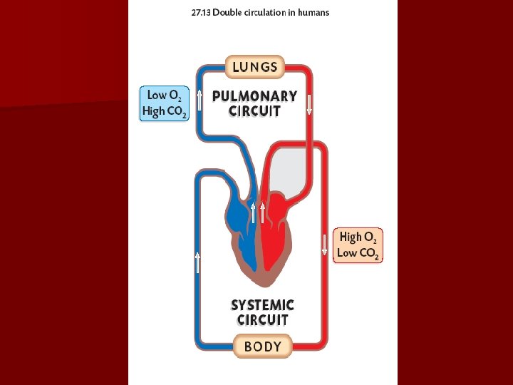
Double Circulation n Heart is really a double pump. The 2 sides of the heart are divided by the septum. n Why is this necessary? n Answer= n Hence humans have a 2 -circuit circulatory system. The Pulmonary Circuit & The Systemic Circuit
Double Circulation n Pulmonary circuit Right ventricle pumps de-oxygenated blood around the pulmonary circuit. This circuit is short so the walls of the ventricle are thin
Double Circulation n Systemic Circuit The Left ventricle pumps oxygenated blood to head, arms, trunk and legs. This is a much longer route so the walls are much thicker and stronger.
Double Vs Single Circulation n Allows oxygen rich and poor blood to be kept separate. n Ensures the blood pressure is high enough to reach all parts of the body. n Single circulation system can only produce low pressure, this restricts activities of animal i. e. worm
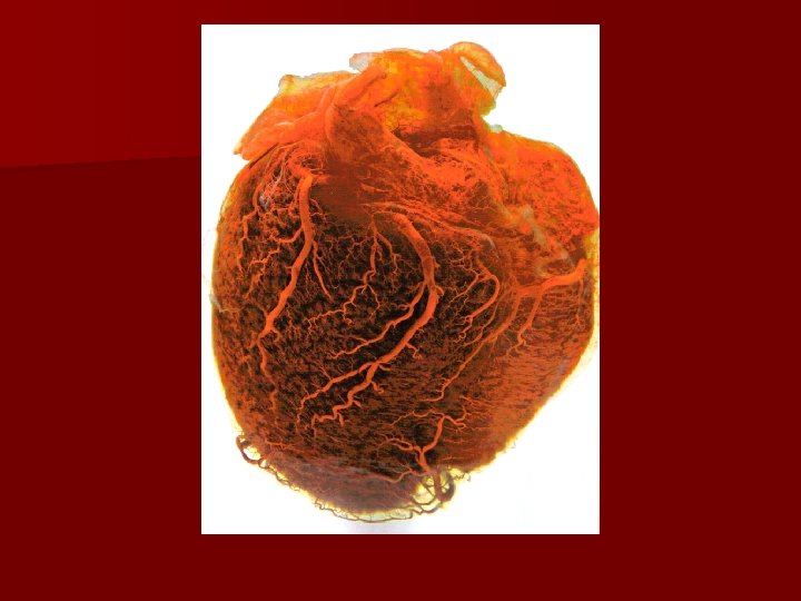
Blood supply to the Heart itself n supplied by the coronary arteries These are connected by the aorta n coronary atrium n Blockage veins drain blood into the right of the coronary arteries is a common cause for heart attacks
What Controls the Heartbeat n The pacemaker or SA (Sino-atrial node), located in the wall at the top of the right atrium n It sends out a regular electrical impulse which causes the atria to contract, followed by the ventricles. n Frequency of these impulses are controlled by the brain & can speed up or slow down.
Pulmonary artery Superior vena cava Pacemaker Inferior vena cava Pulmonary vein Aorta
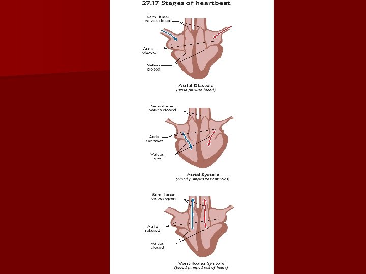
Stages of heartbeat Diastole = Chambers relax Systole = Chambers contract 1. Blood enters heart (Atrial Diastole) both Ventricles & Atria are relaxed. All valves are closed.
Stages of Heartbeat 2. Blood is pumped from atria to ventricles (Atrial systole) PM cause atria to contract (how? ), this pumps blood into ventricle. The tri & bi-cuspid valves open while the venae cava & pulmonary veins close. Semi-lunar valves remain closed.
Stages of heartbeat 3. Blood leaves the heart (Ventricular Systole) Atria relax, impulses from AV (atrio-ventricular) nodes cause ventricles to contract. This forces blood out through the pulmonary artery & Aorta The pressure forces open semi-lunar valves & closes the tri & bi-cuspid valves Ventricles relax, closing the semi-lunar valves.
Sound of Heartbeat n “Lub-dub” n Caused sound by valves being forced shut n Lub due to bi & tri-cuspid valves closing n Dub due to semi-lunar valves snapping shut
Pulse n When left (thick) ventricle contracts, pressure of blood forced in the aorta causes it to expand & then contract n This forms a wave down the arteries called pulse n Average adult pulse rate is 72 beats/min Most people between 60 -100
Blood Pressure n This is the force exerted by the blood against the walls of the blood vessels n Human blood pressure is measured by finding the amount of pressure needed to stop the flow of blood n This produces 2 readings 1. When there is a pulse 2. When no pulse
Blood Pressure n Typical blood pressure in adults is 120/80 mm of mercury. These values rise with age. n If the lower value goes over 95 mm of Hg the person suffers from high blood pressure n High blood pressure is caused by blockages in arteries.
Effects of smoking on Heart n Tabacco contains 400 harmful chemicals, including nicotine (more addictive then heroin) n Nicotine pressure n Carbon n In causes increased heart rate & blood monoxide reduces O 2 carried by blood Ireland 7000 people a yr die form smoking
Effects of exercise on Heart n When we exercise our muscles get bigger & stronger this is the same for our heart n Exercise weight n Most improves circulation and reduces body beneficial exercise is aerobic exercise e. g. Walking, jogging, running, swimming and Dancing so get your feet moving.
Effects of Diet on Heart n 3 main factors = salt, fat & being overweight n Harmful fats found in red meat & dairy, they contain Cholesterol which forms blockages n 1 packet of processed foods contains your entire days intake of salt n Obesity attacks causes high blood pressure & heart