
Chapter 38 Circulation Circulatory System Accepts oxygen nutrients


- Slides: 40
Chapter 38: Circulation
Circulatory System • Accepts oxygen, nutrients, and other substances from the respiratory and digestive systems and delivers them to cells • Accepts carbon dioxide and wastes from cells and delivers them to respiratory and urinary systems for disposal
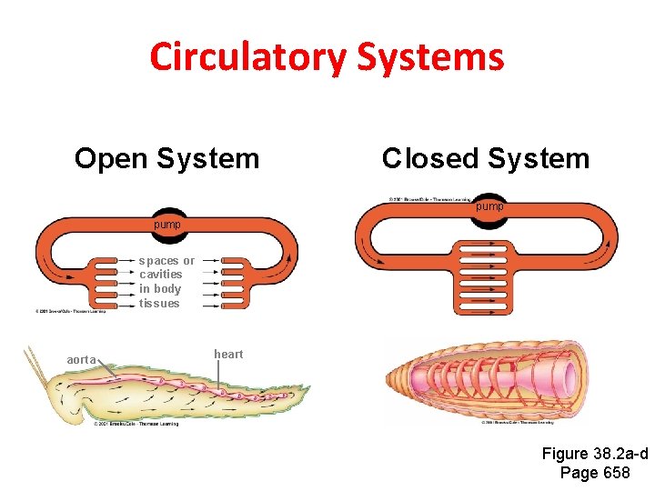
Circulatory Systems Open System Closed System pump spaces or cavities in body tissues aorta heart Figure 38. 2 a-d Page 658
Velocity of Flow Varies • Volume of blood flowing through vessels always has to equal heart’s output • Flow velocity is highest in large-diameter transport vessels • Flow velocity is slowest in capillary beds; blood spreads out into many vessels with greater total cross-sectional area
Vertebrate Systems • Fish – Two-chambered heart pumps blood through one circuit • Amphibians – Heart pumps blood through two partially separate circuits • Birds and mammals – Four-chambered heart pumps blood through two entirely separate circuits
Flow Analogy river in river out lake 1 2 3 1 1 2 2 3 3 Figure 38. 3 d Page 658
Functions of Blood • Transports oxygen and nutrients to cells • Carries carbon dioxide and wastes away from cells • Helps stabilize internal p. H • Carries infection-fighting cells • Helps equalize temperature
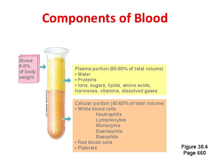
Components of Blood 6 -8% of body weight Plasma portion (50 -60% of total volume) • Water • Proteins • Ions, sugars, lipids, amino acids, hormones, vitamins, dissolved gases Cellular portion (40 -50% of total volume) • White blood cells Neutrophils Lymphocytes Monocytes Eosinophils Basophils • Red blood cells • Platelets Figure 38. 4 Page 660
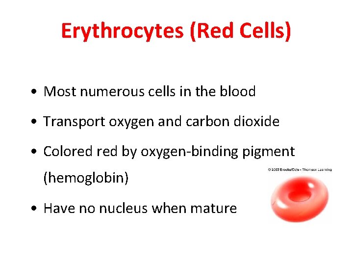
Erythrocytes (Red Cells) • Most numerous cells in the blood • Transport oxygen and carbon dioxide • Colored by oxygen-binding pigment (hemoglobin) • Have no nucleus when mature
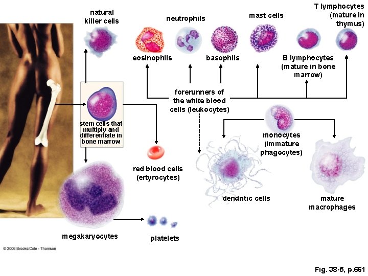
Leukocytes (White Cells) • Function in housekeeping and defense • Cell types Basophils Eosinophils B lymphocytes Neutrophils T lymphocytes Macrophages NK cells Dendritic cells Mast cells
natural killer cells mast cells neutrophils eosinophils basophils T lymphocytes (mature in thymus) B lymphocytes (mature in bone marrow) forerunners of the white blood cells (leukocytes) stem cells that multiply and differentiate in bone marrow monocytes (immature phagocytes) red blood cells (ertyrocytes) dendritic cells megakaryocytes mature macrophages platelets Fig. 38 -5, p. 661
Blood Cell Development • Stem cells in bone marrow are unspecialized cells that retain the capacity to divide • Some daughter cells of stem cells differentiate to form blood cells • Body must continually replace blood cells
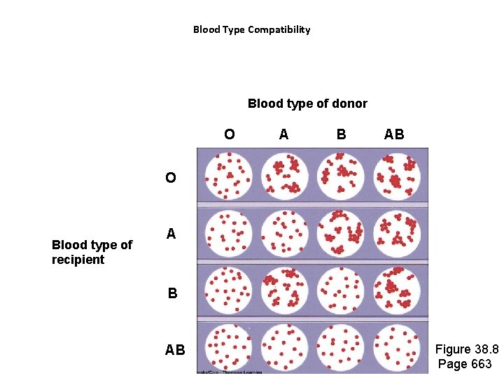
Blood Type in Transfusions • Require that donor and recipient have same blood type • If bloods of incompatible types are mixed, recipient’s immune system will attack and destroy donor cells • This is an agglutination reaction
ABO Blood Type • Type A red cells have one type of marker at surface • Type B red cells have a different type of marker • Type AB cells have both markers • Type O cells have neither marker
Blood Type Compatibility Blood type of donor O A B AB O Blood type of recipient A B AB Figure 38. 8 Page 663
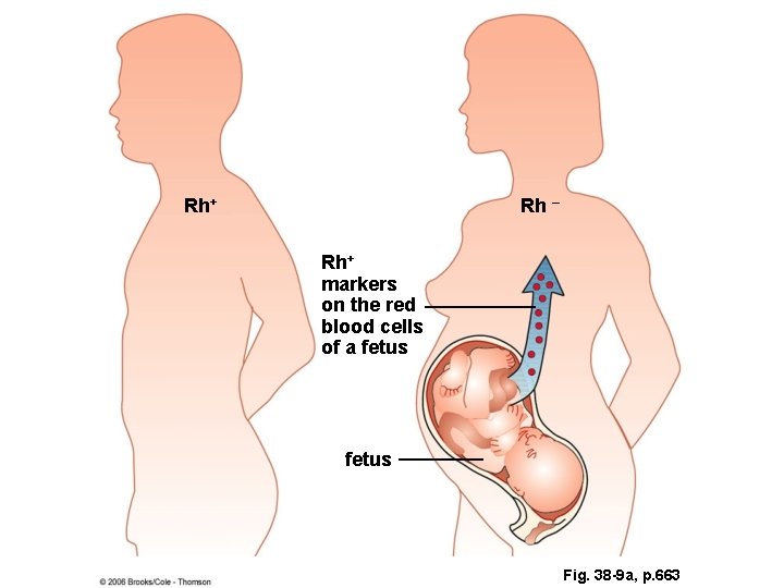
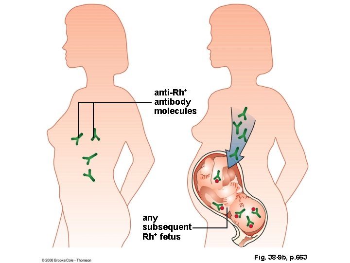
Rh Blood Type • Based on presence or absence of Rh marker on red cells • Can cause problems during pregnancy – If mother is Rh negative, – has previously carried Rh positive child, – is carrying fetus that is Rh positive • Mother’s antibodies can attack fetal cells
Rh+ Rh – Rh+ markers on the red blood cells of a fetus Fig. 38 -9 a, p. 663
anti-Rh+ antibody molecules any subsequent Rh+ fetus Fig. 38 -9 b, p. 663
Human Heart Is a Double Pump • Partition separates heart into left and right sides • Each pumps blood through a different circuit
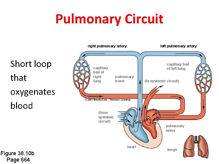
Pulmonary Circuit right pulmonary artery Short loop that oxygenates blood capillary bed of right lung left pulmonary artery capillary bed of left lung pulmonary trunk (to systemic circuit) (from systemic circuit) pulmonary veins heart Figure 38. 10 b Page 664 lungs
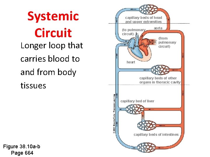
Systemic Circuit Longer loop that carries blood to and from body tissues capillary beds of head and upper extremities (to pulmonary circuit) aorta (from pulmonary circuit) heart capillary beds of other organs in thoracic cavity capillary bed of liver capillary beds of intestines Figure 38. 10 a-b Page 664
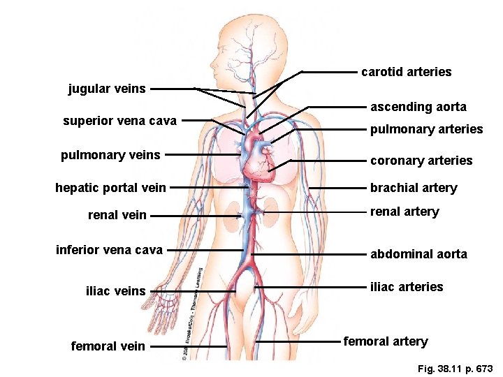
Major Vessels carotid arteries jugular veins superior vena cava pulmonary veins hepatic portal vein renal vein inferior vena cava iliac veins femoral vein ascending aorta pulmonary arteries coronary arteries brachial artery renal artery abdominal aorta iliac arteries femoral artery Fig. 38. 11 p. 673
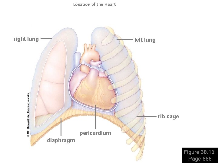
Location of the Heart right lung left lung rib cage pericardium diaphragm Figure 38. 13 Page 666
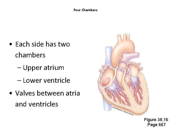
Four Chambers • Each side has two chambers – Upper atrium – Lower ventricle • Valves between atria and ventricles Figure 38. 16 Page 667
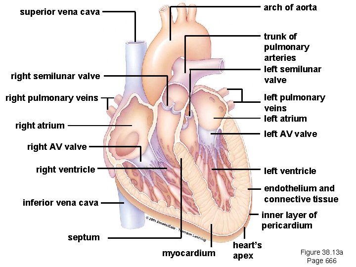
arch of aorta superior vena cava trunk of pulmonary arteries left semilunar valve right semilunar valve left pulmonary veins left atrium right pulmonary veins right atrium left AV valve right ventricle left ventricle endothelium and connective tissue inferior vena cava inner layer of pericardium septum Heart Anatomy myocardium heart’s apex Figure 38. 13 a Page 666
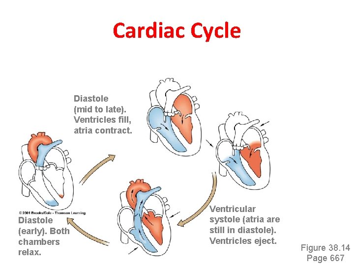
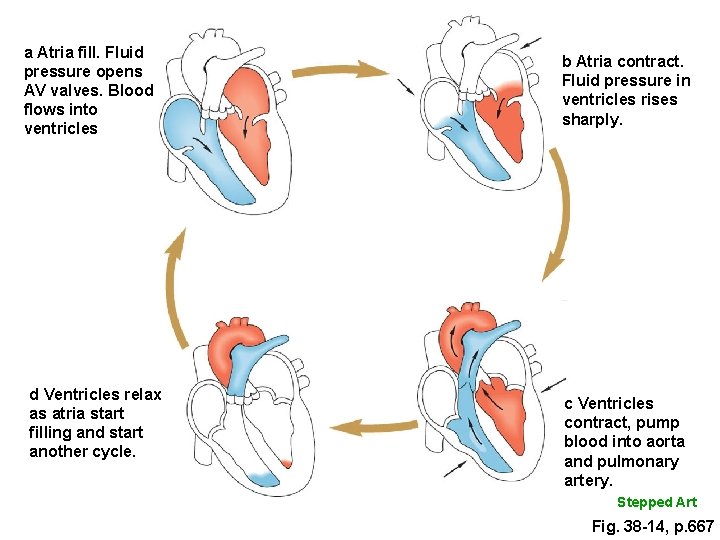
Cardiac Cycle Diastole (mid to late). Ventricles fill, atria contract. Diastole (early). Both chambers relax. Ventricular systole (atria are still in diastole). Ventricles eject. Figure 38. 14 Page 667
a Atria fill. Fluid pressure opens AV valves. Blood flows into ventricles d Ventricles relax as atria start filling and start another cycle. b Atria contract. Fluid pressure in ventricles rises sharply. c Ventricles contract, pump blood into aorta and pulmonary artery. Stepped Art Fig. 38 -14, p. 667
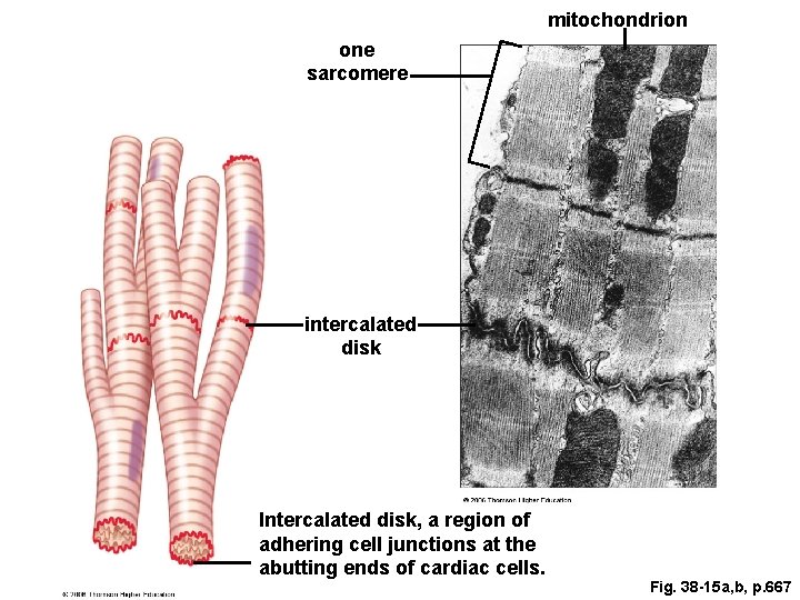
mitochondrion one sarcomere intercalated disk Intercalated disk, a region of adhering cell junctions at the abutting ends of cardiac cells. Fig. 38 -15 a, b, p. 667
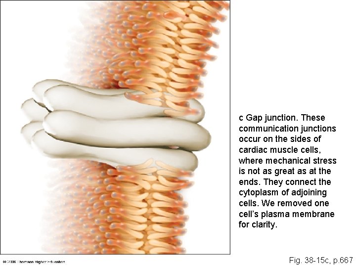
c Gap junction. These communication junctions occur on the sides of cardiac muscle cells, where mechanical stress is not as great as at the ends. They connect the cytoplasm of adjoining cells. We removed one cell’s plasma membrane for clarity. Fig. 38 -15 c, p. 667
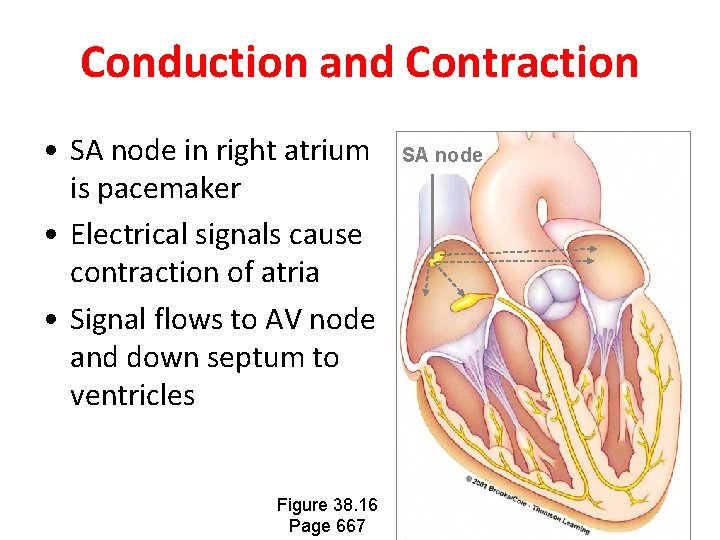
Conduction and Contraction • SA node in right atrium is pacemaker • Electrical signals cause contraction of atria • Signal flows to AV node and down septum to ventricles Figure 38. 16 Page 667 SA node
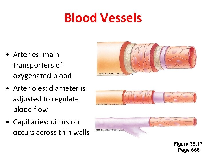
Blood Vessels • Arteries: main transporters of oxygenated blood • Arterioles: diameter is adjusted to regulate blood flow • Capillaries: diffusion occurs across thin walls Figure 38. 17 Page 668
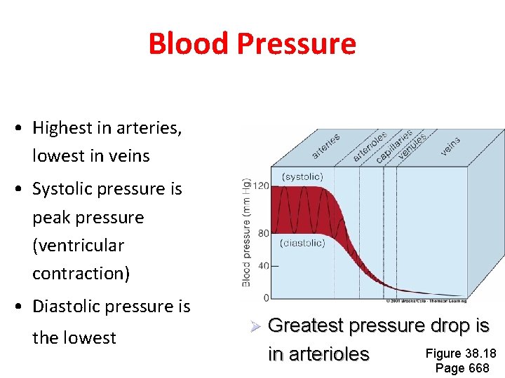
Blood Pressure • Highest in arteries, lowest in veins • Systolic pressure is peak pressure (ventricular contraction) • Diastolic pressure is the lowest Ø Greatest pressure drop is Figure 38. 18 in arterioles Page 668
Controlling Blood Pressure • Cardiac output adjusted by controls over rate and strength of heartbeat • Total resistance is controlled by vasoconstriction of arterioles • Baroreceptor response is main short-term control of blood pressure
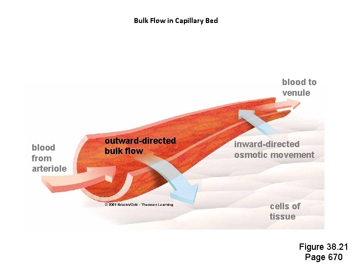
Bulk Flow in Capillary Bed blood to venule blood from arteriole outward-directed bulk flow inward-directed osmotic movement cells of tissue Figure 38. 21 Page 670
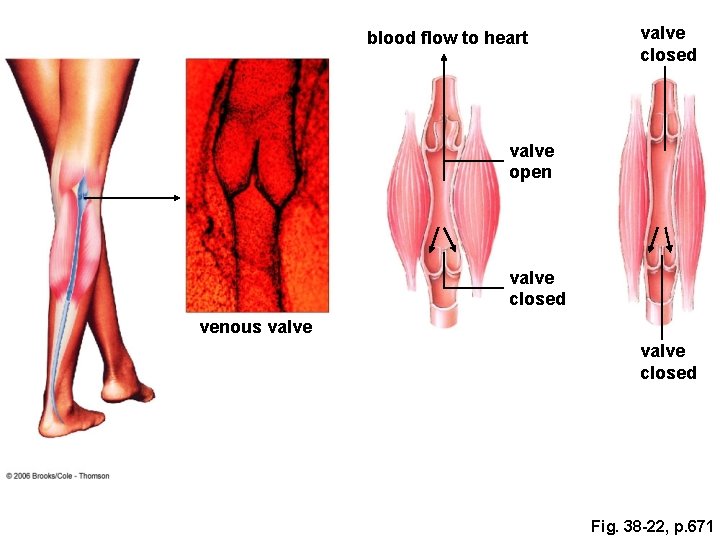
blood flow to heart valve closed valve open valve closed venous valve closed Fig. 38 -22, p. 671
Hypertension • Blood pressure above 140/90 • Tends to be genetic • May also be influenced by diet • Contributes to atherosclerosis • “Silent killer” - few outward signs
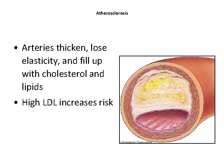
Atherosclerosis • Arteries thicken, lose elasticity, and fill up with cholesterol and lipids • High LDL increases risk
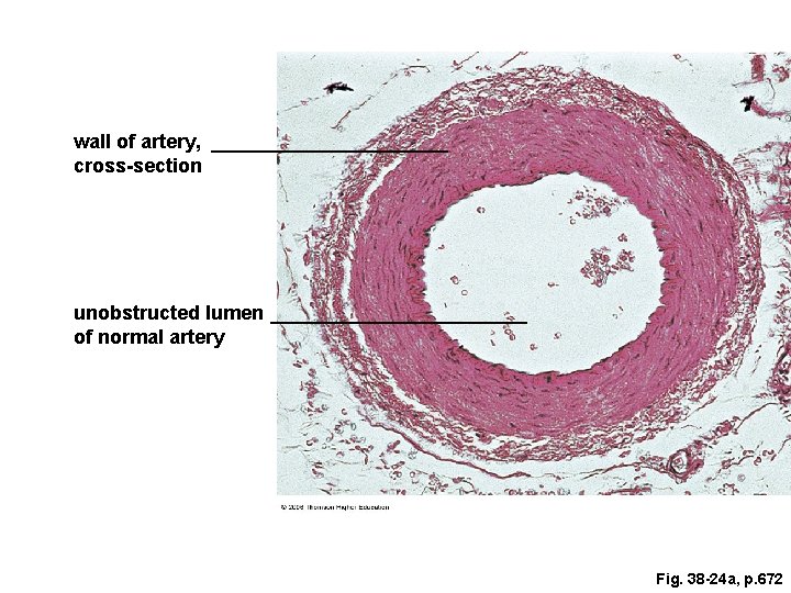
wall of artery, cross-section unobstructed lumen of normal artery Fig. 38 -24 a, p. 672
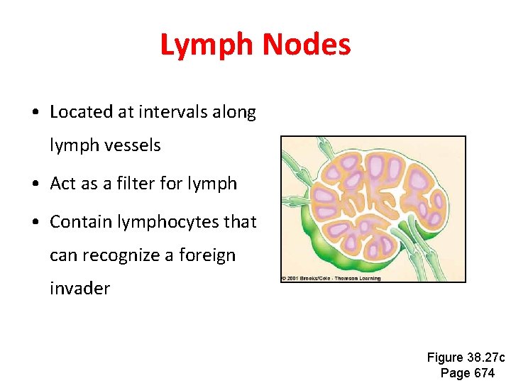
Lymph Nodes • Located at intervals along lymph vessels • Act as a filter for lymph • Contain lymphocytes that can recognize a foreign invader Figure 38. 27 c Page 674
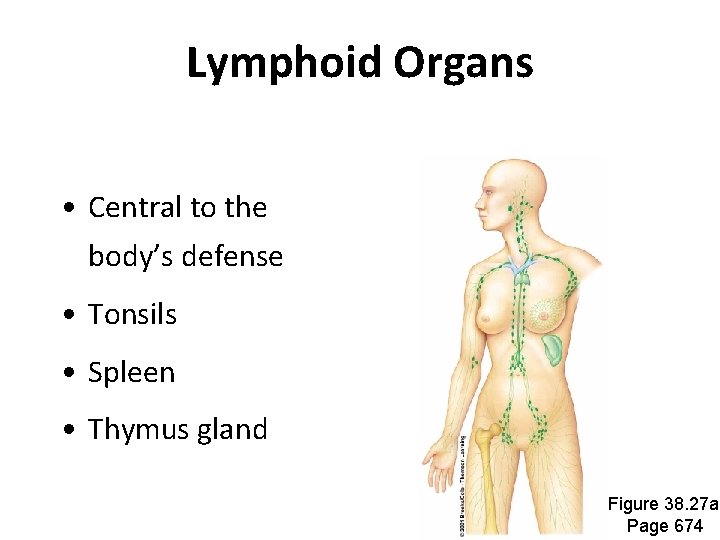
Lymphoid Organs • Central to the body’s defense • Tonsils • Spleen • Thymus gland Figure 38. 27 a Page 674