Directions If you havent taken the mink test


 • Erupt from 6 months to 2")


 Blood from heart")


 Lower esophageal Sphincter (esophagus/stomach) Pyloric Sphincter (Stomach/Small Intestines) Ileocecum Sphincter")


- Slides: 70
Directions • If you haven’t taken the mink test or the unit 08 muscle test yet, you will do that today. • Go sit in the back and wait for the test to be handed out. • If you have taken the test, you will be finishing the reading guide on chapter 15 that we started a few weeks ago with the substitute. • Get an i. Pad and a book. • Download the reading guide from my website. • Daily updates 2/23/2016 (you’ll need to scroll down) • Finish the reading guide. • You should already have it started in your notes.
Write Unit 09 The Digestive System
Write 15. 1 Introduction
Write Introduction • Digestion: • The mechanical and chemical break down of foods • The absorption of nutrients.
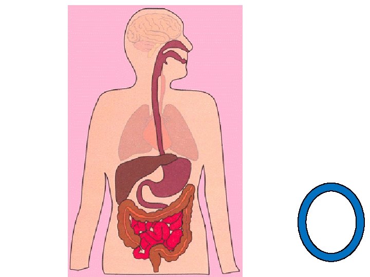
Write Introduction • Digestive System • Alimentary Canal and Accessory Organs • Alimentary Canal • Single tube that runs from mouth to anus • Mouth, pharynx, esophagus, stomach, small intestine large intestine and anal canal
Write Introduction Alimentary Canal Organs Accessory Organs 1. Salivary Glands Mouth 2. Pharynx 2. Liver 3. Esophagus 3. Gall Bladder 4. Stomach 4. Pancreas 5. Small Intestines 6. Large Intestines 7. Anal Canal The digestive system is a tube, open at both ends. It supplies body cells with nutrients that can be used to make energy and build polymers that the body needs.
Listen
Listen
Watch Digestive System Video • https: //www. youtube. com/watch? v=_QYwsc. ALNng
Write 15. 2 General Characteristics of the Alimentary Canal
Watch General Characteristics of the Alimentary Canal • The tube that makes up your alimentary canal is made up of four layers of tissue. Mucosa Tissue Types Submucosa Epithelium, Loose connective tissue, tissue smooth muscle tissue Muscular Layer Serosa Smooth muscle tissue Epithelial tissue Structures found Glands within the membrane Glands, blood vessels, Cicrular and lymphatic vessels and longitudinal fibers nerves None Function Nourishes and removes waste from surrounding tissue Secretes serous fluid Protection Secretion Absorption Peristalsis
Listen
Listen
Write Movements of the Tube • The motor functions of the alimentary canal are of two basic types: • Mixing movements • Rhythmic smooth muscle contractions • Propelling movements • Peristalsis • Contraction behind the food • Dilation in front of the food
Watch Peristalsis Video • https: //www. youtube. com/watch? v=Ujr 0 UAby. PS 4
Write 15. 3 Mouth
Write Structures and Functions of the Mouth Structure Function Cheeks Skin, fat and muscle Expression and chewing Lips Muscle, sensory receptors Judge temperature and texture Tongue Muscle Move food Papillae of the tongue Rough projections Friction for handling food Taste buds Frenulum Membranous fold Connects tongue to floor of mouth Palate Hard and soft parts Closes opening to nasal cavity and pharynx Uvula Muscular arch Closes opening to nasal cavity and pharynx Palatine tonsils Lymphatic tissue Help body fight infection Pharyngeal tonsils (adenoid) Lymphatic tissue Help body fight infection
Write Teeth types • Primary teeth (deciduous) • Erupt from 6 months to 2 -4 years old • 20 total • Not permanent (fall out) • Secondary Teeth • 6 years until 20 years • 32 total • Permanent
Listen
Write Teeth Types • Incisors • Bite off large chunks of food • Cuspids/canine • Grasp and tear food • Bicuspids and Molars • Grinding food
Listen
Draw
Write 15. 4 Salivary Glands
Write Salivary Glands • Glands that secrete Saliva • Two Secretory cells • Serous • Secrete watery fluid with amylase • Amylase • Enzyme that breaks down carbohydrates • Mucous Cells • Secrete mucus • Binds and lubricates food
Write Major Salivary Glands Parotid Submandibular Sublingual Size Largest Medium Smallest Location Anterior/inferior to Floor of mouth ear Floor of the mouth Secretion Watery fluid + amylase mucous More viscous than parotid
Write 15. 5 Pharynx and Esophagus
Listen
Write Pharynx and Esophagus • Pharynx • Cavity posterior to the mouth • Connects nasal and oral cavities to the esophagus • Structures of the Pharynx • Nasopahrynx • Provides air from nasal cavity • Oropharynx • Passageway for food from mouth • Movement of air to and from nasopharynx • Laryngopharynx • Passage way to esophagus • Epiglottis • Membrane that blocks trachea when swallowing
Write Swallowing Mechanism 1. Soft Palate/uvula rises preventing food from entering the nasal cavity. 2. Epiglottis blocks trachea 3. Tongue presses against soft palate separating oral cavity from pharynx 4. Muscle contraction pulls pharynx upwards towards the food 5. Lower portion of pharynx relax and open esophagus 6. Peristalsis https: //www. youtube. com/watch? v=p. Nc. V 6 y. Afq-g
Write Esophagus • Straight collapsible tube • Food passageway • Lower esophageal sphincter • Prevent stomach acid from going up into the esophagus • Heartburn Video • https: //www. youtube. com/watch? v=NTUCt. Dqc. Ykg
Write 15. 6 Stomach
Listen
Write Stomach • J Shaped wrinkly Pouch • 1 liter capacity • Mixes and digests food
Draw and Label
Parts of the Stomach • Cardiac Region • Fundic Region • Body Region • Pyloric Canal
Write Gastric Secretions • Gastric Glands • Composed of 3 cell types • Mucous cells • Secrete mucous • Chief Cells • Digestive enzymes • Parietal Cells • Hydrochloric Acid (HCl) • All three fluids together = Gastric Juice
Write Important Enzymes • Pepsinogen: Pepsinogen • Inactive precursor to pepsin • Pepsin: Pepsin • Digests proteins Pepsinogen + Hydrochloric Acid = Pepsin
Write Gastric Juice Regulation • Gastric Juice is regulated nervously and hormonally • Smell and taste can stimulate • Hormones: • Gastrin: Gastrin Increases gastric juice production • Choleocystokinenin: Choleocystokinenin stops stomach contractions while food empties into small intestine.
Listen
Write Mixing and emptying action • Chyme: Food + gastric Juice • Movement of food: • Liquids • Fastest • Carbohydrates • Faster • Proteins • Medium • Fats • Slow
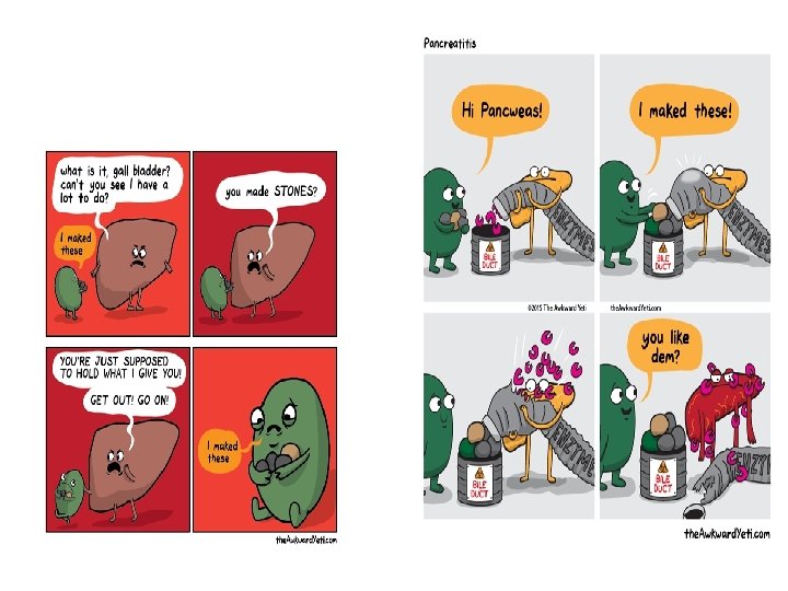
Write 15. 7 Pancreas
Listen
Write Pancreas • Pancreas • Function: Secretion • Endocrine Secretions: Insulin • Exocrine Secretion: Pancreatic Juice • Enzymes that digest • Carbohydrates • Pancreatic amylase • Fats • Pancreatic lipase • Nucleic acids • Nucleases • Proteins • Trypsin • Chymotrypsin • carboxypeptidase
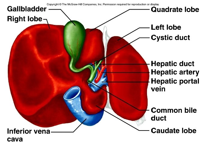
Write 15. 8 Liver
Write Liver • Functions: • • Maintain blood glucose levels Converts proteins and carbohydrates into fat Protein metabolism Storage of vitamins Helps destroy damaged red blood cells Removes toxins (ex. Alcohol) Bile production*
Listen
Detox Diets Article • http: //www. theguardian. com/lifeandstyle/2014/de c/05/detox-myth-health-diet-science-ignorance
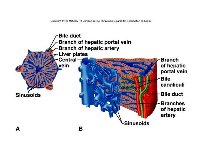
Liver Structure • Hepatic Lobules • Functional unit of the liver • Central vein • Hepatic cells radiating out • Hepatic Sinusoids • Attach to hepatic portal vein
https: //www. youtube. com/watch? v=P 5_Bxsbm. Xc. A
Blood from small intestine – Portal Vein - (High nutrient/low oxygen) Blood from heart – hepatic artery - (Low Nutrient/high oxygen)
Bile Production • Bile • Yellowish green liquid produced by liver cells • Composed of: • Bile salts: Digest fats • Bile pigments: products of red blood cell breakdown • Bilirubin – why your poop is brown • Cholesterol • Electrolytes
Gall Bladder • Function: Bile storage tank
Small Intestines
Structure: Duodenum Jejunum Ileum
Structure: • Duodenum • 25 cm • C shaped • Jejunum • 2. 5 m • Ilelum • 3. 5 m
Small Intestine Structure • Intestinal Villi • Tiny projections • Most numerous on duodenum • Greatly increase absorption
Small Intestine secretions • Enzymes • Peptidase • Proteins amino acids • Sucrase, maltase and lactase • Carbohydrates monosaccharides • Intestinal lipase • Fats fatty acids and glycerol
15. 10 Large Intestines
Structure
Structure ASCENDING COLON SIGMOID COLON RECTUM CECUM DESCENDING COLON TRANSVERSE COLON
Large Intestines Function • Secretes Mucus • Absorb Water and electrolytes • Intestinal Flora • Bacteria that break down what your body could not • Example: Fiber • Create Vitamins like B 12 and K • Excretes intestinal gas (farts)
Sphincters • Sphincter: Ring of muscles that closes an opening • Upper esophageal • Gateway between mouth and esophagus • Lower esophageal or cardiac sphincter • Gate way between esophagus and stomach • Pyloric Sphincter • Gateway between stomach and small intestines • Ileocecal • Gateway between small intestines and large intestines • Anal • Sphincter at the end of the rectum
Upper esophageal Sphincter (Mouth/pharynx) Lower esophageal Sphincter (esophagus/stomach) Pyloric Sphincter (Stomach/Small Intestines) Ileocecum Sphincter (Small/Large Intestines) Anal Sphincter (Anus – outside)
Digestive System Activity • • Glue 3 pieces of printer paper together Cut pieces of string according to length on your handout Glue string onto the printer paper in the proper order Draw in the accessory organs in the correct locations • Salivary Glands (3), Liver, Gall Bladder, Pancreas • Label the organs of the alimentary canal and the accessory organs • Mouth or Tongue, Esophagus, Stomach, Small Intestines (Duodenum, Jejunum, Ileum), Large Intestines(Cecum, Ascending Colon, Transverse Colon, Descending Colon, Sigmoid Colon), Rectum, Liver, Gall Bladder, Pancreas, Salivary Glands (Parotid, Submandibular, Sublingual) • Read the rest of your handout • *Please use the dark red string for the small intestines. • That way I won’t run out of the other colors of string! • Extra credit: Label the sphincters of the alimentary canal. • Upper esophageal sphincter, lower esophageal sphincter, pyloric sphincter, ileocecum sphincter, anal sphincter
Close read • Please make sure you have a pencil or pen for your school-wide close read post test!