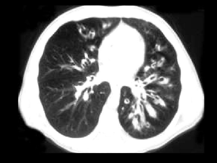
Congenital cystic adenomatoid malformation Findings Multilocular low attn


- Slides: 123
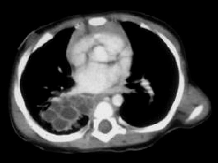
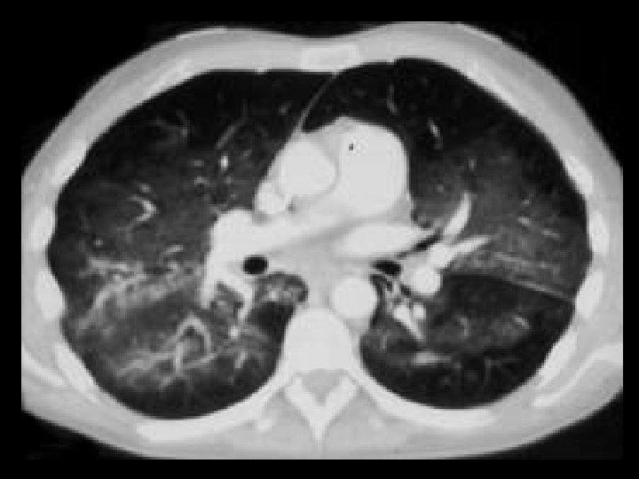
Congenital cystic adenomatoid malformation • Findings: – Multilocular low attn lesion in the RLL with thin enhancing septa • ddx: – Pulmonary abscess
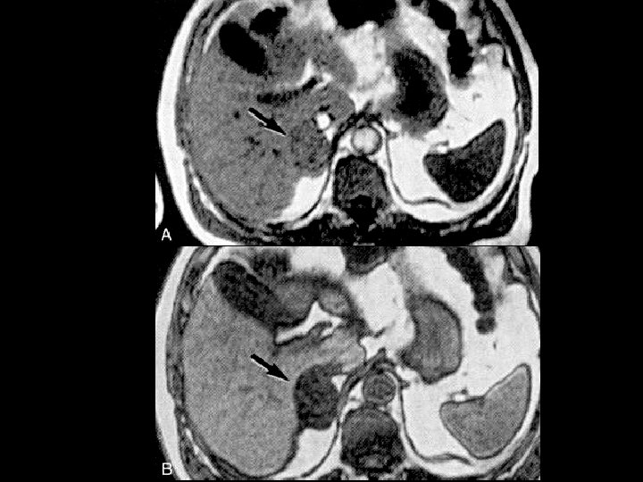
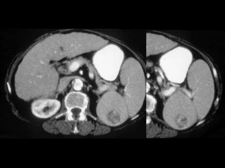
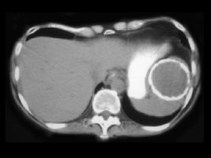
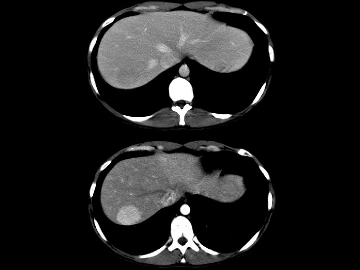
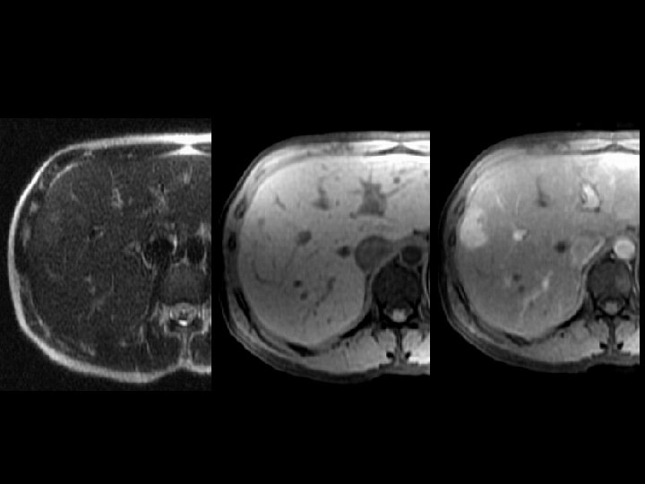
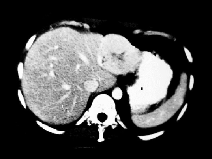
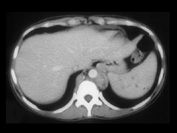
Adrenal adenoma • Findings: – Large right adrenal lesion – Isointense to liver on in -phase scan – Hypointense to liver (signal loss) on out-ofphase scan • ddx: – NONE! – This is an Aunt Minnie!
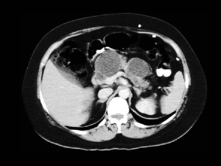
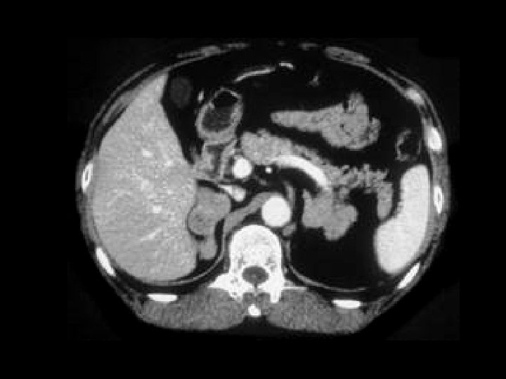
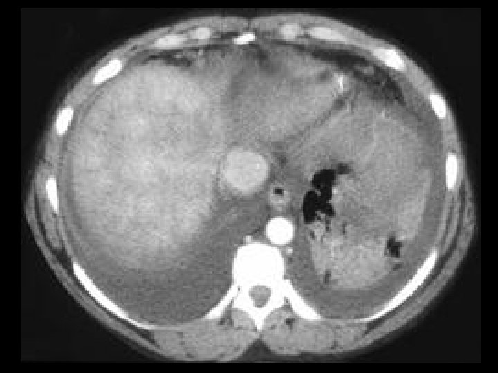
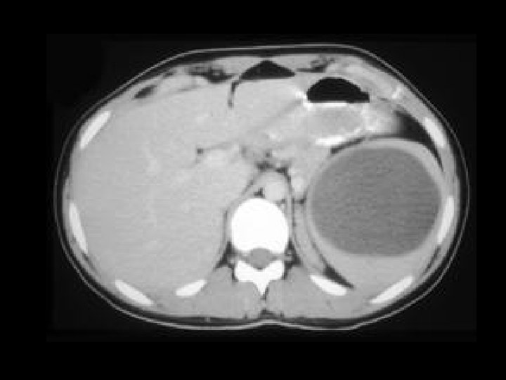
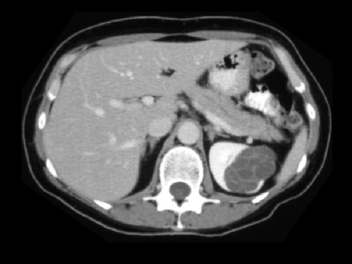
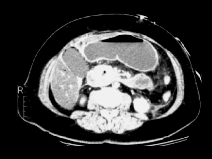
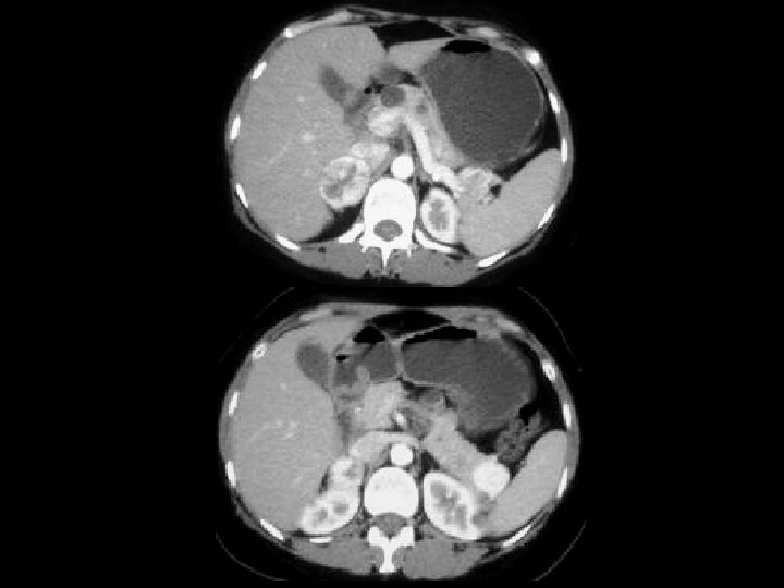
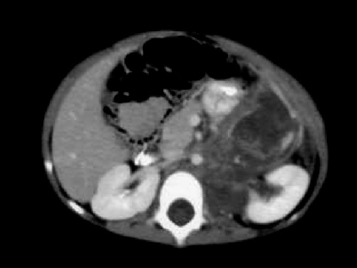
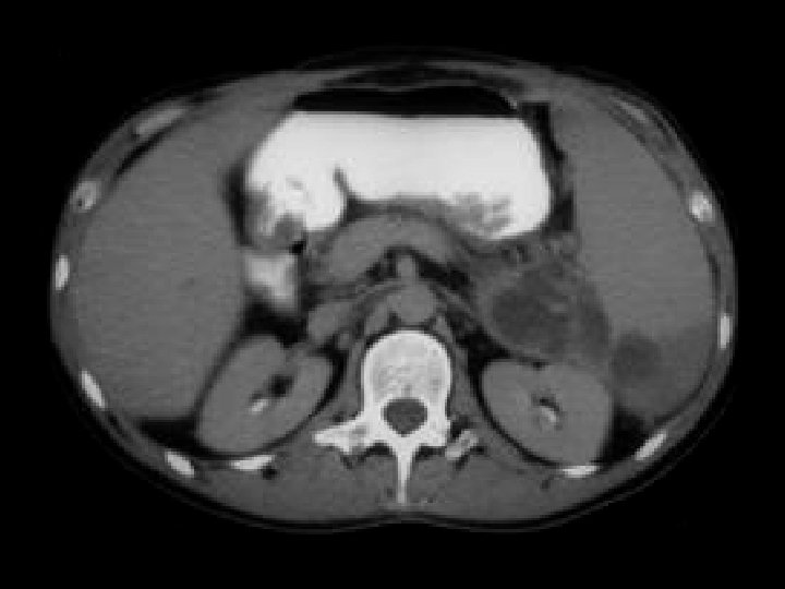
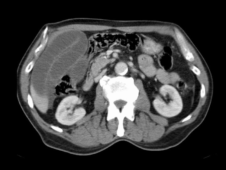
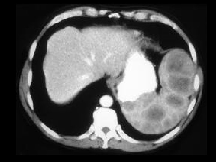
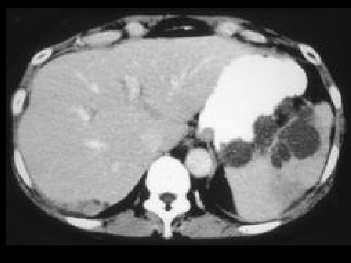
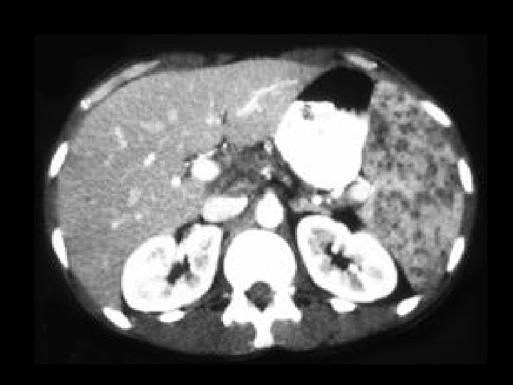
Pancreatic pseudocysts • Findings: – Multiple large pancreatic cystic lesions • ddx: – Von-Hippel Lindau – Cystic pancreatic neoplasm
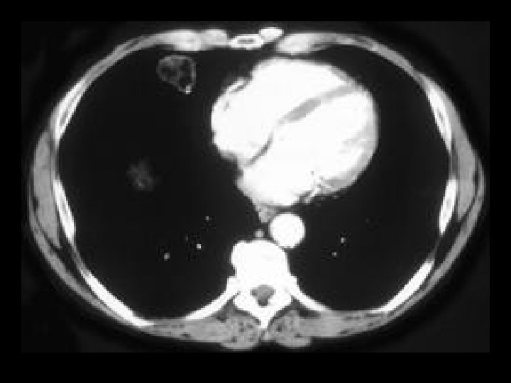
Pulmonary hamartoma • Findings: – Solitary pulmonary fatcontaining nodule in the RML – May exhibit “popcorn” calcification • ddx: – NONE! – This is an Aunt Minnie!
Adrenal macronodular hyperplasia • Findings: – Bilateral lobular enlargement of the adrenal glands. • ddx: – Cushing’s syndrome – Metastases – Infection • Tuberculosis • Histoplasmosis
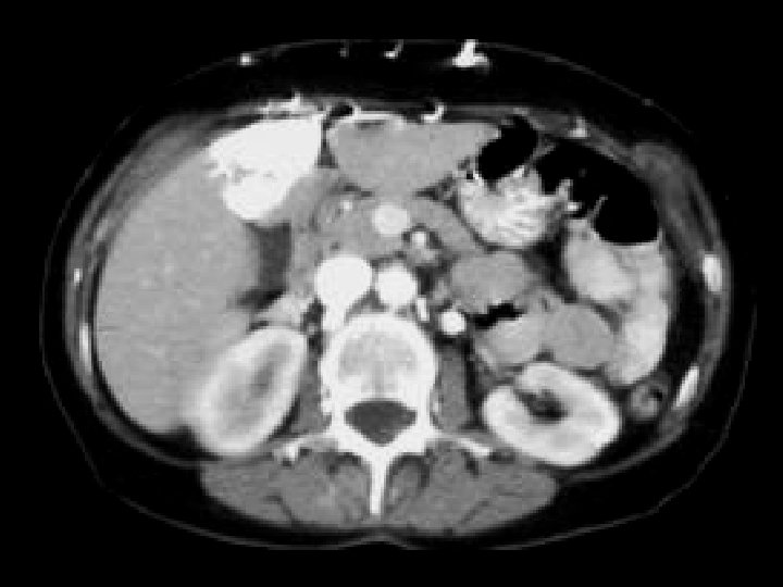
Von-Hippel Lindau • Findings: – Numerous bilateral renal cyst – Solid enhancing right renal mass = RCC • ddx: – NONE! – This is an Aunt Minnie!
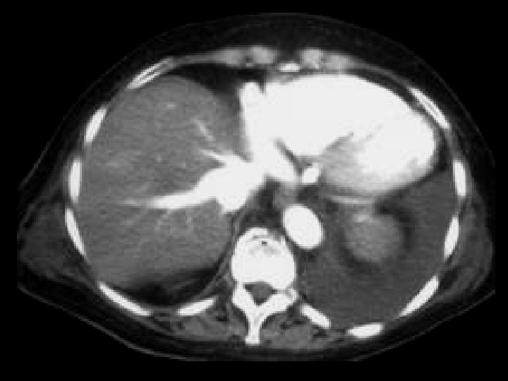
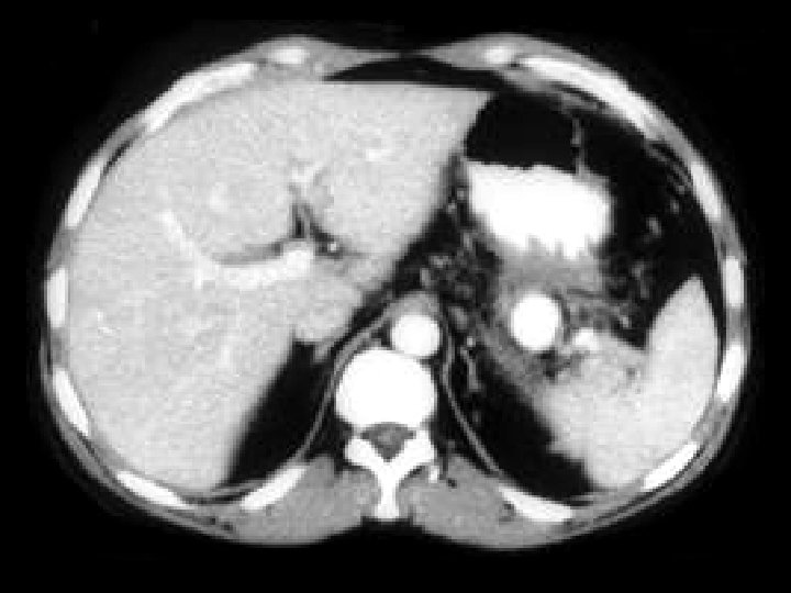
SVC obstruction • Findings: – Contrast-enhanced CT shows intense enhancement of the medial segment of the left lobe = “liver hot spot” sign – Collateral vessels seen anteriorly in the soft tissues • ddx: – NONE! – This is an Aunt Minnie!
Adrenal metastasis • Findings: – Large heterogeneous adrenal mass – Size alone makes it a surgical lesion • ddx: – Adenoma – Adenocarcinoma – Adrenal cortical carcinoma
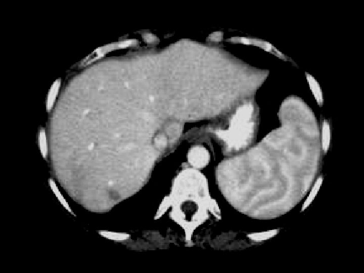
Splenic infarct • Findings: – Large spleen containing a low attenuation geographic lesion extending from the hilus to the periphery • ddx: – NONE! – This is an Aunt Minnie!
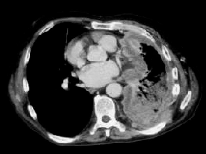
Malignant mesothelioma • Findings: – Encasement and compression of left lung by enhancing soft tissue mass – Invasion of the posterolateral chest wall and mediastinum • ddx: – Metastases • Breast • Thymus
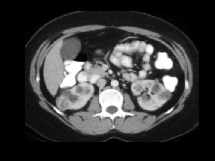
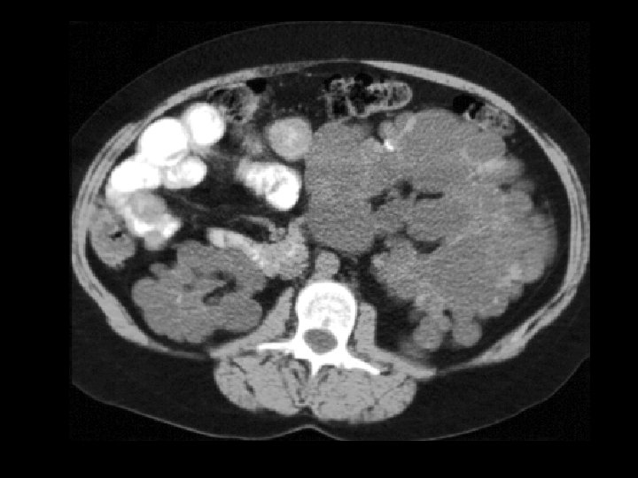
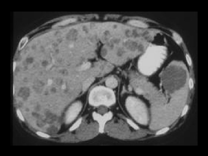
ADPKD • Findings: – Enlarged bilateral kidneys containing innumerable cysts – May also see hepatic cysts – Cysts complicated by hemorrhage or infection – NO increase risk of RCC • ddx: – NONE! – This is an Aunt Minnie!
Splenic hemangioma • Findings: – Low attenuation splenic lesion – Most common solid lesion of the spleen – Imaging and enhancement characteristics often unlike hepatic hemangiomas • ddx: – NONE! – This is an Aunt Minnie!
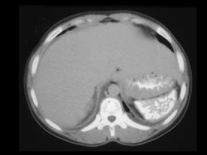
Nutmeg liver due to heart failure • Findings: – CT scan shows ascites and paradoxical enhancement of the liver during the arterial phase • ddx: – Hepatitis – Cirrhosis – Budd-Chiari
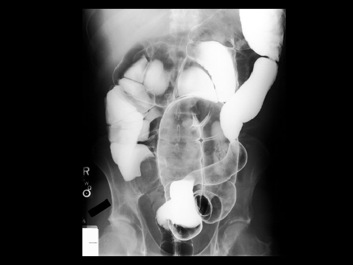
Appendix mucocele • Findings: – Smooth round filling defect at the cecal base – CT shows dilated tubular structure filled with low density material • ddx: – Mucinous adenocarcinoma of the appendix – ? Tubo-ovarian abscess
Calcified splenic cyst • Findings: – Large low density splenic lesion – Thick calcified wall • ddx: – Posttraumatic (false) – Epidermoid (true) – Chronic hydatid disease
Pneumacystis pneumonia • Findings: – Bilateral ground glass opacity emanating from the hila – Normal underlying parenchyma • ddx: – Hypersensitivity pneumonitis – Usual interstitial pneumonia – Cryptogenic organizing pneumonia – Hemorrhage
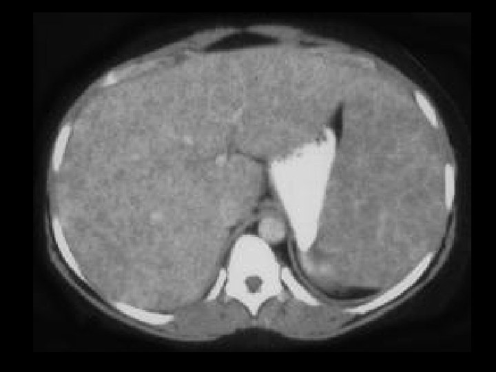
Focal nodular hyperplasia • Findings: – Intensely enhancing lesion in the hepatic dome in the arterial phase – Quick washout in the portal-venous phase • ddx: – Hypervascular tumor – Metastasis
Non-calcified splenic cyst • Findings: – Large homogeneous low density splenic lesion – No calcified wall • ddx: – Epidermoid cyst (true) – Pancreatic pseudocyst – Hydatid disease – Abscess
Right heart dysfunction • Findings: – Opacification of the hepatic veins during the arterial phase – Hepatic parenchyma unremarkable • ddx: – NONE! – This is an Aunt Minnie!
Cystic renal mass • Findings: – Low density renal mass with numerous thin septations – Partially exophytic, partially extending to the sinus • ddx: – Cystic RCC – Multilocular cystic nephroma – Abscess – Xanthogranulomatous pyelonephritis (focal)
Splenic arter aneurysm • Findings: – Dense round lesion medial to the splenic hilum – Same density as the aorta • Causes – Portal hypertension – Pancreatitis – FMD • ddx: – Hypervascular mass of stomach or pancreas
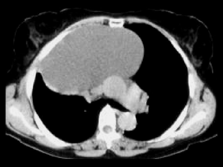
Thymic cyst • Findings: – Large cystic mass in the anterior mediastinum – High attenuation along the periphery = calcification or rim enhancement • ddx: – Cystic teratoma
Emphysematous cholecystitis • Findings: – Distended gallbladder with gas within the wall – Increased risk in pts. with DM – Surgical emergency • ddx: – NONE! – This is an Aunt Minnie!
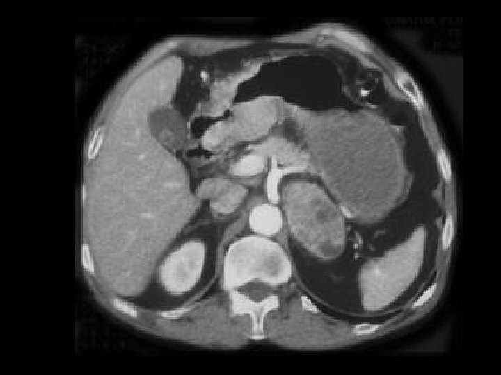
Islet cell tumor • Findings: – Enhancing mass in the pancreatic tail = gastrinoma – Pancreatic cysts • Associations: – Z-E syndrome – VHL – MEN I • ddx: – Hypervascular metastasis
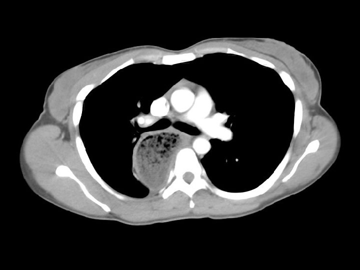
Neuroblastoma • Findings: – Large retroperitoneal mass encases the aorta and extends to the renal hilum – Peripheral enhancement and several foci of high density (? Ca 2+) • ddx: – Lymphoma – Metastatic LAN
Focal nodular hyperplasia • Findings: – Isointense T 1 and T 2 hepatic mass = “stealth” lesion – Intense enhancement in the arterial phase • ddx: – NONE! – This is an Aunt Minnie!
Lymphoma • Findings: – Innumerable low density lesions of the liver and one in the spleen • ddx: – Metastatic disease – Fungal abscesses
Dissecting pancreatic pseudocyst • Findings: – Lobular low density subcapsular lesion of the posterior spleen • ddx: – Lymphoma – Abscess
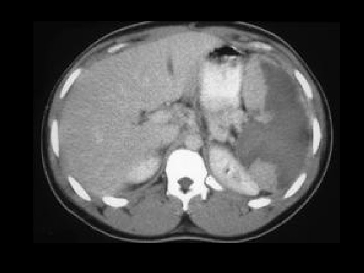
Hepatosplenic sarcoidosis • Findings: – Hepatosplenomegaly – Diffuse infiltration by innumerable tiny low density lesions • ddx: – Microabscesses • TB, histo, CMV, PCP – Bacterial microabscesses – Metastatic disease – Kaposi’s sarcoma
Moire spleen • Findings: – Normal swirling enhancement pattern of the spleen during the arterial phase • ddx: – NONE! – This is an Aunt Minnie!
Adenomyosis of the gallbladder • Findings: – Diffuse gallbladder wall thickening – “ring down” artifact from the non-dependent wall – Abnl thickening of the smooth muscle layer – May be focal or diffuse – No long term consequence • ddx: – NONE! – This is an Aunt Minnie!
Focal nodular hyperplasia of the liver • Findings: – Single mass in the lateral segment of the left lobe – Intense arterial enhancement EXCEPT for a central “scar” • ddx: – NONE! – This is an Aunt Minnie!
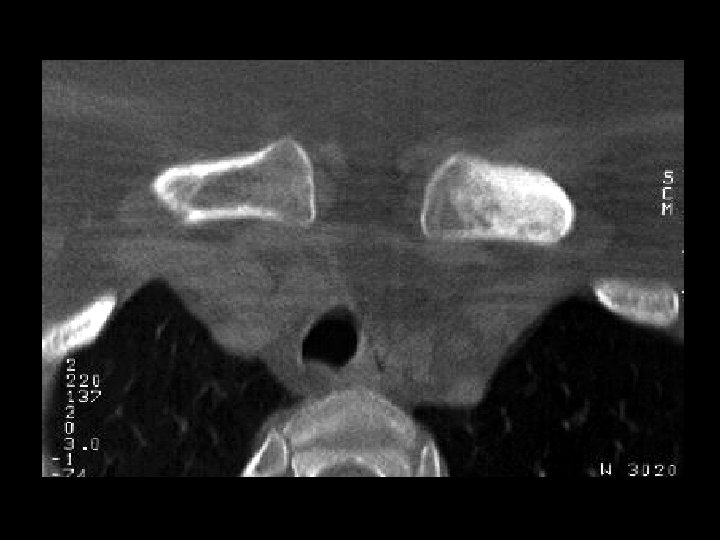
Osteitis condensans • Findings: – Focal increased bone density of the medial head of the left clavicle – No periosteal reaction or soft tissue mass – Rare idiopathic disorder • ddx: – Sclerotic metastasis – Radiation necrosis – Osteomyelitis
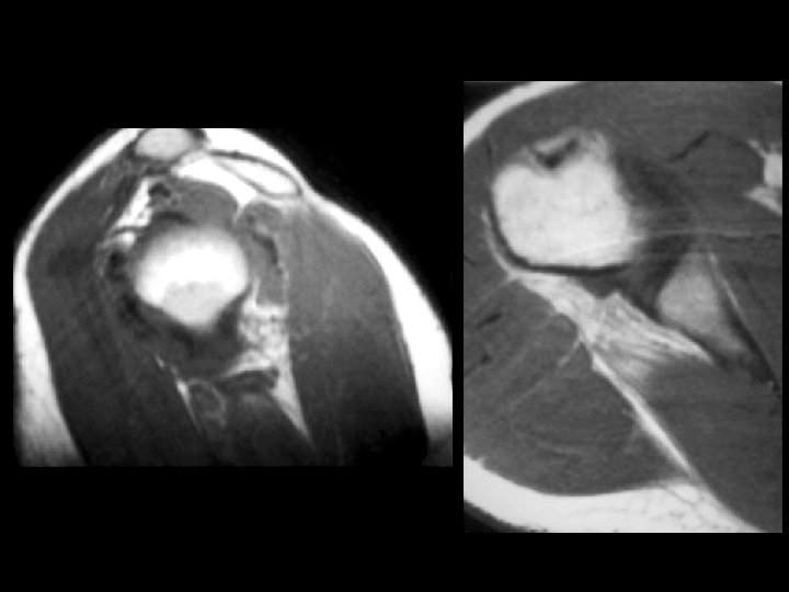
Atrophy of the teres minor • Findings: – High T 1 signal at the attachment of the teres minor = fatty muscular atrophy – Axillary nerve impingement in the quadralateral space • ddx: – NONE! – This is an Aunt Minnie! S I S T
Achalasia • Findings: – Dilated and debrisfilled esophagus • ddx: – Pseudoachalasia • Obstructing tumor • Benign or malignant stricture • Chaga’s disease – Posterior mediastinal abscess
Bladder TCC • Findings: – Diffuse wall thickening of the bladder • ddx: – Underdistension – Muscular hypertrophy from outlet obstruction – Cystitis
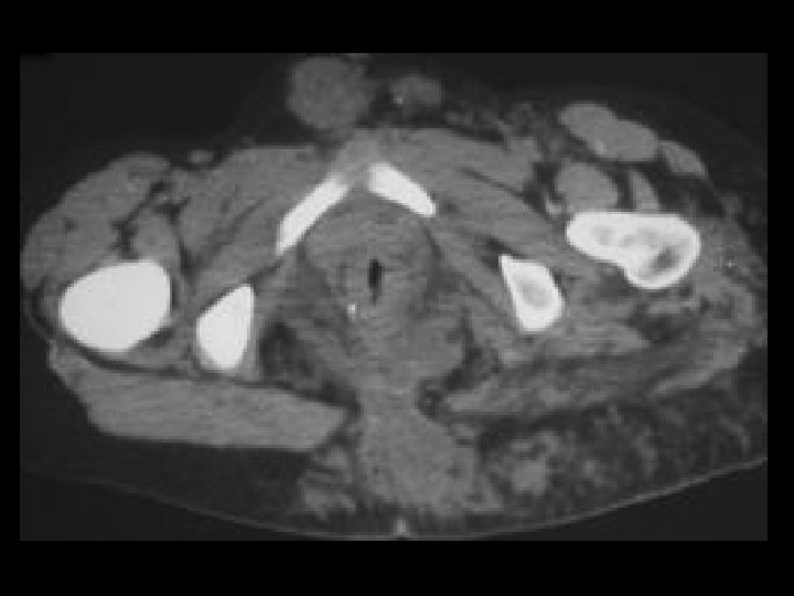
Cervical carcinoma • Findings: – Fluid-filled endometrial cavity – Suggestion of soft tissue fullness in LUS – Incidental left rectus sheath hematoma • ddx: – Normal menses – Cervical stenosis – Obstructing metastasis
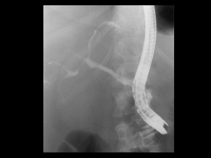
Cholangiocarcinoma • Findings: – ERCP shows diffuse irregular narrowing of the CBD and intrahepatic biliary tree • ddx: – Scerlosing cholangitis – Ascending cholangitis – AIDS cholangiopathy
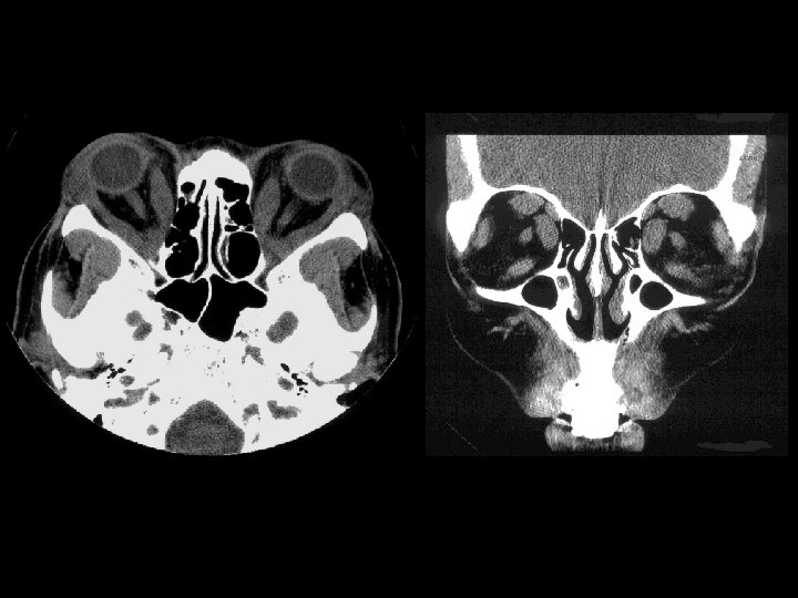
Thyroid opthalmopathy • Findings: – Bilateral enlargement of the extraoccular muscles except the lateral rectus = “I ’ M S L ow” – Sparing of the tendon – Usually NO eye pain • ddx: – Pseudotumor • Involves tendons • Pts have eye pain
Closed loop SBO • Findings: – Dilated fluid-filled small bowel loops localized in the RUQ – Medial displacement of the colon • ddx: – Internal hernia – Small bowel volvulus
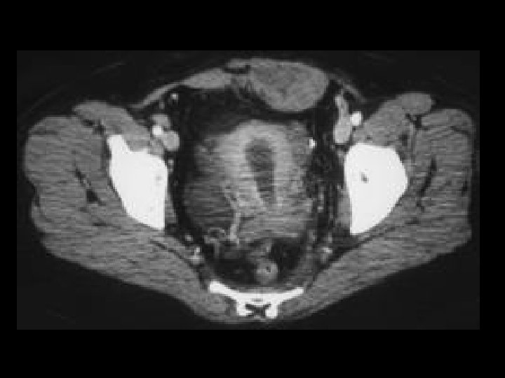
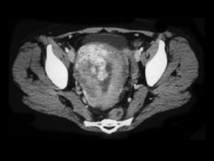
Endometrial carcinoma • Findings: – Irregular enhancing mass extending from the endomentrial cavity to the myometrium • ddx: – Leiomyosarcoma – Metastasis – Gestational trophoblastic disease (if pt pregnant)
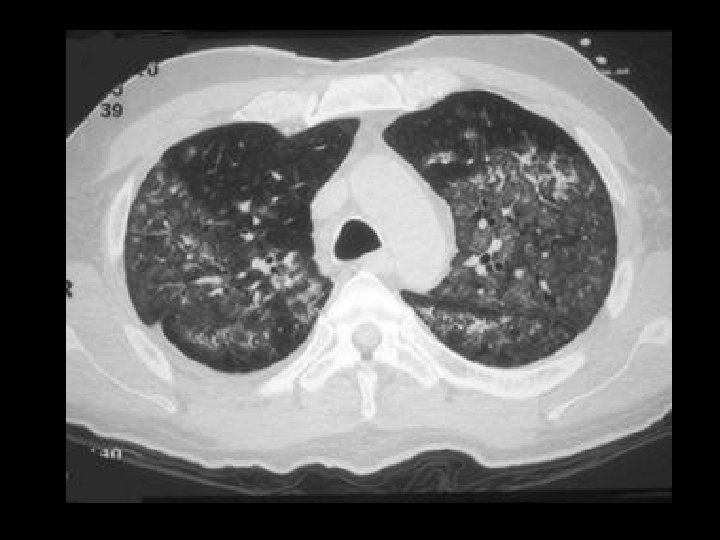
Pulmonary alveolar proteinosis • Findings: – Diffuse ground glass opacities of the secondary pulmonary lobules = “crazy paving” – Scatter thickening of the interlobular septa – Increased risk of Nocardia infection • Rx: BAL • ddx: – NONE! – This is an Aunt Minnie!
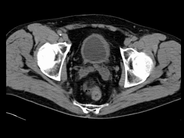
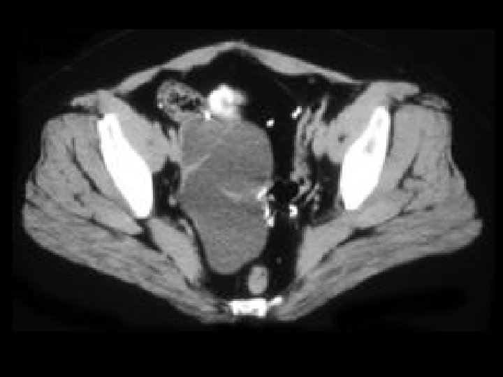
Ovarian cystadenocarcinoma • Findings: – Large multilocular cystic lesion with fine septa in the right hemipelvis • ddx: – Ovarian cystadenoma – Tubo-ovarian abscess – Mucocele of the appendix
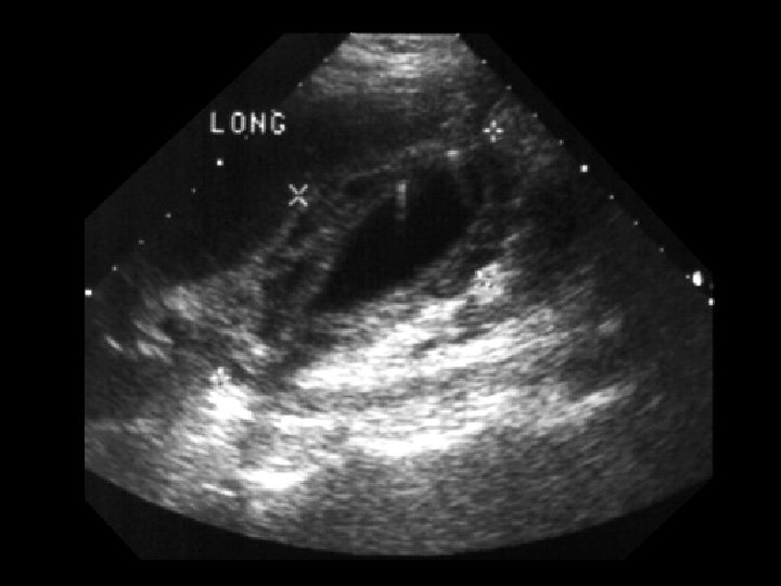
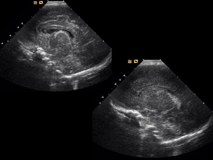
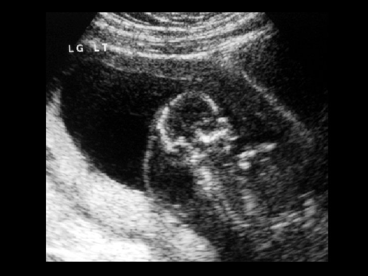
Normal head US • Findings: – Sagittal images through the anterior fontanel show normal appearance of the caudo-thalamic groove – Normal choroid plexus in the lateral ventricle
Diffuse bronchiectasis • Findings: – Thick-walled tubular structures extending from the hila to the periphery – Accompanying pulmonary artery = “signet ring” sign • ddx: – Immotile ciliary syndrome – Chronic infections (immune deficiency)
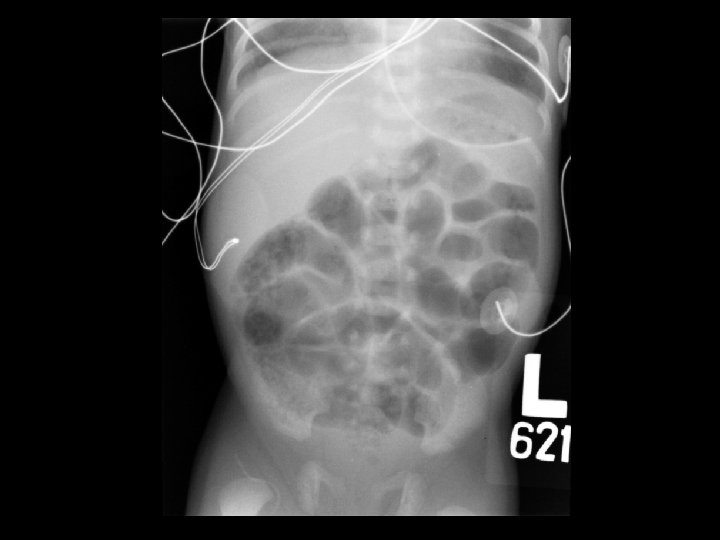
Early necrotizing enterocolitis • Findings: – Mildly distended bowel loops with “soap bubble” appearance in the RLQ – No evidence of pneumatosis or perforation • Rx: bowel rest; surgery if free gas • ddx: – NONE! – This is an Aunt Minnie!
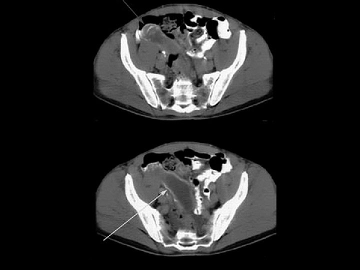
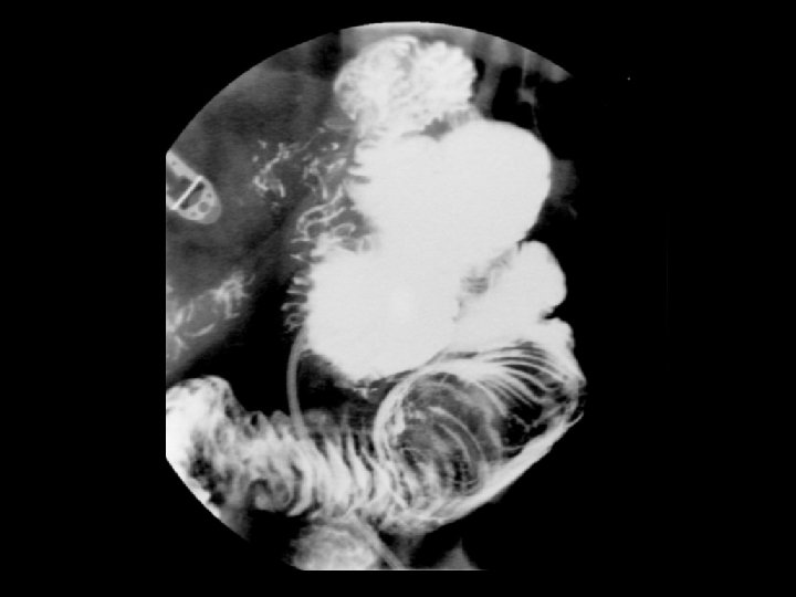
Intussusception • Findings: – Low density intraluminal mass within a small bowel loop – Contrast surrounding this mass produced the “coil spring” appearance – Associated with a lead point in adults: • Lipoma • Carcinoma • lymphoma • Meckel’s diverticulum
Gaucher spleen • Findings: – Enlarged spleen containing numerous low density lesions with mild peripheral enhancement • ddx: – Lymphoma – Metastases – Abscesses
Klippel Trauneny Webber syndrome • Findings: – Unilateral muscular and soft tissues enlargement – Numerous vessels within the soft tissues • ddx: – NONE! – This is an Aunt Minnie!
Metastatic ovarian carcinoma • Findings: – Numerous low density round lesions within the anterior spleen and within the gastrosplenic ligament • ddx: – Dissecting pancreatic pseudocyst
Sickle cell spleen • Findings: – Small, densely calcified spleen • ddx: – NONE! – This is an Aunt Minnie!
Splenic tuberculosis & retrocrural LAN • Findings: – Several punctate low density lesions within an otherwise unremarkable spleen – Large enhancing lymph nodes surrouding the aorta in the retrocrural space • ddx: – Lymphoma – MAI
Splenic microabscesses • Findings: – Enlarged spleen contains innumerable low density lesions • ddx: – Fungal – Bacterial – Metastases – Lymphoma – Kaposi’s sarcoma – Gamma-Gandy bodies (portal hypertension) – Sarcoidosis
Allergic bronchopulmonary aspergillosis • Findings: – Tubular branching opacities of the left upper lobe emanating from the hilum = “finger-in-glove” appearance • ddx: – Pulmonary AVM – Rule out malignancy!
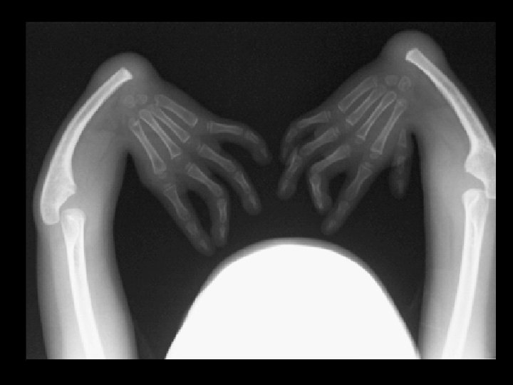
Thrombocytopenia-absent radius syndrome • • Findings: – Absent bilateral radii and left thumb – Prone to bleeding due to thrombocytopenia ddx: – Many other causes of radial dysplasia • Holt Oram • VATER • Fanconi syndrome
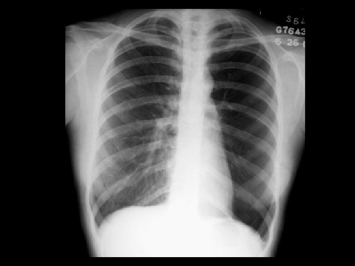
Swyer-James syndrome • Findings: – Hyperlucent left lung – Diminished pulmonary parenchymal and vascular markings – Expiration view shows air trapping • ddx: – Endobronhial foreign body – Pneumothorax – Congenital lobar emphysema – Pulomary artery hypoplasia
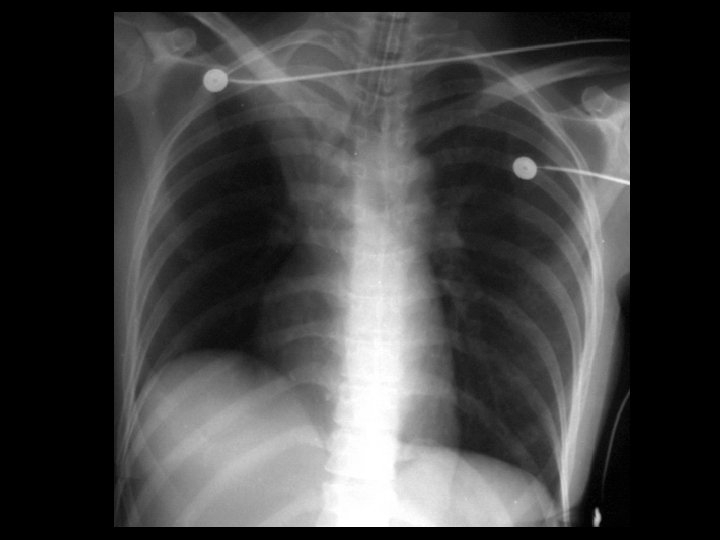
RUL collapse • Findings: – Wedge-shaped opacity of the right apex – Upper displacement of the minor fissure – Right sided mediastinal shift – Elevation of the right hemidiaphragm • ddx: – NONE! – This is an Aunt Minnie!
Cystic hygroma • Findings: – 2 nd trimester fetal US shows a large cystic structure along the posterior neck – Recommend amnio • Causes: – Isoloated (nl karyotype) – Generalized hydrops = lymphagiectasia (fatal) – Turner’s / Noonan’s – Trisomy 21, 18, 13
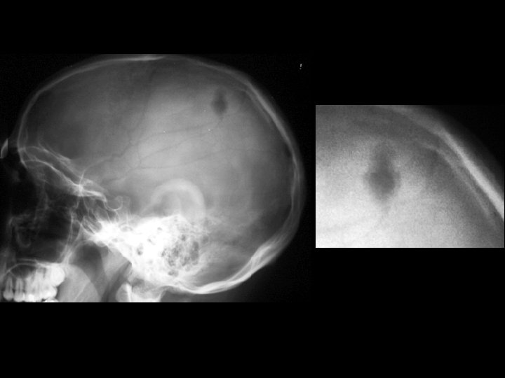
Langerhan’s cell histiocytosis • Findings: – Single geographic lytic lesion of the skull involving inner and outer table – No sclerotic margin but surrounding sclerotic reaction • ddx: – Metastasis – Osteomyelitis – Multiple myeloma (multiple) – Epidermoid (sclerotic rim)
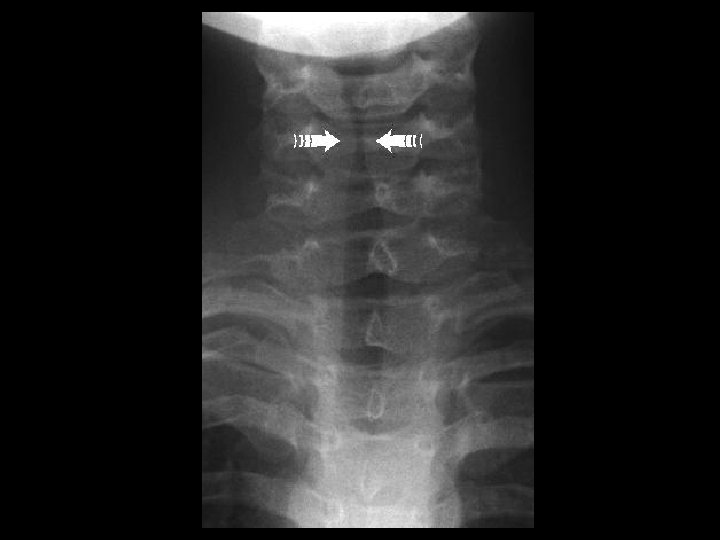
Laryngotracheobronchitis • Findings: – Narrowing of the subglottic airway = “steeple sign” – a. k. a. Croup – viral infxn (parainfluenza) but may be bacterial (S. aureus) – Check lateral film for acute epiglottitis (25% have subglottic narrowing) • ddx: – epiglotitis – membranous croup
Pulmonary sling • Findings: – Soft tissue opacity between the trachea and esophagus – Normal appearance on the frontal radiograph – Pt. may present with wheezing • ddx: – Lymphadenopathy – Esophageal mass