Congenital heart disease A congenital heart malformation occurs


. -Isolated or in association")


- Slides: 29
Congenital heart disease
A congenital heart malformation occurs in about 1% of live births. -There is an overall MALE predominance. -Although (ASD , PDA occur commonly in females). -Aetiology is unknown.
Causes may involve: 1 -Maternal rubella infection. 2 -Maternal Alcohol abuse. 3 -Maternal drug treatment and radiation. 4 -Genetic and chromosomal abnormalities.
Symptoms and signs: -Central cyanosis (because of right-to-left shunting of blood or because of complete mixing of systemic and pulmonary blood flow). -Pulmonary hypertension (from large left to right shunt as a result persistently elevated pulmonary flow leading to development of pulmonary artery vascular resistance. Eisenmenger reaction). -Clubbing of fingers. -Paradoxical embolism of thrombus from the systemic veins to systemic arterial system.
-Reduced growth is common in children with cyanotic heart disease. -Syncope is common when severe right or left ventricular outflow tract obstruction is present. -Squatting is the position adopted by children with Fallot’s tetralogy (It results in obstruction of venous return of desaturated blood an increase in the peripheral systemic vascular resistance, this leads to reduced right to left shunt and improved cerebral oxygenation.
% Occurrence in first degree relatives VSD ASD PDA Fallot’s tetralogy y stenosis 7% Coarctation of aorta Aortic stenosis Others 39% 10% 6% 4% 2% 4% 4% 7% 6% 15% 2% 4%
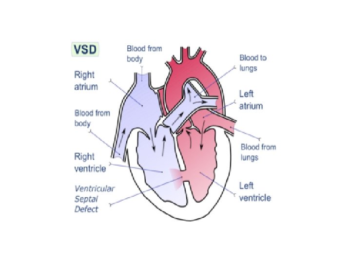
VSD Most common congenital malformation (1 in 500 live births). -Isolated or in association with other anomalies. -Left vent pressure is higher than the right; therefore blood moves from left to right and pulmonary flow increases, leading to obliterative pul vascular changes causing pul arterial pr to equal sys pressure (Eisenmenger syndrom), consequently the shunt is reduced or reversed becoming right to left and central cyanosis may develop.
Clinical features: -Loud systolic murmur in an asymptomatic pt if small VSD these usually close spontaneously. -Moderate VSDs produce fatigue and dyspnoea Physical signs include cardiac enlargement and prominent apex beat and often a palpable systolic thrill at the lower left sternal edge, and a loud pan-systolic murmur is heard at same position. -Large VSDs are associated with increasing pul hypertension, the murmur may be soft because the flow across the VSD is small.
Investigations: -ECG -Chest X-ray In large defects CXR shows prominent pul artery , and cardiomegaly ECG shows signs of both right and left ventricular hypertrophy -2 D Echocardiography
Treatment: Moderate and large VSDs should be surgically repaired before the development of severe pulmonary hypertension. Infective endocarditis prophylaxis should be adviced (e. g. Dental procedures: Oral Amoxycilin 3 g 1 hour before procedure)
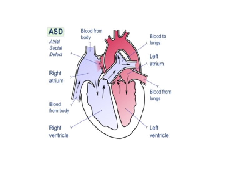
ASD Is often first diagnosed in adults Is more common in women There are two types of ASDs (ostium secundum and ostium primum) Ostium secundum is the common form. Left to right shunt of blood at the level of atria occurs and because the Rt ventricle is easily distended and the pul vascular resistance is low, there will be a great increase in Rt heart output. -Above 30 yrs there may be increase in pul vas resistance which gives rise to pul hypertension. Aterial fibrillation is common at this age.
Clinical features: -Most children with ASDs are asymptomatic, although prone to pulmonary infection. -Dyspnoea and weakness -Pailpitations due to atrial arrythmias -Rt heart failure -Splitting of the 2 nd heart sound is wide and fixed -Loud ejection systolic pulmonary flow murmur.
Investigations: Chest X-ray prominent pul artery and pulmonary plethora, there might be Rt vent hypertrophy. ECG Rt bundle branch block (because of dilatation of the right vent) and right axis deviation. Echo Rt vent hypertrophy and pul artery dilatation Flow disturbance by Doppler Cardiac catheterization not commonly used
Treatment: A significant ASD should be repaired before the age of 10 yr or as soon as possible if diagnosed in adulthood. There is a good result from surgery unless hypertension has developed.
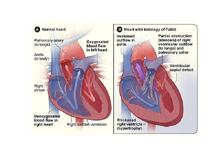
Fallot’s tetralogy Is the most common cyanotic congenital heart abnormality in children who survive beyond the neonatal period, it consists of: 1. VSD 2. Rt ventricular outflow obstruction 3. Positioning of aorta above the VSD (‘overriding aorta’) 4. Rt. Ventricular hypertrophy The combination leads to a high Rt ventricular pressure and right to left shunting of blood through the VSD, thus patient is centrally cyanosed.
Clinical features: -Children may present with dyspnoea or fatigue or with hypoxic episodes (Fallot’s spells), i. e. deep cyanosis and possible syncope on exertion, squatting is common. -Systolic ejection murmur at the second left intercostal space close to the sternum. -Central cyanosis -Clubbing of fingers and polycythemia are abvious after about 1 yr -Growth retardation.
Investigations: Chest X-ray Large Rt vent and small pul artery. ECG Rt vent hypertrophy. Echo Discontinuity between aorta and the anterior wall of the vent septum. Cardiac catheterization To evaluate the size and degree of the Rt vent outflow obstruction.
Treatment: Complete surgical correction is possible even in infancy.
Cardiac Catheterization
Is an invasive diagnostic procedure in which radiopaque arterial and venous catheters are introduced into selected vesseles of the right and left side of the heart, guided by fluoroscopy. Most commonly, the catheters are inserted percutaneously through blood vessels or via a cutdown. Pressures and oxygen saturation levels in the four heart chambers are measured.
Indications: ØCAD, coronary artery patency, extent of atherosclerosis and determine whether revascularization procedures including CABG, may be of benefit to the patient. ØDiagnosis of Pulmonary hypertension through checking the PAWP (PACP). ØTreat stenotic heart valves via percutaneous ballon valvuloplasty.
During the procedure: -Pt has one or more IV lines (for fluids, sedatives, heparin, and other medications) -BP and ECG monitoring (dysrhythmias may develop as catheters are positioned in coronary arteries or during injection of contrast agents) -Resuscitation equipments readily available, and staff must be prepared to provide advanced cardiac life support measures as necessary. -Assessing pt for any allergies to contrast agents or iodine containing substances (eg, seafood).
Before the procedure: -Assessing patients for any allergies to contrast agents or iodine containing substances (eg, seafood). (prepare antihistamines or methylprednisolone) -Blood tests (BUN, creatinine, INR, PT, a. PTT, Hb, PCV, plates and electrolyte levels). -Preventive strategies for high risk patients (elderly, DM, HF, hypotension, dehydration, preexisting renal disease) who are at risk from contrast agent-induced nephropathy (defined as an increase in the baseline serum creatinine by 25% or more within 2 days of the procedure)
Preventive measures for contrast agent- induced nephropathy: Includes preprocedure and postprocedure hydration with IV infusions of saline or sodium bicarbonate and antioxidant acetylcystine (Mucomyst)
Nursing responsibilities before catheterization: >The patient is instructed to fast for 8 to 12 hr, if performed as an outpatient procedure , a friend, relative or family member must transport pt. >Patient told about expected duration of the procedure, involve lying on a hard table for less two hours >The patient is reassured that IV medications will be given to maintain comfort >Patient informed about sensations during procedure (eg, palpitations, need to void sensation) >Patient encouraged to express fears and anxiety
Nursing responsibilities after catheterization: <Observe catheter access site for bleeding or hematoma formation, distal pulses assess. Every 15 min for 1 hr and then every 1 to 2 hrs until pulses are stable. <Temp, color, capillary refill, pain and numbness of the affected extremity. <Cardiac monitoring for arrythmias or vasovagal reaction(hypotesion-bradycardia-nausea) <Bed rest maintaned for 2 -4 hrs, affected leg straight and head of bed elevated above 30’ and analgesia prescribed <Monitoring for any signs nephropathy. <Applying pt safety standards