DIGESTION Core Course No ZOOA P 4 T


")

 Contents of normal gastric juice (fasting state).")


 (Source: ibid)")


, which")
 and has")


 of a very alkaline")


 are liberated by the")


 in the GI tract.")


 Despite the predominance of absorptive mechanisms, secretion")


: exhibits macroheterogeneity; secreted by cells in the mucosa of")
")


: 42 amino acid residues; produced")


: • It is found in nerves in the")


- Slides: 74
DIGESTION Core Course No. ZOOA – P 4 T, Group-A, Topic No. 2
GI Tract. . . its speciality The lumen of the gastrointestinal tract is functionally contiguous with the outside of the body. The intestine - substantial surface area, which is important for its absorptive function Gut is an unusual organ in that it becomes colonized, almost from birth, with a large number of commensal bacteria (particularly in the colon, or large intestine). This relationship is mutually beneficial – metabolism + protection form pathogens
Secretions include…. • Throughout the length of the intestine, glandular structures deliver secretions into the lumen, particularly in the stomach and mouth. • Also important in the process of digestion are secretions from the pancreas and the biliary system of the liver.
Why such functional divisions?
Organization of the wall of the intestine into functional layers. (Adapted from Barrett KE: Gastrointestinal Physiology. Mc. Graw-Hill, 2006. ) Connective Tissue layer Smooth Muscle
1. Stem Cells – located towards the base of the crypt 2. The gastrointestinal epithelium is one of the most rapidly dividing tissues in the body. 3. These microvilli are endowed with a dense glycocalyx (the brush border) that probably protects form digestive enzymes 4. These so called “brush border hydrolases” perform the final steps of digestion for specific nutrients.
GASTROINTESTINAL SECRETIONS
Salivary Glands SGs when maximally stimulated, secreting their own weight in saliva every minute
Acini to Duct to Mouth
Is saliva important? • It has a number of organic constituents that serve to initiate digestion (particularly of starch, mediated by amylase) • Protects the oral cavity from bacteria (such as Immunoglobulin A and lysozyme). • Serves to lubricate the food bolus (aided by mucins). • Hypotonic compared with plasma and alkaline; the latter feature is important to neutralize any gastric secretions that reflux into the esophagus.
• • Swallowing Keeps the mouth moist Solvent for the molecules that stimulate the taste buds Aids speech by facilitating movements of the lips and tongue Keeps the mouth and teeth clean The buffers in saliva help maintain the oral p. H at about 7. 0 Also help neutralize gastric acid and relieve heartburn when gastric juice is regurgitated into the esophagus xerostomia
As the rate of secretion increases, there is less time for Na. Cl to be extracted and the tonicity of the saliva rises, but it always stays somewhat hypotonic with respect to plasma. Overall, the three pairs of salivary glands that drain into the mouth supply 1000 to 1500 m. L of saliva per day.
Regulation of Salivary Secretion (Adapted from Barrett KE: Gastrointestinal Physiology. Mc. Graw-Hill, 2006. )
Entry to Stomach
(Source: ibid) Contents of normal gastric juice (fasting state).
Structure of a gastric gland from the fundus and body of the stomach. (Adapted from Barrett KE: Gastrointestinal Physiology. Mc. Graw. Hill, 2006. )
Chemical digestion is now operative… • The parietal cells, which secrete hydrochloric acid and intrinsic factor • The chief cells, which produce pepsinogens and gastric lipase • The acid secreted by parietal cells serves to sterilize the meal and also to begin the hydrolysis of dietary macromolecules. • Intrinsic factor is important for the later absorption of vitamin B 12 or Cyanocobalamin • Pepsinogen is the precursor of pepsin, which initiates protein digestion. • Lipase similarly begins the digestion of dietary fats.
Cyanocobalamin (vitamin B 12) (Source: ibid)
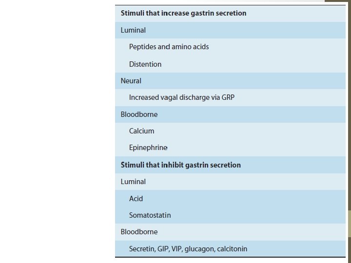
Regulation of gastric acid and pepsin secretion by soluble mediators and neural input • Gastrin is a hormone that is released by G cells in the antrum of the stomach both in response to a specific neurotransmitter released from enteric nerve endings, known as gastrin releasing peptide (GRP, or bombesin), and also in response to the presence of oligopeptides in the gastric lumen. • Gastrin is then carried through the bloodstream to the fundic glands, where it binds to receptors not only on parietal (and likely, chief cells) to activate secretion, but also on so-called enterochromaffin-like cells (ECL cells) that are located in the gland, and release histamine. • Histamine is also a trigger of parietal cell secretion, via binding to H 2 histamine receptors. Finally, parietal and chief cells can also be stimulated by acetylcholine, released from enteric nerve endings in the fundus.
Regulation of gastric acid and pepsin secretion by soluble mediators and neural input Gastrin is released from G cells in the antrum and travels through the circulation to influence the activity of ECL cells and parietal cells. The specific agonists of the chief cell are not well understood. Gastrin release is negatively regulated by luminal acidity via the release of somatostatin from antral D cells. (Source: ibid)
Termination of Gastric secretion • During the cephalic phase, the secretion is predominantly activated by vagal input that originated from dorsal vagal complex (brain), which coordinates input from higher centers. • Vagal outflow to the stomach then releases GRP and acetylcholine, thereby initiating secretory function. • However, before the meal enters the stomach, there are few additional triggers and thus the amount of secretion is limited. • Once the meal is swallowed, on the other hand, meal constituents trigger substantial release of gastrin and the physical presence of the meal also distends the stomach and activates stretch receptors, which provoke a “vago-vagal” as well as local reflexes that further amplify secretion. • The meal also buffers gastric acidity that would otherwise serve as a feedback inhibitory signal to shut off secretion secondary to the release of somatostatin, which inhibits both G and ECL cells as well as secretion by parietal cells themselves
The Dorsal Vagal Complex in brain
Gastric parietal cells are highly specialized for their unusual task of secreting concentrated acid. The cells are packed with mitochondria that supply energy to drive the apical H, K-ATPase, or proton pump, that moves H+ ions out of the parietal cell against a concentration gradient of more than a million-fold. Composite diagram of a parietal cell, showing the resting state (lower left) and the active state (upper right). (Source: ibid)
Amplification of the apical surface area is accompanied by an increased density of H+, K+ –ATPase molecules at this site. Note that acetylcholine (ACh) and gastrin signal via calcium, whereas histamine signals via c. AMP. (Source: ibid)
The Molecular Mechanisms of Gastric Secretion • At rest, the proton pumps are sequestered within the parietal cell in a series of membrane compartments known as tubulovesicles. • When the parietal cell begins to secrete, on the other hand, these vesicles fuse with invaginations of the apical membrane known as canaliculi, thereby substantially amplifying the apical membrane area and positioning the proton pumps to begin acid secretion • The apical membrane also contains potassium channels, which supply the K+ ions to be exchanged for H+, and Cl– channels that supply the counterion for HCl secretion. • The secretion of protons is also accompanied by the release of equivalent numbers of bicarbonate ions into the bloodstream, which as we will see, are later used to neutralize gastric acidity once its function is complete
Ion transport proteins in parietal cells Protons are generated in the cytoplasm via the action of carbonic anhydrase II (C. A. II). Bicarbonate ions are exported from the basolateral pole of the cell either by vesicular fusion or via a chloride/bicarbonate exchanger. (Source: ibid)
The synergistic action…. • The three agonists of the parietal cell—gastrin, histamine, and acetylcholine—each bind to distinct receptors on the basolateral membrane. • Gastrin and acetylcholine promote secretion by elevating cytosolic free calcium concentrations, whereas histamine increases intracellular cyclic adenosine 3', 5'-monophosphate (c. AMP). • The net effect of these second messengers are the transport and morphological changes described above. However, it is important to be aware that the two distinct pathways for activation are synergistic, with a greater than additive effect on secretion rates when histamine plus gastrin or acetylcholine, or all three, are present simultaneously. • The physiologic significance of this synergism is that high rates of secretion can be stimulated with relatively small changes in availability of each of the stimuli. Synergism is also therapeutically significant because secretion can be markedly inhibited by blocking the action of only one of the triggers.
Now this is the turn of the pancreas…. .
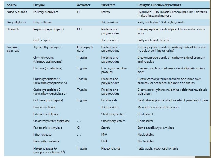
THE LIST IS PRETTY LONG
Composition of normal human pancreatic juice.
The portion of the pancreas that secretes pancreatic juice is a compound alveolar gland resembling the salivary glands. Granules containing the digestive enzymes (zymogen granules) are formed in the cell and discharged by exocytosis from the apexes of the cells into the lumens of the pancreatic ducts.
The small duct radicles coalesce into a single duct (pancreatic duct of Wirsung), which usually joins the common bile duct to form the ampulla of Vater. The ampulla opens through the duodenal papilla, and its orifice is encircled by the sphincter of Oddi. Some individuals have an accessory pancreatic duct (duct of Santorini) that enters the duodenum more proximally.
Pancreatic juice: Its composition • The pancreatic juice is alkaline (Table ) and has a high HCO 3– content (approximately 113 m. Eq/L vs. 24 m. Eq/L in plasma). • About 1500 m. L of pancreatic juice is secreted per day. • Bile and intestinal juices are also neutral or alkaline, and these three secretions neutralize the gastric acid, raising the p. H of the duodenal contents to 6. 0 to 7. 0. • By the time the chyme reaches the jejunum, its p. H is nearly neutral, but the intestinal contents are rarely alkaline. • Note: an equivalent is the number of moles of an ion in a solution, multiplied by the valence of that ion. If 1 mol of Na. Cl and 1 mol of Ca. Cl 2 dissolve in a solution, there is 1 equiv Na, 2 equiv Ca, and 3 equiv Cl in that solution. (The valence of calcium is 2, so for that ion you have 1 mole and 2 equivalents. ) m. Eq = (mg × valence) ÷ molar mass
It may result a threat…. • The potential danger of the release into the pancreas of a small amount of trypsin is apparent; the resulting chain reaction would produce active enzymes that could digest the pancreas. • It is therefore not surprising that the pancreas normally contains a trypsin inhibitor. • Another enzyme activated by trypsin is phospholipase A 2. • This enzyme splits a fatty acid off phosphatidylcholine (PC), forming lyso-PC. Lyso-PC damages cell membranes. It has been hypothesized that in acute pancreatitis, a severe and sometimes fatal disease, phospholipase A 2 is activated in the pancreatic ducts, with the formation of lyso-PC from the PC that is a normal constituent of bile. This causes disruption of pancreatic tissue and necrosis of surrounding fat.
Lyso-PC Phosphatidylcholine Phospholipase A 2
Regulation of Secretion of pancreatic juice • Secretion (hormone mediated) of a very alkaline pancreatic juice that is rich in HCO 3– and poor in enzymes. The effect on duct cells is due to an increase in intracellular c. AMP. Secretin also stimulates bile secretion. • CCK acts on the acinar cells to cause the release of zymogen granules and production of pancreatic juice rich in enzymes but low in volume. Its effect is mediated by phospholipase C • As the volume of pancreatic secretion increases, its Cl– concentration falls and its HCO 3– concentration increases. Although HCO 3– is secreted in the small ducts, it is reabsorbed in the large ducts in exchange for Cl– • The magnitude of the exchange is inversely proportionate to the rate of flow. • Like CCK, acetylcholine acts on acinar cells via phospholipase C to cause discharge of zymogen granules, and stimulation of the vagi (Xth Cr. Nrv. ) causes secretion of a small amount of pancreatic juice rich in enzymes.
Ion transport pathways present in pancreatic duct cells CA, carbonic anhydrase; NHE-1, sodium/hydrogen exchanger-1; NBC, sodiumbicarbonate cotransporter.
Effect of a single dose of secretin on the composition and volume of the pancreatic juice in humans.
Composition of Bile
These are different Bile Acids
The bile salts have a number of important actions: 1. reduce surface tension and, in conjunction with phospholipids and monoglycerides 2. responsible for the emulsification of fat preparatory to its digestion and absorption in the small intestine 3. They are amphipathic, i. e. they have both hydrophilic and hydrophobic domains; one surface of the molecule is hydrophilic because the polar peptide bond and the carboxyl and hydroxyl groups are on that surface, whereas the other surface is hydrophobic.
Lipid digestion and passage to intestinal mucosa. Fatty acids (FA) are liberated by the action of pancreatic lipase on dietary triglycerides and, in the presence of bile salts (BS), form micelles (the circular structures), which diffuse through the unstirred layer to the mucosal surface. (Adapted from Thomson ABR: Dietschy JM, Gotto AM Jr, Ontko JA [editors]: American Physiological Society, 1978. )
Their hydrophilic portions face out and their hydrophobic portions face in. Above a certain concentration, called the critical micelle concentration, all bile salts added to a solution form micelles. Lipids collect in the micelles, with cholesterol in the hydrophobic center and amphipathic phospholipids and monoglycerides lined up with their hydrophilic heads on the outside and their hydrophobic tails in the center. The micelles play an important role in keeping lipids in solution and transporting them to the brush border of the intestinal epithelial cells, where they are absorbed
90% to 95% of the bile salts are absorbed from the small intestine. Once they are deconjugated, they can be absorbed by nonionic diffusion, but most are absorbed in their conjugated forms from the terminal ileum by an extremely efficient Na+–bile salt cotransport system powered by basolateral Na+–K+ ATPase. The absorbed bile salts are transported back to the liver in the portal vein and reexcreted in the bile The remaining 5– 10% of the bile salts enter the colon and are converted to the salts of deoxycholic acid and lithocholic acid. Lithocholate is relatively insoluble and is mostly excreted in the stools; only 1% is absorbed. However, deoxycholate is absorbed.
Some more information… • Normal rate of bile salt synthesis is 0. 2 to 0. 4 g/d. • The total bile salt pool of approximately 3. 5 g recycles repeatedly via the enterohepatic circulation; it has been calculated that the entire pool recycles twice per meal and six to eight times per day. • When bile is excluded from the intestine, up to 50% of ingested fat appears in the feces. • A severe malabsorption of fat-soluble vitamins also results.
INTESTINAL FLUID & ELECTROLYTE TRANSPORT
What happens initially…. • Water moves passively into and out of the gastrointestinal lumen, driven by electrochemical gradients established by the active transport of ions and other solutes. • In the period after a meal, much of the fluid reuptake is driven by the coupled transport of nutrients, such as glucose, with sodium ions. • In the period between meals, absorptive mechanisms center exclusively around electrolytes. • In both cases, secretory fluxes of fluid are largely driven by the active transport of chloride ions into the lumen, although absorption still predominates overall.
The fluid quantity includes…. . • The intestines are presented each day with about 2000 m. L of ingested fluid plus 7000 m. L of secretions from the mucosa of the gastrointestinal tract and associated glands. • 99% of this fluid is reabsorbed, with a daily fluid loss of only 200 m. L in the stools.
Daily water turnover (m. L) in the GI tract.
Absorption of Salts further…. .
In the small intestine, secondary active transport of Na+ is important in bringing about absorption of glucose, some amino acids, and other substances such as bile acids Conversely, the presence of glucose in the intestinal lumen facilitates the reabsorption of Na+ In the period between meals, when nutrients are not present, sodium and chloride are absorbed together from the lumen by the coupled activity of a sodium/hydrogen exchanger (NHE) and chloride/bicarbonate exchanger in the apical membrane, in a socalled electroneutral mechanism
Water then follows to maintain an osmotic balance. In the colon, moreover, an additional electrogenic mechanism for sodium absorption is expressed, particularly in the distal colon. In this mechanism, sodium enters across the apical membrane via an ENa. C (epithelial sodium) channel that is identical to that expressed in the distal tubule of the kidney This underpins the ability of the colon to desiccate the stool and ensure that only a small portion of the fluid load used daily in the digestion and absorption of meals is lost from the body. Following a low-salt diet, increased expression of ENa. C in response to aldosterone increases the ability to reclaim sodium from the stool.
The cystic fibrosis transmembrane conductance regulator (CFTR) Despite the predominance of absorptive mechanisms, secretion also takes place continuously throughout the small intestine and colon to adjust the local fluidity of the intestinal contents as needed for mixing, diffusion, and movement of the meal and its residues along the length of the gastrointestinal tract. Cl – normally enters enterocytes from the interstitial fluid via Na+–K+– 2 Cl– cotransporters in their basolateral membranes
• Cl– is then secreted into the intestinal lumen via channels that are regulated by various protein kinases. • The cystic fibrosis transmembrane conductance regulator (CFTR) channel that is defective in the disease of cystic fibrosis is quantitatively most important, and is activated by protein kinase A and hence by c. AMP • Water moves into or out of the intestine until the osmotic pressure of the intestinal contents equals that of the plasma. • The osmolality of the duodenal contents may be hypertonic o hypotonic, depending on the meal ingested, but by the time the meal enters the jejunum, its osmolality is close to that of plasma. • This osmolality is maintained throughout the rest of the small intestine
More than 15 types of hormone-secreting enteroendocrine cells (e. g. G cell, S Cell etc. ) have been identified in the mucosa of the stomach, small intestine, and colon!!!!
Gastrointestinal Hormones • Others manufacture serotonin or histamine and are called enterochromaffin or enterochromaffin-like (ECL) cells, respectively • Gastrins: Secreted from G cells; exhibits macroheterogeneity; three major forms: G 34, G 17, and G 14 gastrins; stimulation of gastric acid and pepsin secretion and stimulation of the growth of the mucosa of the stomach and small and large intestines. • Acid in the antrum inhibits gastrin secretion, partly by a direct action on G cells and partly by release of somatostatin, a relatively potent inhibitor of gastrin secretion. • In conditions such as pernicious anemia in which the acidsecreting cells of the stomach are damaged, gastrin secretion is chronically elevated
Old Melodies….
GASTRIN
Gastrointestinal Hormones • Cholecystokinin (CCK): exhibits macroheterogeneity; secreted by cells in the mucosa of the upper small intestine; the stimulation of pancreatic enzyme secretion, the contraction of the gallbladder (the action for which it was named), and relaxation of the sphincter of Oddi, which allows both bile and pancreatic juice to flow into the intestinal lumen. • There also two protein releasing factors that activate CCK secretion, known as CCK-releasing peptide and monitor peptide, which derive from the intestinal mucosa and pancreas, respectively. • Because the bile and pancreatic juice that enter the duodenum in response to CCK further the digestion of protein and fat, and the products of this digestion stimulate further CCK secretion, a sort of positive feedback operates in the control of the secretion of this hormone.
Cholecystokinin (CCK)
Structures of some of the hormonally active polypeptides secreted by cells in the human GI tract. Homologous amino acid residues are enclosed by the lines that generally cross from one polypeptide to another. Arrows indicate points of cleavage to form smaller variants. Tys, tyrosine sulfate. All gastrins occur in unsulfated (gastrin I) and sulfated (gastrin II) forms. Glicentin, an additional member of the secretin family, is a Cterminally extended relative of glucagon.
Gastrointestinal Hormones • Secretin: secreted by S cells that are located deep in the glands of the mucosa of the upper portion of the small intestine; only a single form is isolated • The secretion of secretin is increased by the products of protein digestion and by acid bathing the mucosa of the upper small intestine. • It increases the secretion of HCO 3 - by the duct cells of the pancreas and biliary tract, thus causing the secretion of a watery, alkaline pancreatic juice. • It also augments the action of CCK in producing pancreatic secretion of digestive enzymes. • It decreases gastric acid secretion and may cause contraction of the pyloric sphincter. • FINALLY…. Secretin causes alkaline pancreatic juice to flood into the duodenum, neutralizing the acid from the stomach and thus inhibiting further secretion of the hormone
Secretin
Gastrointestinal Hormones • GIP (Gastric Inhibitory Peptide/Glucose-dependent Insulinotropic Polypeptide): 42 amino acid residues; produced by K cells in the mucosa of the duodenum and jejunum. • Its secretion is stimulated by glucose and fat in the duodenum, and because in large doses it inhibits gastric secretion and motility, it was named gastric inhibitory peptide • GIP stimulates insulin secretion. • The integrated action of gastrin, CCK, secretin, and GIP in facilitating digestion and utilization of absorbed nutrients is summarized hereafter
GIP Structure
Integrated action of gastrointestinal hormones in regulating digestion and utilization of absorbednutrients. The dashed arrows indicate inhibition. The exact identity of the hormonal factor or factors from the intestine that inhibit(s) gastric acid secretion and motility is unsettled, but it may be peptide YY.
Gastrointestinal Hormones Vasoactive intestinal peptide (VIP): • It is found in nerves in the gastrointestinal tract and thus is not itself a hormone, despite its similarities to secretin. • VIP contains 28 amino acid residues • It is also found in blood with a half-life of about 2 minutes. • In the intestine, it markedly stimulates intestinal secretion of electrolytes and hence of water. • Its other actions include relaxation of intestinal smooth muscle, including sphincters; dilation of peripheral blood vessels; and inhibition of gastric acid secretion.
VIP Structure
Motilin A. Motilin has 22 amino acid residues B. Secreted by enterochromaffin cells and Mo cells in the stomach small intestine, and colon. C. It acts on G protein-coupled receptors on enteric neurons in the duodenum and colon and on injection produces contraction of smooth muscle in the stomach and intestines D. The antibiotic erythromycin binds to motilin receptors, and derivatives of this compound may be of value in treating patients in whom gastrointestinal motility is decreased.
Principal peptides found in the enteric nervous system.
Schematic of the splanchnic circulationunder fasting conditions. Note that even during fasting, the liver receives the majority of its blood supply via the portal vein.
Thank You…. . Reference: Guyton & Hall Textbook of Medical Physiology