YEDTEPE NVERSTES GZ HASTANES DISORDERS OF THE CONJUNCTIVA



- Slides: 76
YEDİTEPE ÜNİVERSİTESİ GÖZ HASTANESİ DISORDERS OF THE CONJUNCTIVA AND CORNEA Yrd. Doç. Dr. VİLDAN ÖZTÜRK
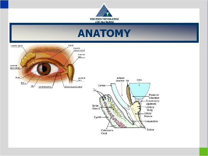
YEDİTEPE ÜNİVERSİTESİ GÖZ HASTANESİ ANATOMY
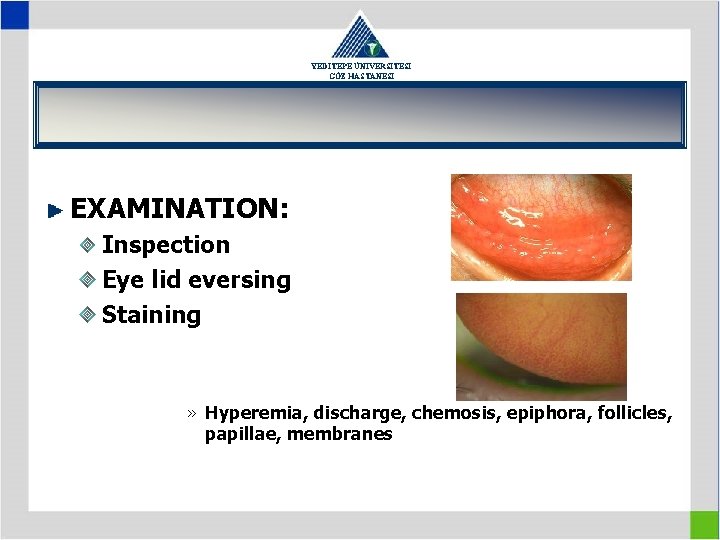
YEDİTEPE ÜNİVERSİTESİ GÖZ HASTANESİ EXAMINATION: Inspection Eye lid eversing Staining » Hyperemia, discharge, chemosis, epiphora, follicles, papillae, membranes
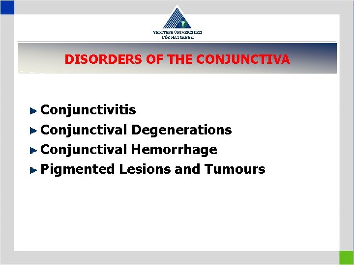
YEDİTEPE ÜNİVERSİTESİ GÖZ HASTANESİ DISORDERS OF THE CONJUNCTIVA Conjunctivitis Conjunctival Degenerations Conjunctival Hemorrhage Pigmented Lesions and Tumours
YEDİTEPE ÜNİVERSİTESİ GÖZ HASTANESİ CONJUNCTIVITIS Bacterial Conjunctivitis Viral Conjuntivitis Chlamydial Conjunctivitis Allergic Conjunctivitis in Mucocutaneous Diseases
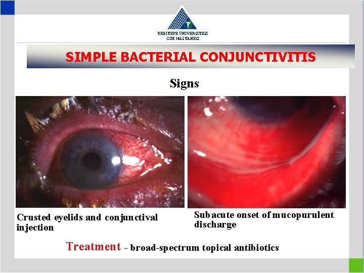
YEDİTEPE ÜNİVERSİTESİ GÖZ HASTANESİ SIMPLE BACTERIAL CONJUNCTIVITIS Signs Crusted eyelids and conjunctival injection Subacute onset of mucopurulent discharge Treatment - broad-spectrum topical antibiotics
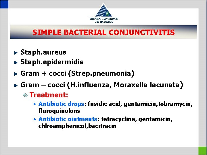
YEDİTEPE ÜNİVERSİTESİ GÖZ HASTANESİ SIMPLE BACTERIAL CONJUNCTIVITIS Staph. aureus Staph. epidermidis Gram + cocci (Strep. pneumonia) Gram – cocci (H. influenza, Moraxella lacunata) Treatment: • Antibiotic drops: fusidic acid, gentamicin, tobramycin, fluroquinolons • Antibiotic ointments: tetracycline, gentamicin, chlroamphenicol, bacitracin
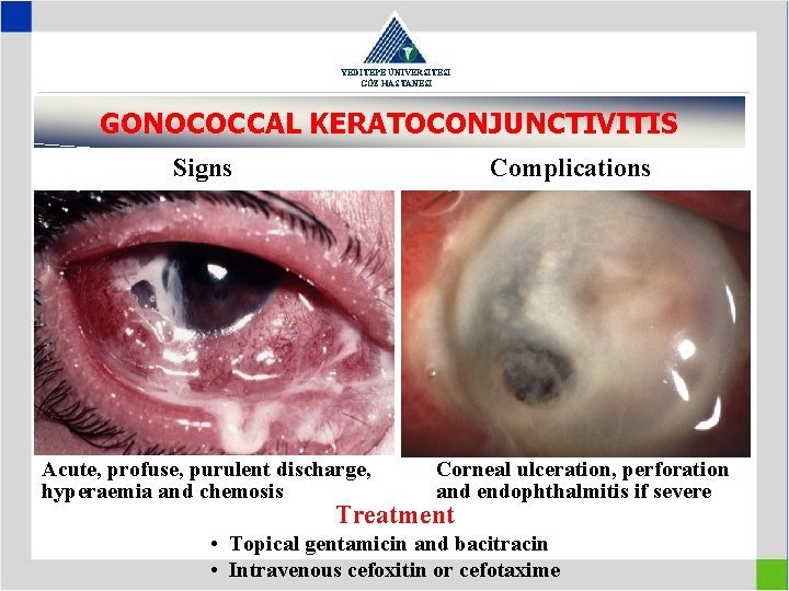
YEDİTEPE ÜNİVERSİTESİ GÖZ HASTANESİ GONOCOCCAL KERATOCONJUNCTIVITIS Signs Complications Acute, profuse, purulent discharge, hyperaemia and chemosis Corneal ulceration, perforation and endophthalmitis if severe Treatment • Topical gentamicin and bacitracin • Intravenous cefoxitin or cefotaxime
YEDİTEPE ÜNİVERSİTESİ GÖZ HASTANESİ VIRAL CONJUNCTIVITIS • Adenoviral keratoconjunctivitis • Molluscum contagiosum conjunctivitis • Herpes simplex conjunctivitis
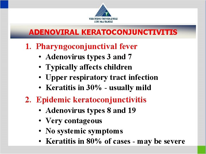
YEDİTEPE ÜNİVERSİTESİ GÖZ HASTANESİ ADENOVIRAL KERATOCONJUNCTIVITIS 1. Pharyngoconjunctival fever • • Adenovirus types 3 and 7 Typically affects children Upper respiratory tract infection Keratitis in 30% - usually mild 2. Epidemic keratoconjunctivitis • • Adenovirus types 8 and 19 Very contageous No systemic symptoms Keratitis in 80% of cases - may be severe
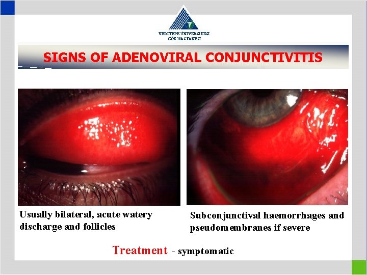
YEDİTEPE ÜNİVERSİTESİ GÖZ HASTANESİ SIGNS OF ADENOVIRAL CONJUNCTIVITIS Usually bilateral, acute watery discharge and follicles Subconjunctival haemorrhages and pseudomembranes if severe Treatment - symptomatic
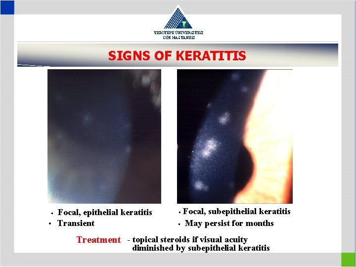
YEDİTEPE ÜNİVERSİTESİ GÖZ HASTANESİ SIGNS OF KERATITIS Focal, epithelial keratitis • Transient • Focal, subepithelial keratitis • May persist for months • Treatment - topical steroids if visual acuity diminished by subepithelial keratitis
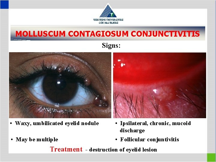
YEDİTEPE ÜNİVERSİTESİ GÖZ HASTANESİ MOLLUSCUM CONTAGIOSUM CONJUNCTIVITIS Signs: • Waxy, umbilicated eyelid nodule • May be multiple • Ipsilateral, chronic, mucoid discharge • Follicular conjuntivitis Treatment - destruction of eyelid lesion
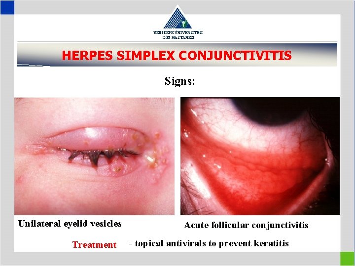
YEDİTEPE ÜNİVERSİTESİ GÖZ HASTANESİ HERPES SIMPLEX CONJUNCTIVITIS Signs: Unilateral eyelid vesicles Treatment Acute follicular conjunctivitis - topical antivirals to prevent keratitis
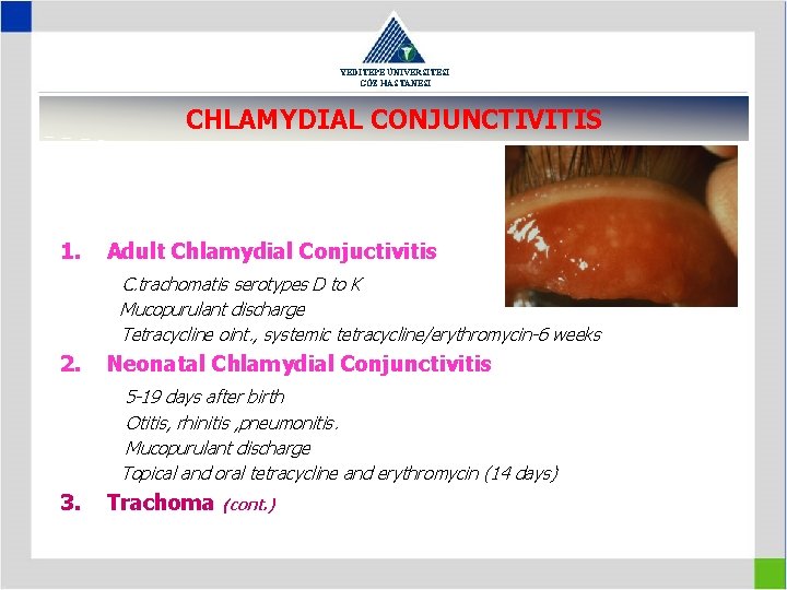
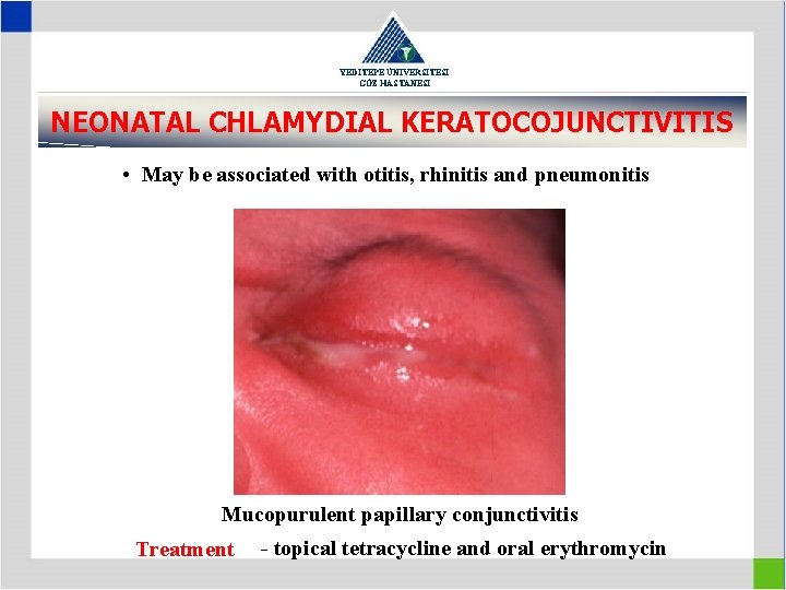
YEDİTEPE ÜNİVERSİTESİ GÖZ HASTANESİ CHLAMYDIAL CONJUNCTIVITIS 1. Adult Chlamydial Conjuctivitis C. trachomatis serotypes D to K Mucopurulant discharge Tetracycline oint. , systemic tetracycline/erythromycin-6 weeks 2. Neonatal Chlamydial Conjunctivitis 5 -19 days after birth Otitis, rhinitis , pneumonitis. Mucopurulant discharge Topical and oral tetracycline and erythromycin (14 days) 3. Trachoma (cont. )
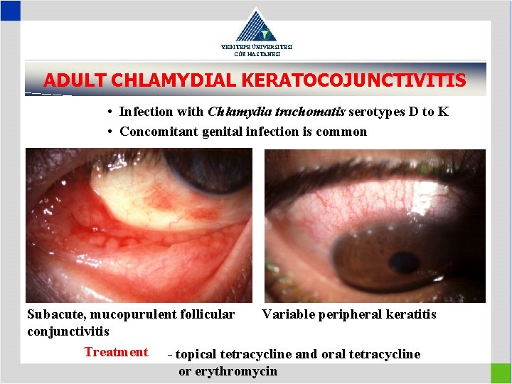
YEDİTEPE ÜNİVERSİTESİ GÖZ HASTANESİ ADULT CHLAMYDIAL KERATOCOJUNCTIVITIS • Infection with Chlamydia trachomatis serotypes D to K • Concomitant genital infection is common Subacute, mucopurulent follicular Variable peripheral keratitis conjunctivitis Treatment - topical tetracycline and oral tetracycline or erythromycin
YEDİTEPE ÜNİVERSİTESİ GÖZ HASTANESİ • Presents CHLAMYDIAL between 5 and 19 KERATOCOJUNCTIVITIS days after birth NEONATAL • May be associated with otitis, rhinitis and pneumonitis Mucopurulent papillary conjunctivitis Treatment - topical tetracycline and oral erythromycin
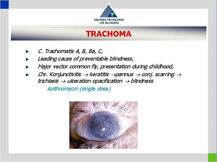
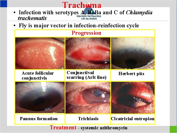
YEDİTEPE ÜNİVERSİTESİ GÖZ HASTANESİ TRACHOMA C. Trachomatis A, B, Ba, C, Leading cause of preventable blindness, Major vector common fly, presentation during childhood, Chr. Konjunctivitis keratitis pannus conj. scarring trichiasis ulceration opacification blindness Azithromycin (single dose)
Trachoma • Infection with serotypes A, B, Ba and C of Chlamydia trachomatis • Fly is major vector in infection-reinfection cycle Progression YEDİTEPE ÜNİVERSİTESİ GÖZ HASTANESİ Acute follicular conjunctivis Pannus formation Conjunctival scarring (Arlt line) Trichiasis Herbert pits Cicatricial entropion Treatment - systemic azithromycin
YEDİTEPE ÜNİVERSİTESİ GÖZ HASTANESİ ALLERGIC CONJUNCTIVITIS 1. Allergic rhinoconjunctivitis 2. Vernal keratoconjunctivitis 3. Atopic keratoconjunctivitis 4. Giant papillary conjunctivitis
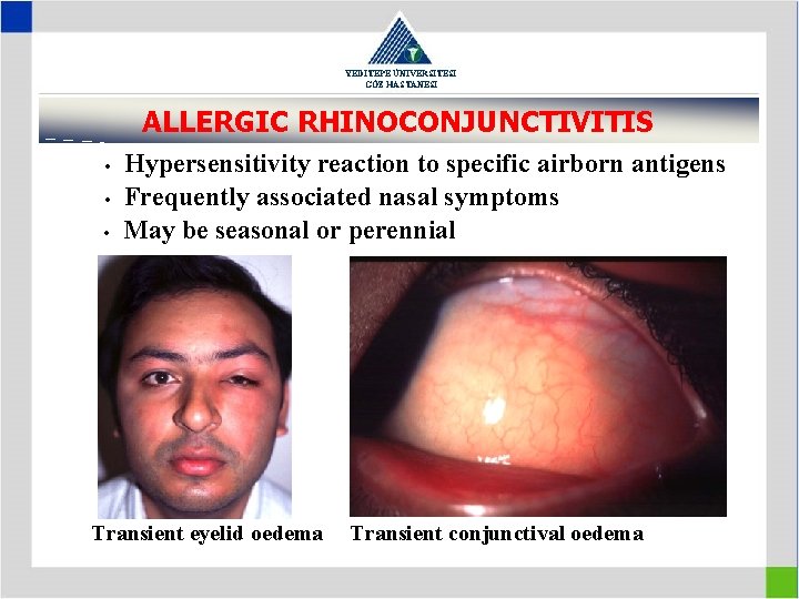
YEDİTEPE ÜNİVERSİTESİ GÖZ HASTANESİ ALLERGIC RHINOCONJUNCTIVITIS • • • Hypersensitivity reaction to specific airborn antigens Frequently associated nasal symptoms May be seasonal or perennial Transient eyelid oedema Transient conjunctival oedema
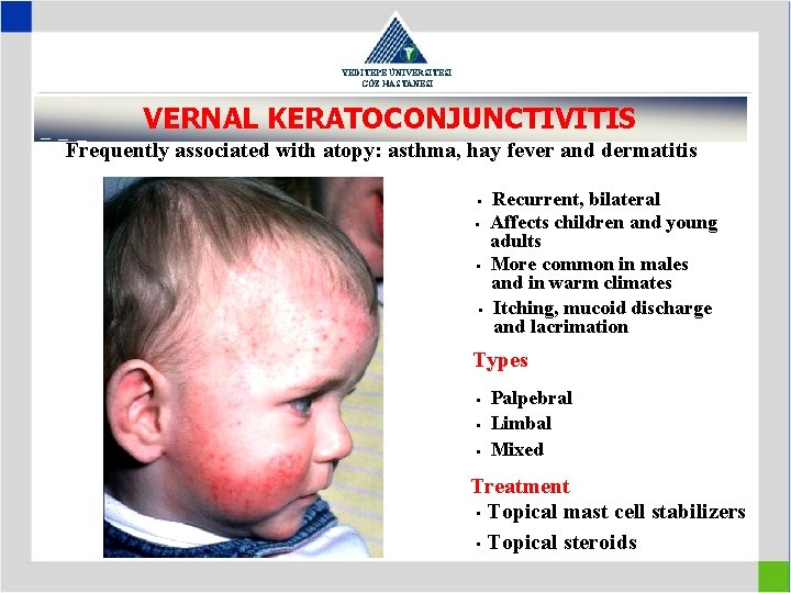
YEDİTEPE ÜNİVERSİTESİ GÖZ HASTANESİ VERNAL KERATOCONJUNCTIVITIS Frequently associated with atopy: asthma, hay fever and dermatitis • • Recurrent, bilateral Affects children and young adults More common in males and in warm climates Itching, mucoid discharge and lacrimation Types • • • Palpebral Limbal Mixed Treatment • Topical mast cell stabilizers • Topical steroids
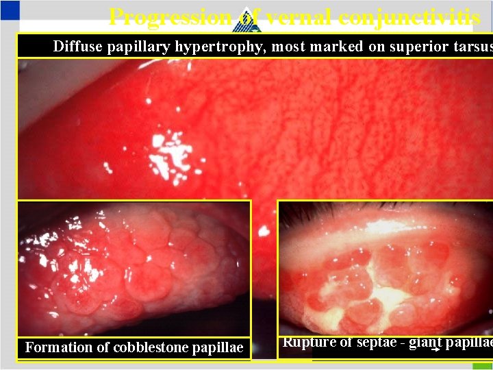
Progression of vernal conjunctivitis Diffuse papillary hypertrophy, most marked on superior tarsus YEDİTEPE ÜNİVERSİTESİ GÖZ HASTANESİ Formation of cobblestone papillae Rupture of septae - giant papillae
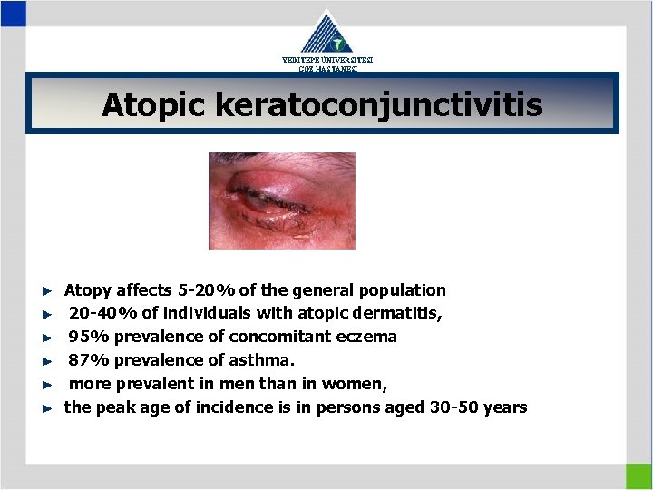
YEDİTEPE ÜNİVERSİTESİ GÖZ HASTANESİ Atopic keratoconjunctivitis Atopy affects 5 -20% of the general population 20 -40% of individuals with atopic dermatitis, 95% prevalence of concomitant eczema 87% prevalence of asthma. more prevalent in men than in women, the peak age of incidence is in persons aged 30 -50 years
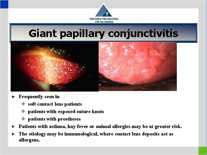
YEDİTEPE ÜNİVERSİTESİ GÖZ HASTANESİ Giant papillary conjunctivitis Frequently seen in soft contact lens patients with exposed suture knots patients with prostheses Patients with asthma, hay fever or animal allergies may be at greater risk. The etiology may be immunological, where contact lens deposits act as allergens.
YEDİTEPE ÜNİVERSİTESİ GÖZ HASTANESİ IMMUNOBULLOUS DISEASES 1. Cicatricial pemphigoid 2. a rare chronic autoimmune subepithelial blistering disease characterized by erosive skin lesions of the mucous membranes and skin that results in scarring of at least some sites of involvement 2. Stevens-Johnson syndrome 3. Toxic epidermal necrolysis (Lyell disease) 4. Epidermolysis bullosa 5. Pemphigus vulgaris 6. Linear Ig. A bullous dermatosis
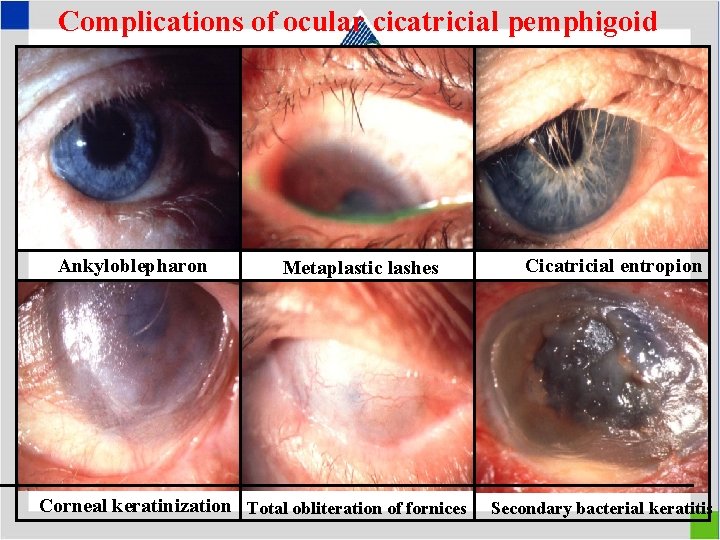
Complications of ocular cicatricial pemphigoid YEDİTEPE ÜNİVERSİTESİ GÖZ HASTANESİ Ankyloblepharon Metaplastic lashes Corneal keratinization Total obliteration of fornices Cicatricial entropion Secondary bacterial keratitis
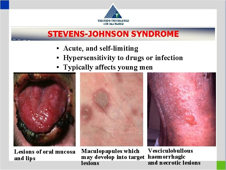
YEDİTEPE ÜNİVERSİTESİ GÖZ HASTANESİ STEVENS-JOHNSON SYNDROME • Acute, and self-limiting • Hypersensitivity to drugs or infection • Typically affects young men Lesions of oral mucosa Maculopapules which Vesciculobullous may develop into target haemorrhagic and lips and necrotic lesions
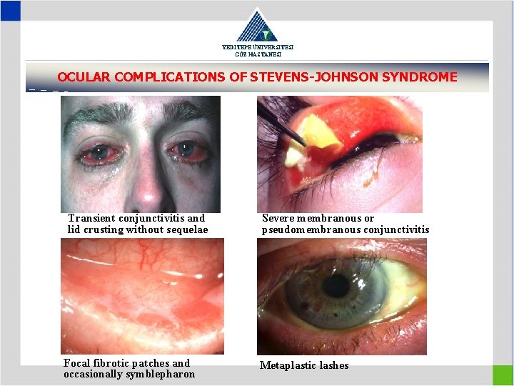
YEDİTEPE ÜNİVERSİTESİ GÖZ HASTANESİ OCULAR COMPLICATIONS OF STEVENS-JOHNSON SYNDROME Transient conjunctivitis and lid crusting without sequelae Focal fibrotic patches and occasionally symblepharon Severe membranous or pseudomembranous conjunctivitis Metaplastic lashes
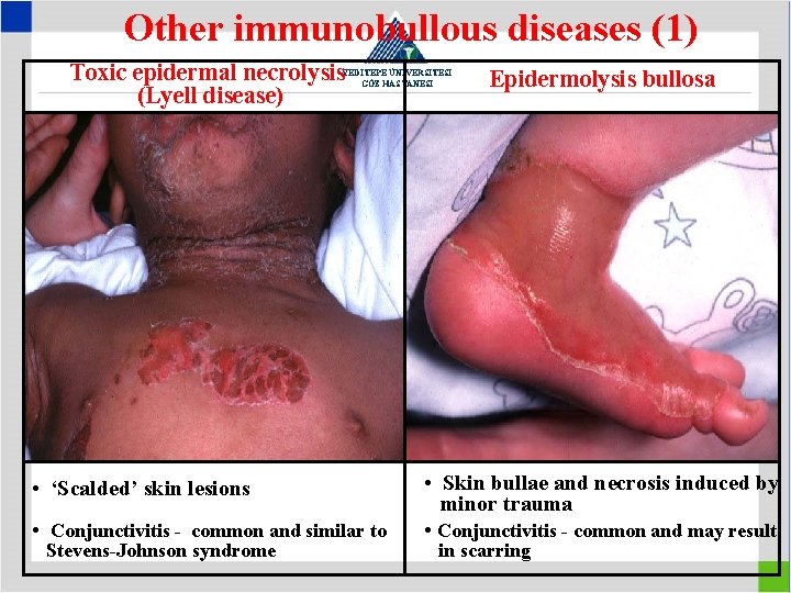
Other immunobullous diseases (1) Toxic epidermal necrolysis (Lyell disease) YEDİTEPE ÜNİVERSİTESİ GÖZ HASTANESİ • ‘Scalded’ skin lesions • Conjunctivitis - common and similar to Stevens-Johnson syndrome Epidermolysis bullosa • Skin bullae and necrosis induced by minor trauma • Conjunctivitis - common and may result in scarring
YEDİTEPE ÜNİVERSİTESİ GÖZ HASTANESİ Pingeucula, pterygium
YEDİTEPE ÜNİVERSİTESİ GÖZ HASTANESİ Pingeucula, pterygium benign, reactive, proliferative lesion of the conjunctiva yellow-white thickening with increased vascularity sunlight and other environmental exposure are predisposing elements Pingeucula limited to the area of the conjunctiva and pterygium encroaching on the cornea histologically, these lesions are characterized by degeneration of collagen in the substantia propria of bulbar conjunctiva (elastoid and basophilic degeneration epithelium is alternately thinned (atrophy) or thickened (proliferative) and shows no atypia
YEDİTEPE ÜNİVERSİTESİ GÖZ HASTANESİ Conjunctival Tumours / Non-pigmented tumours Conjunctival papilloma 1) Pedunculated papilloma -Human papilloma virus types 6 and 11 -In childhood or early adult life -Signs: may be multiple, ocassionally bilateral, mostly palpebral conj. , fornix and caruncle -Treatment: -High rate of spontaneous resolution of small lesions -Large lesions→ cryotherapy, excision
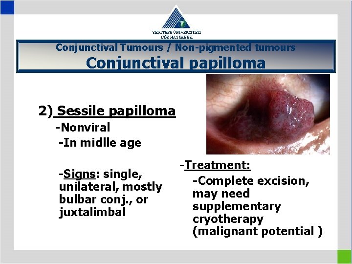
YEDİTEPE ÜNİVERSİTESİ GÖZ HASTANESİ Conjunctival Tumours / Non-pigmented tumours Conjunctival papilloma 2) Sessile papilloma -Nonviral -In midlle age -Signs: single, unilateral, mostly bulbar conj. , or juxtalimbal -Treatment: -Complete excision, may need supplementary cryotherapy (malignant potential )
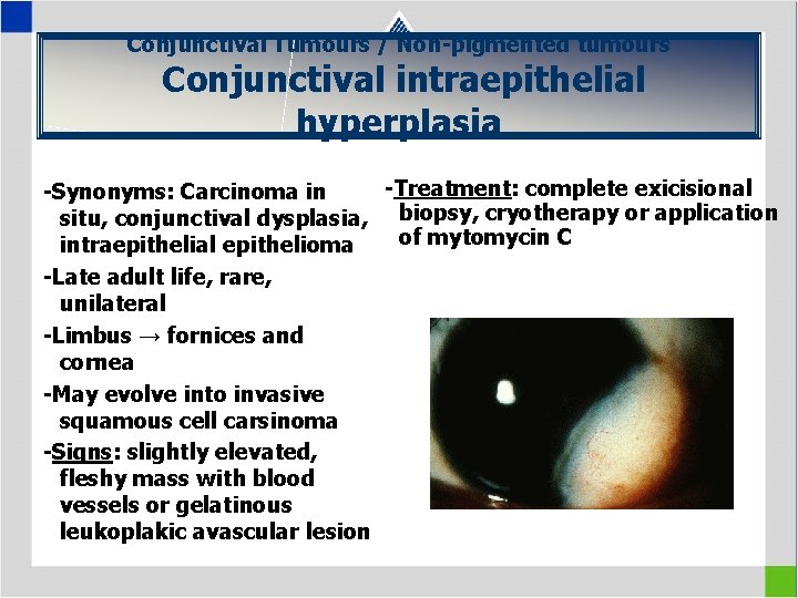
Conjunctival Tumours / Non-pigmented tumours Conjunctival intraepithelial hyperplasia YEDİTEPE ÜNİVERSİTESİ GÖZ HASTANESİ -Treatment: complete exicisional -Synonyms: Carcinoma in situ, conjunctival dysplasia, biopsy, cryotherapy or application of mytomycin C intraepithelial epithelioma -Late adult life, rare, unilateral -Limbus → fornices and cornea -May evolve into invasive squamous cell carsinoma -Signs: slightly elevated, fleshy mass with blood vessels or gelatinous leukoplakic avascular lesion
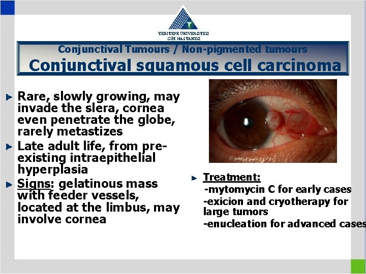
YEDİTEPE ÜNİVERSİTESİ GÖZ HASTANESİ Conjunctival Tumours / Non-pigmented tumours Conjunctival squamous cell carcinoma Rare, slowly growing, may invade the slera, cornea even penetrate the globe, rarely metastizes Late adult life, from preexisting intraepithelial hyperplasia Signs: gelatinous mass with feeder vessels, located at the limbus, may involve cornea Treatment: -mytomycin C for early cases -exicion and cryotherapy for large tumors -enucleation for advanced cases
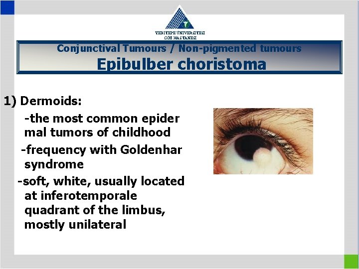
YEDİTEPE ÜNİVERSİTESİ GÖZ HASTANESİ Conjunctival Tumours / Non-pigmented tumours Epibulber choristoma 1) Dermoids: -the most common epider mal tumors of childhood -frequency with Goldenhar syndrome -soft, white, usually located at inferotemporale quadrant of the limbus, mostly unilateral
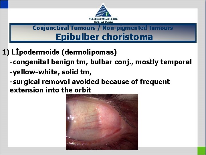
YEDİTEPE ÜNİVERSİTESİ GÖZ HASTANESİ Conjunctival Tumours / Non-pigmented tumours Epibulber choristoma 1) Lİpodermoids (dermolipomas) -congenital benign tm, bulbar conj. , mostly temporal -yellow-white, solid tm, -surgical removal avoided because of frequent extension into the orbit
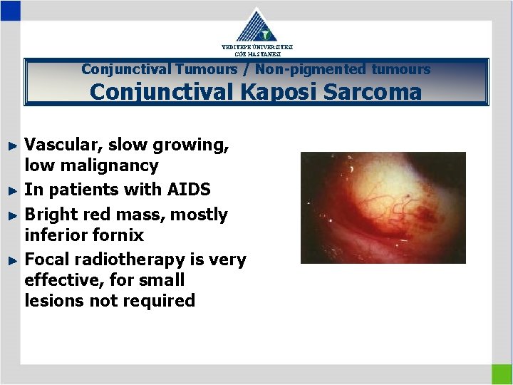
YEDİTEPE ÜNİVERSİTESİ GÖZ HASTANESİ Conjunctival Tumours / Non-pigmented tumours Conjunctival Kaposi Sarcoma Vascular, slow growing, low malignancy In patients with AIDS Bright red mass, mostly inferior fornix Focal radiotherapy is very effective, for small lesions not required
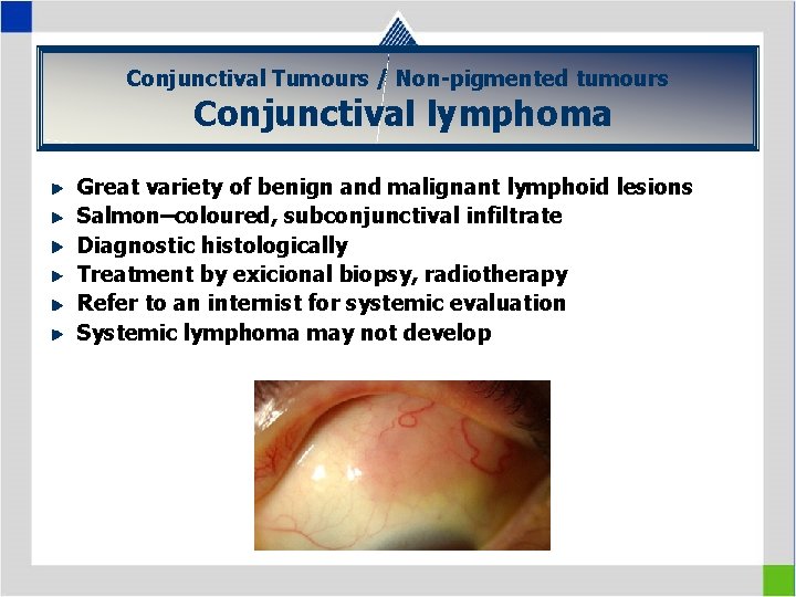
Conjunctival Tumours / Non-pigmented tumours YEDİTEPE ÜNİVERSİTESİ GÖZ HASTANESİ Conjunctival lymphoma Great variety of benign and malignant lymphoid lesions Salmon–coloured, subconjunctival infiltrate Diagnostic histologically Treatment by exicional biopsy, radiotherapy Refer to an internist for systemic evaluation Systemic lymphoma may not develop
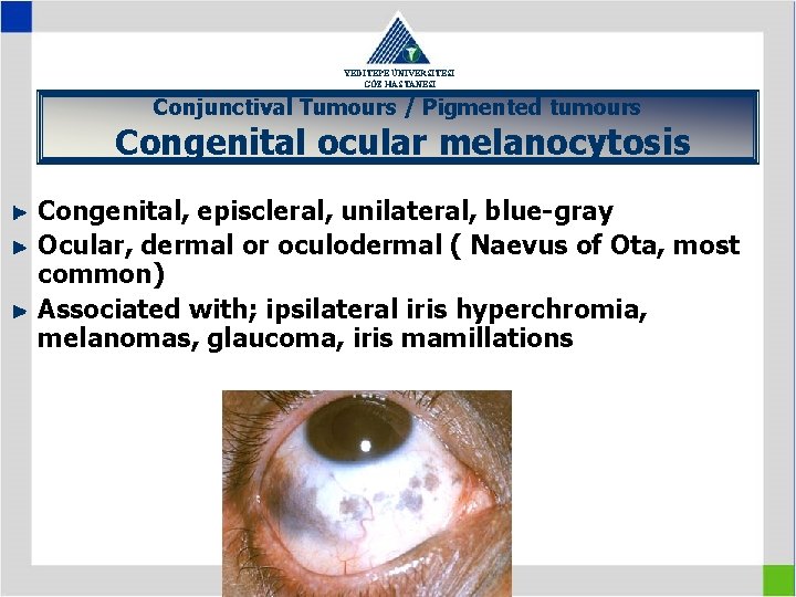
YEDİTEPE ÜNİVERSİTESİ GÖZ HASTANESİ Conjunctival Tumours / Pigmented tumours Congenital ocular melanocytosis Congenital, episcleral, unilateral, blue-gray Ocular, dermal or oculodermal ( Naevus of Ota, most common) Associated with; ipsilateral iris hyperchromia, melanomas, glaucoma, iris mamillations
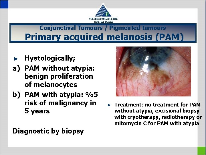
YEDİTEPE ÜNİVERSİTESİ GÖZ HASTANESİ Conjunctival Tumours / Pigmented tumours Primary acquired melanosis (PAM) Hystologically; a) PAM without atypia: benign proliferation of melanocytes b) PAM with atypia: %5 risk of malignancy in 5 years Diagnostic by biopsy Treatment: no treatment for PAM without atypia, excisional biopsy with cryotherapy, radiotherapy or mitomycin C for PAM with atypia
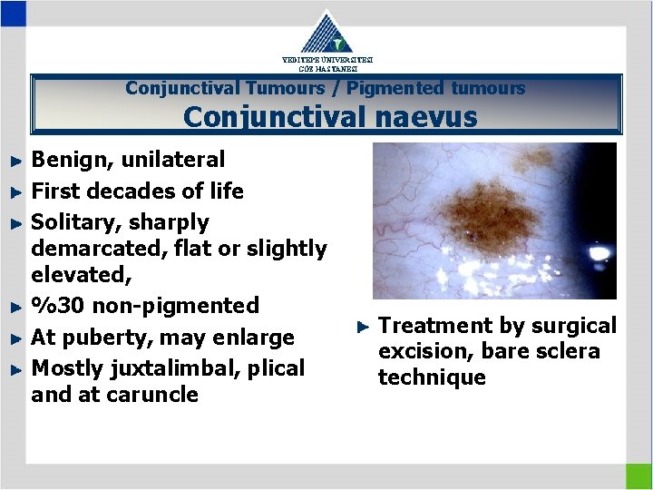
YEDİTEPE ÜNİVERSİTESİ GÖZ HASTANESİ Conjunctival Tumours / Pigmented tumours Conjunctival naevus Benign, unilateral First decades of life Solitary, sharply demarcated, flat or slightly elevated, %30 non-pigmented At puberty, may enlarge Mostly juxtalimbal, plical and at caruncle Treatment by surgical excision, bare sclera technique
YEDİTEPE ÜNİVERSİTESİ GÖZ HASTANESİ Conjunctival Tumours / Pigmented tumours Conjunctival melanoma (1) Usually in 6 th decade %2 of all eye Usually limbal, solitary, black malignancies or gray nodule, may be non% 50 -75 arises from pigmented, fixed to sclera PAM with atypia %20 arises from naevus Least common is novo
YEDİTEPE ÜNİVERSİTESİ GÖZ HASTANESİ Conjunctival Tumours / Pigmented tumours Conjunctival melanoma (2) Differential diagnosis: -large naevus at puberty -ciliary body melanoma with extraocular extension -melanocytoma; congenital, black, can not be moved over the globe -pyogenic granuloma
YEDİTEPE ÜNİVERSİTESİ GÖZ HASTANESİ Conjunctival Tumours / Pigmented tumours Conjunctival melanoma (3) Treatment: -Exicional biopsy often with supplemental cryotherapy unless intraocular or orbital involvement is present -Exenteration for extensive and aggressive disease, does not improve the survival
YEDİTEPE ÜNİVERSİTESİ GÖZ HASTANESİ CORNEAL DİSEASES 1. 2. 3. 4. Corneal Infections (KERATITIS) Ectatic Corneal Diseases Dystrophies and Degenerations Corneal Surgery
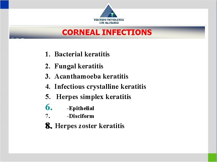
YEDİTEPE ÜNİVERSİTESİ GÖZ HASTANESİ CORNEAL INFECTIONS 1. Bacterial keratitis 2. 3. 4. 5. 6. 7. Fungal keratitis Acanthamoeba keratitis Infectious crystalline keratitis Herpes simplex keratitis -Epithelial -Disciform 6. Herpes zoster keratitis 8.
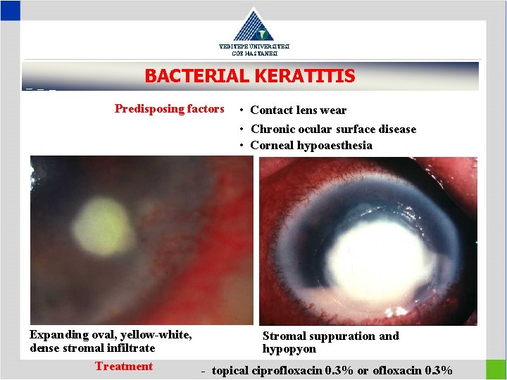
YEDİTEPE ÜNİVERSİTESİ GÖZ HASTANESİ BACTERIAL KERATITIS Predisposing factors • Contact lens wear • Chronic ocular surface disease • Corneal hypoaesthesia Expanding oval, yellow-white, Stromal suppuration and dense stromal infiltrate hypopyon Treatment - topical ciprofloxacin 0. 3% or ofloxacin 0. 3%
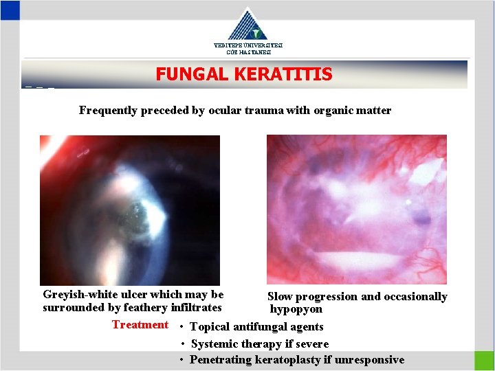
YEDİTEPE ÜNİVERSİTESİ GÖZ HASTANESİ FUNGAL KERATITIS Frequently preceded by ocular trauma with organic matter Greyish-white ulcer which may be Slow progression and occasionally surrounded by feathery infiltrates hypopyon Treatment • Topical antifungal agents • Systemic therapy if severe • Penetrating keratoplasty if unresponsive
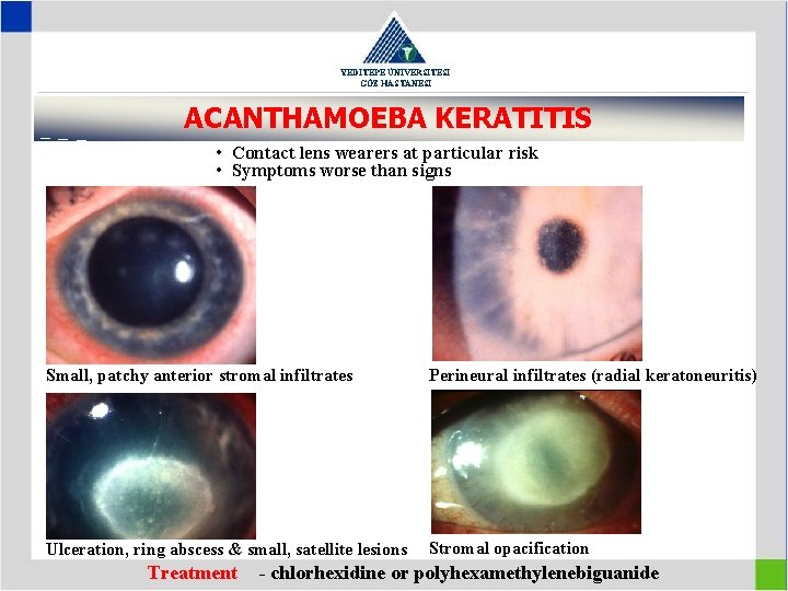
YEDİTEPE ÜNİVERSİTESİ GÖZ HASTANESİ ACANTHAMOEBA KERATITIS • Contact lens wearers at particular risk • Symptoms worse than signs Small, patchy anterior stromal infiltrates Perineural infiltrates (radial keratoneuritis) Ulceration, ring abscess & small, satellite lesions Stromal opacification Treatment - chlorhexidine or polyhexamethylenebiguanide
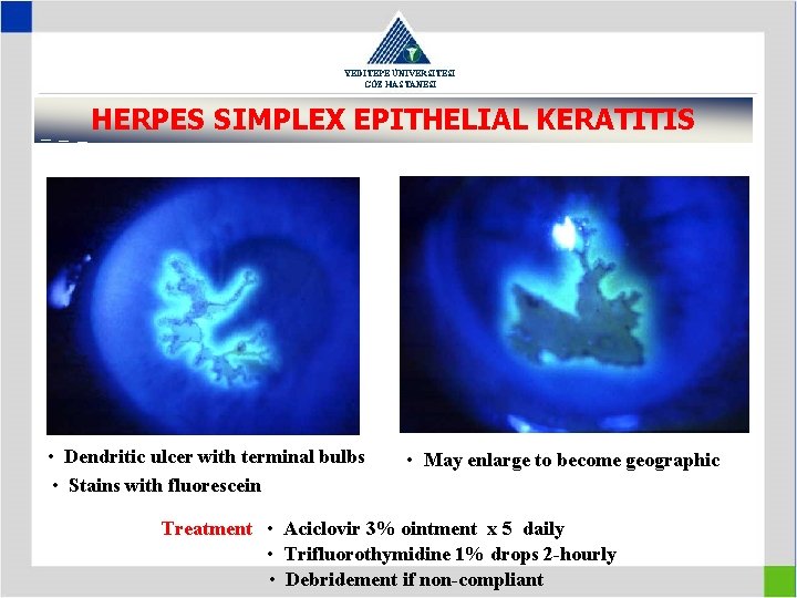
YEDİTEPE ÜNİVERSİTESİ GÖZ HASTANESİ HERPES SIMPLEX EPITHELIAL KERATITIS • Dendritic ulcer with terminal bulbs • Stains with fluorescein • May enlarge to become geographic Treatment • Aciclovir 3% ointment x 5 daily • Trifluorothymidine 1% drops 2 -hourly • Debridement if non-compliant
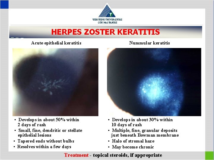
YEDİTEPE ÜNİVERSİTESİ GÖZ HASTANESİ HERPES ZOSTER KERATITIS Acute epithelial keratitis • Develops in about 50% within 2 days of rash • Small, fine, dendritic or stellate epithelial lesions • Tapered ends without bulbs • Resolves within a few days Nummular keratitis • Develops in about 30% within 10 days of rash • Multiple, fine, granular deposits just beneath Bowman membrane • Halo of stromal haze • May become chronic Treatment - topical steroids, if appropriate
YEDİTEPE ÜNİVERSİTESİ GÖZ HASTANESİ CORNEAL ECTASIAS 1. Keratoconus 2. Keratoglobus 3. Pellucid marginal degeneration
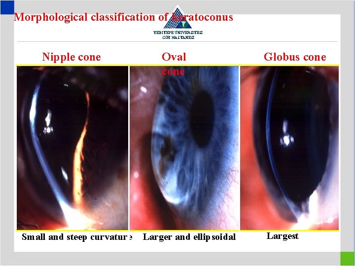
Morphological classification of keratoconus YEDİTEPE ÜNİVERSİTESİ GÖZ HASTANESİ Nipple cone Small and steep curvature Oval cone Larger and ellipsoidal Globus cone Largest
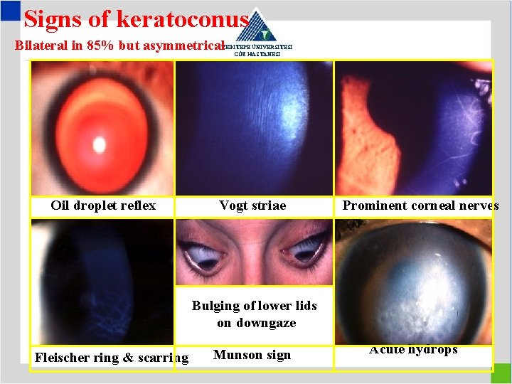
Signs of keratoconus Bilateral in 85% but asymmetrical YEDİTEPE ÜNİVERSİTESİ GÖZ HASTANESİ Oil droplet reflex Vogt striae Prominent corneal nerves Bulging of lower lids on downgaze Fleischer ring & scarring Munson sign Acute hydrops
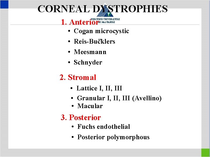
CORNEAL DYSTROPHIES 1. Anterior YEDİTEPE ÜNİVERSİTESİ GÖZ HASTANESİ • • Cogan microcystic. . Reis-Bucklers Meesmann Schnyder 2. Stromal • Lattice I, III • Granular I, III (Avellino) • Macular 3. Posterior • Fuchs endothelial • Posterior polymorphous
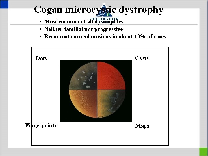
Cogan microcystic dystrophy YEDİTEPE ÜNİVERSİTESİ GÖZ HASTANESİ • Most common of all dystrophies • Neither familial nor progressive • Recurrent corneal erosions in about 10% of cases Dots Cysts Fingerprints Maps
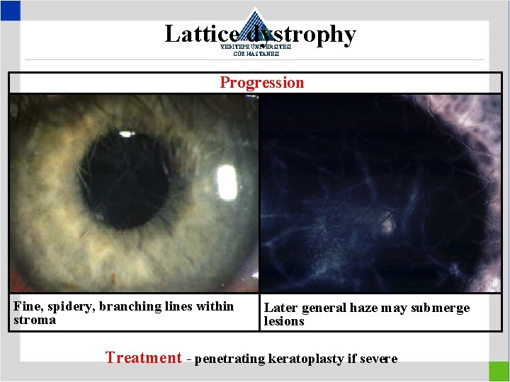
Lattice dystrophy YEDİTEPE ÜNİVERSİTESİ GÖZ HASTANESİ Progression Fine, spidery, branching lines within stroma Later general haze may submerge lesions Treatment - penetrating keratoplasty if severe
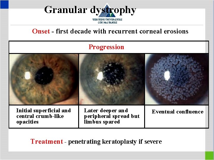
Granular dystrophy YEDİTEPE ÜNİVERSİTESİ GÖZ HASTANESİ Onset - first decade with recurrent corneal erosions Progression Initial superficial and central crumb-like opacities Later deeper and peripheral spread but limbus spared Eventual confluence Treatment - penetrating keratoplasty if severe
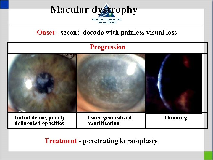
Macular dystrophy YEDİTEPE ÜNİVERSİTESİ GÖZ HASTANESİ Onset - second decade with painless visual loss Progression Initial dense, poorly delineated opacities Later generalized opacification Treatment - penetrating keratoplasty Thinning
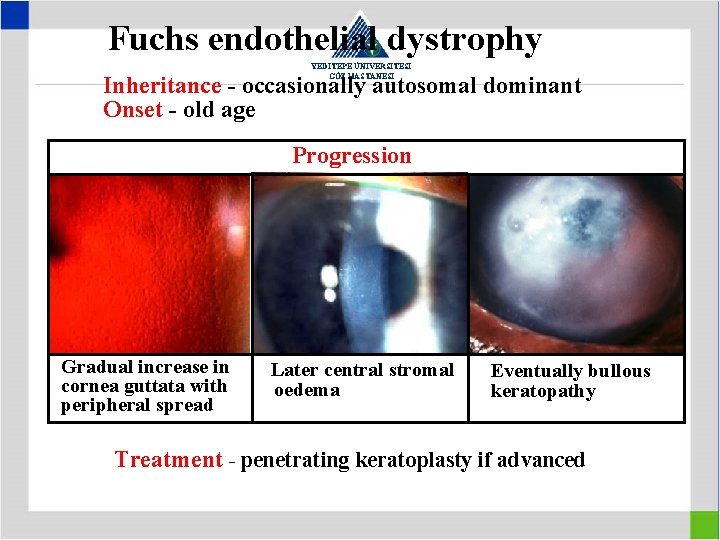
Fuchs endothelial dystrophy YEDİTEPE ÜNİVERSİTESİ GÖZ HASTANESİ Inheritance - occasionally autosomal dominant Onset - old age Progression Gradual increase in cornea guttata with peripheral spread Later central stromal oedema Eventually bullous keratopathy Treatment - penetrating keratoplasty if advanced
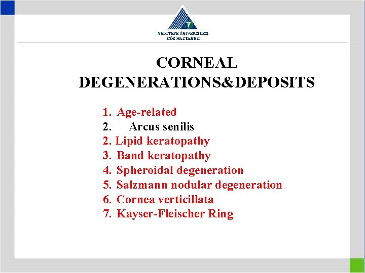
YEDİTEPE ÜNİVERSİTESİ GÖZ HASTANESİ CORNEAL DEGENERATIONS&DEPOSITS 1. Age-related 2. Arcus senilis 2. Lipid keratopathy 3. Band keratopathy 4. Spheroidal degeneration 5. Salzmann nodular degeneration 6. Cornea verticillata 7. Kayser-Fleischer Ring
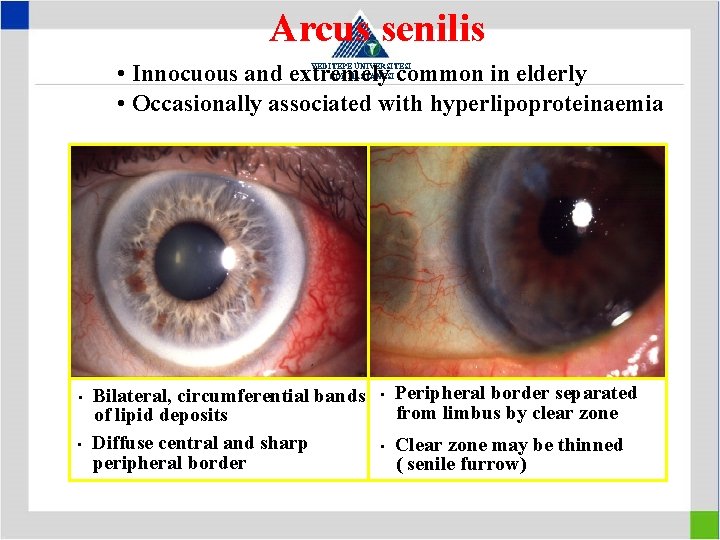
Arcus senilis • Innocuous and extremely common in elderly • Occasionally associated with hyperlipoproteinaemia YEDİTEPE ÜNİVERSİTESİ GÖZ HASTANESİ • • Bilateral, circumferential bands of lipid deposits Diffuse central and sharp peripheral border • • Peripheral border separated from limbus by clear zone Clear zone may be thinned ( senile furrow)
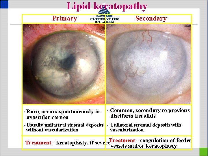
Lipid keratopathy Primary • • YEDİTEPE ÜNİVERSİTESİ GÖZ HASTANESİ Rare, occurs spontaneously in avascular cornea • Usually unilateral stromal deposits without vascularization • Secondary Common, secondary to previous disciform keratitis Unilateral stromal deposits with vascularization Treatment - keratoplasty, if severe. Treatment - coagulation of feeder vessels and/or keratoplasty
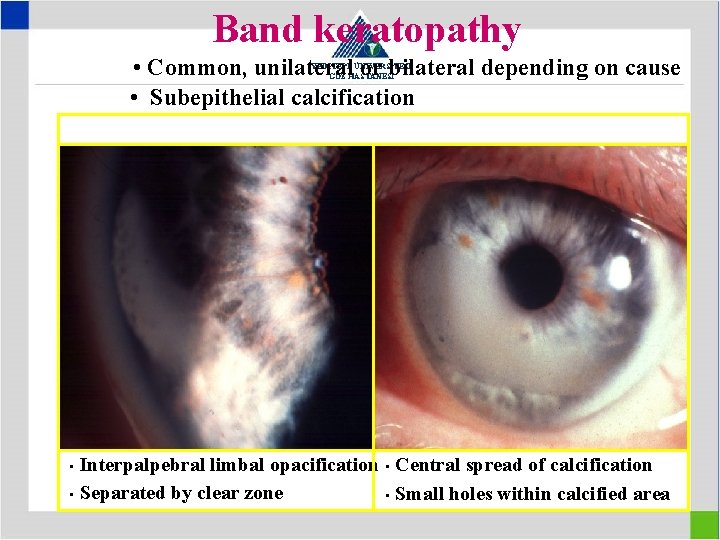
Band keratopathy • Common, unilateral or bilateral depending on cause • Subepithelial calcification YEDİTEPE ÜNİVERSİTESİ GÖZ HASTANESİ Interpalpebral limbal opacification • Central spread of calcification • Separated by clear zone • Small holes within calcified area •
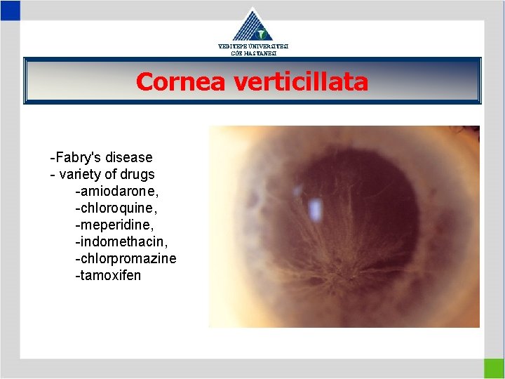
YEDİTEPE ÜNİVERSİTESİ GÖZ HASTANESİ Cornea verticillata -Fabry's disease - variety of drugs -amiodarone, -chloroquine, -meperidine, -indomethacin, -chlorpromazine -tamoxifen
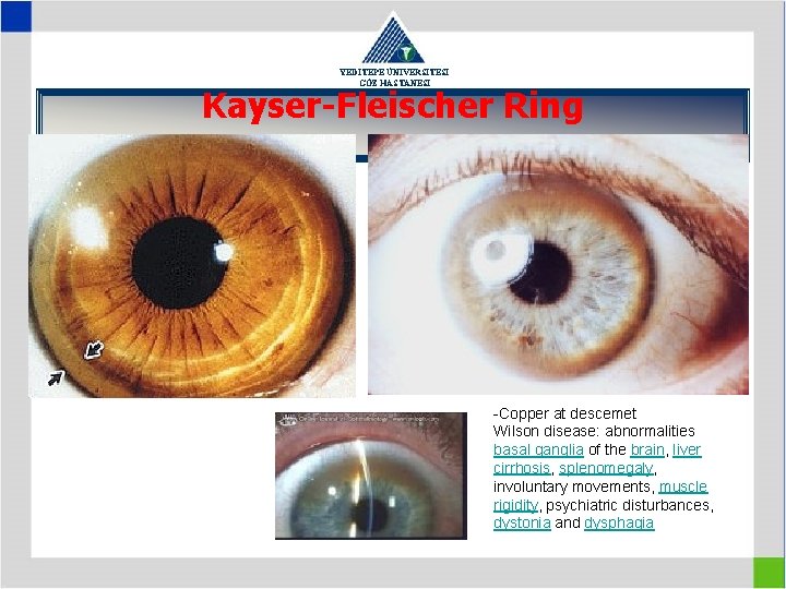
YEDİTEPE ÜNİVERSİTESİ GÖZ HASTANESİ Kayser-Fleischer Ring -Copper at descemet Wilson disease: abnormalities basal ganglia of the brain, liver cirrhosis, splenomegaly, involuntary movements, muscle rigidity, psychiatric disturbances, dystonia and dysphagia
CORNEAL SURGERY YEDİTEPE ÜNİVERSİTESİ GÖZ HASTANESİ 1. Keratoplasty (Penetrating , lamellar) 2. Keratoprosthesis 3. Refractive surgery • Radial keratotomy (RK) • Photorefractive keratectomy (PRK) • Laser assisted subepithelial keratectomy(LASEK) • Epithelial Laser in-situ keratomileusis (Epi. LASIK) • Laser in-situ keratomileusis (LASIK) • Conductive keratoplasty (light-touch) (CK) • Intracorneal ring segments (ICR)
Penetrating Keratoplasty YEDİTEPE ÜNİVERSİTESİ GÖZ HASTANESİ 1. Indications • Optical (e. g. bullous keratopathy, dystrophies) • Tectonic (e. g. severe stromal thinning, descemetocele) • Therapeutic (e. g. severe keratitis) • Cosmetic 2. Adverse prognostic factors • Severe stromal vascularization • Absence of corneal sensation • Progressive conjunctival inflammation (e. g. pemphigoid) • Tear film dysfunction • Glaucoma
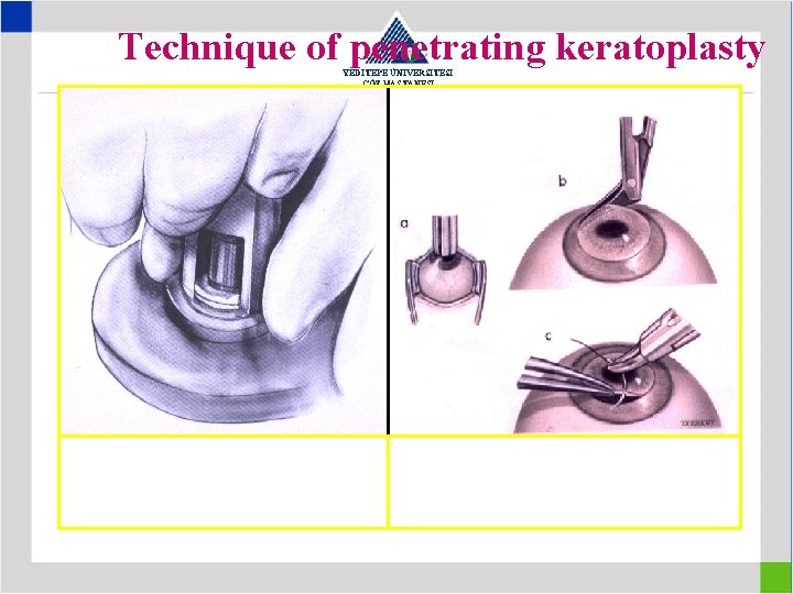
Technique of penetrating keratoplasty YEDİTEPE ÜNİVERSİTESİ GÖZ HASTANESİ Excision of donor tissue a, b - Excision of host tissue c - Fixation of donor tissue
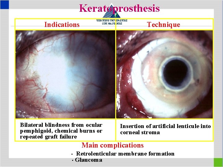
Keratoprosthesis Indications YEDİTEPE ÜNİVERSİTESİ GÖZ HASTANESİ Bilateral blindness from ocular pemphigoid, chemical burns or repeated graft failure Technique Insertion of artificial lenticule into corneal stroma Main complications Retrolenticular membrane formation • Glaucoma •
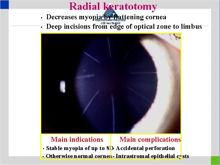
Radial keratotomy Decreases myopia by flattening cornea Deep incisions from edge of optical zone to limbus • YEDİTEPE ÜNİVERSİTESİ GÖZ HASTANESİ • Main indications Main complications Stable myopia of up to 8 D • Accidental perforation • Otherwise normal cornea • Intrastromal epithelial cysts •
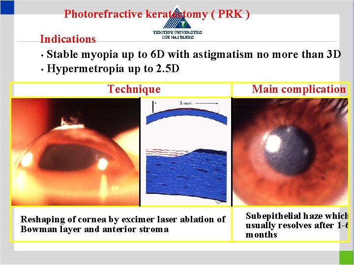
Photorefractive keratectomy ( PRK ) YEDİTEPE ÜNİVERSİTESİ GÖZ HASTANESİ Indications • Stable myopia up to 6 D with astigmatism no more than 3 D • Hypermetropia up to 2. 5 D Technique Reshaping of cornea by excimer laser ablation of Bowman layer and anterior stroma Main complication Subepithelial haze which usually resolves after 1 -6 months
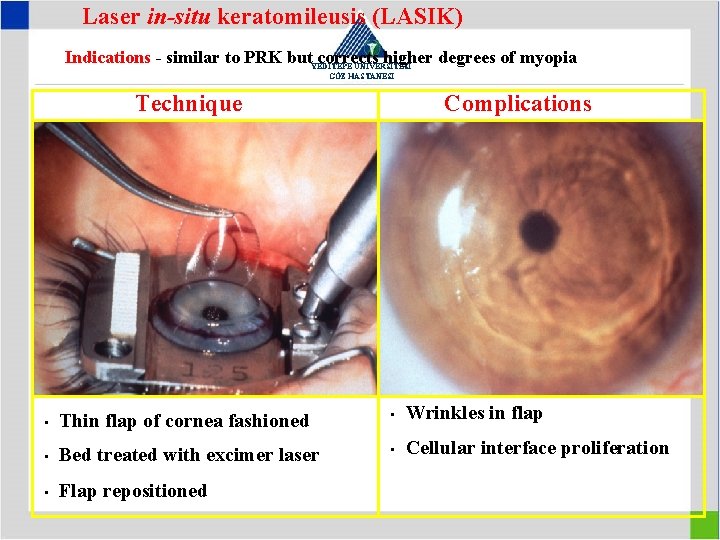
Laser in-situ keratomileusis (LASIK) Indications - similar to PRK but. YEDİTEPE corrects higher degrees of myopia ÜNİVERSİTESİ GÖZ HASTANESİ Technique Complications • Thin flap of cornea fashioned • Wrinkles in flap • Bed treated with excimer laser • Cellular interface proliferation • Flap repositioned
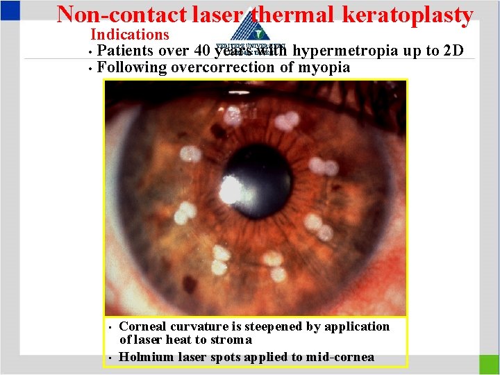
Non-contact laser thermal keratoplasty Indications • Patients over 40 years with hypermetropia up to 2 D • Following overcorrection of myopia YEDİTEPE ÜNİVERSİTESİ GÖZ HASTANESİ • • Corneal curvature is steepened by application of laser heat to stroma Holmium laser spots applied to mid-cornea