IronDeficiency Anemia Majid vafaie Iron deficiency is the


")

. The red cell count(RBC) also decreases. The reticulocyte percentage")

 is normal, and thrombocytosis is often present. Thrombocytopenia")


- Slides: 47
Iron-Deficiency Anemia Majid. vafaie
Iron deficiency is the most widespread and common nutritional disorder in the world. It is estimated that 30% of the global population suffers from iron-deficiency anemia most of them live in developing countries. In the USA, 9% of children ages 12 -36 mo are iron deficient, and 30% of this group have progressed to iron-deficiency anemia.
The incidence of iron-deficiency relates to basic aspects of iron metabolism and nutrition. The body of a full-term newborn infant contains about 0. 5 g of iron, compared to 5 g of iron in adults. This change in quantity of iron from birth to adulthood means that an average of 0. 8 mg of iron must be absorbed each day during the first 15 years of life.
A small additional amount is necessary to balance normal losses of iron by shedding of cells. It is therefore necessary to absorb approximately 1 mg daily to maintain positive iron balance in childhood. Because < 10% of dietary iron usually is absorbed, a dietary intake of 8 -10 mg of iron daily is necessary to maintain iron levels
During infancy when growth is most rapid, the approximately 1 mg/L of iron in bovine and breast milk makes it difficult to maintain body iron. Breast-fed infants have an advantage because they absorb iron 2 -3 times more efficiently than infants fed bovine milk.
ETIOLOGY Most iron in neonates is in circulating hemoglobin. As the relatively high hemoglobin concentration of the newborn infant falls during the first 2 -3 mo of life, considerable iron is reclaimed and stored. These reclaimed stores usually are sufficient for blood formation in the first 6 -9 mo of life in term infants.
Stores are depleted sooner in low-birth weight infants or infants with perinatal blood loss because their iron stores are smaller. Delayed clamping of the umbilical cord can improve iron status and reduce the risk of iron deficiency. Dietary sources of iron are especially important in these infants.
In term infants, anemia caused solely by inadequate dietary iron usually occurs at 9 -24 mo of age and is relatively uncommon thereafter. The usual dietary pattern observed in infants and toddlers with nutritional irondeficiency anemia in developed countries is excessive consumption of bovine milk (low iron content, blood loss from milk protein colitis) in a child who is often overweight. Worldwide, undernutrition is usually responsible for iron deficiency.
Blood loss must be considered as a possible cause in every case of iron-deficiency anemia, particularly in older children. Chronic iron-deficiency anemia from occult bleeding may be caused by a lesion of the gastrointestinal (GI) tract, such as peptic ulcer, Meckel diverticulum, polyp, hemangioma, or inflammatory bowel disease.
Infants can have chronic intestinal blood loss induced by exposure to a heat-labile protein in whole bovine milk. This GI reaction is not related to enzymatic abnormalities in the mucosa, such as lactase deficiency, or to a typical milk allergy. Involved infants characteristically develop anemia that is more severe and occurs earlier than would be expected simply from an inadequate intake of iron
The ongoing loss of blood in the stools can be prevented either by breast-feeding or by delaying the introduction of whole bovine milk in the 1 st year of life and then limiting the quantity of whole bovine milk to <24 oz/24 hr.
Unrecognized blood loss also can be associated with chronic diarrhea and rarely with pulmonary hemosiderosis. In developing countries, infections with hookworm, Trichuris trichiura, Plasmodium, and Helicobacter pylori often contribute to iron deficiency
About 2% of adolescent girls have irondeficiency anemia, due in large part to their adolescent growth spurt and menstrual blood loss. The highest risk of iron deficiency is among teenagers who are or have been pregnant; >30% of these girls have irondeficiency anemia.
CLINICAL MANIFESTATIONS Most children with iron deficiency are asymptomatic and are identified by recommended laboratory screening at 12 months of age or sooner if at high risk. Pallor is the most important clinical sign of iron deficiency but is not usually visible until the hemoglobin falls to 7 -8 g/d. L.
It is most readily noted as pallor of the palms, palmar creases, nail beds, or conjunctivae. Parents often fail to note the pallor because of the typical slow drop over time. Often a visiting friend or relative is the first to notice.
In mild to moderate iron deficiency (i. e. , hemoglobin levels of 6 -10 g/d. L), compensatory mechanisms, including increased levels of (2, 3 -DPG) and a shift of the oxygen dissociation curve, may be so effective that few symptoms of anemia aside from mild irritability are noted.
When the hemoglobin level falls to <5 g/d. L, irritability, anorexia, and lethargy develop, and systolic flow murmurs are often heard. As the hemoglobin continues to fall, tachycardia and high output cardiac failure can occur.
Iron deficiency has nonhematologic systemic effects. The most concerning effects in infants and adolescents are impaired intellectual and motor functions that can occur early in iron deficiency before anemia develops
There is evidence that these changes might not be completely reversible after treatment with iron, increasing the importance of prevention. Pica, the desire to ingest non-nutritive substances, and pagophagia, the desire to ingest ice, are other systemic symptoms of iron deficiency. The pica can result in the ingestion of leadcontaining substances and result in concomitant plumbism
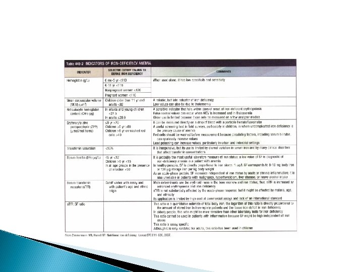
LABORATORY FINDINGS In progressive iron deficiency, a sequence of biochemical and hematologic events occurs Clinically, iron deficiency anemia is not difficult to diagnose. First, tissue iron stores are depleted. This depletion is reflected by reduced serum ferritin, an iron-storage protein, which provides an estimate of body iron stores in the absence of inflammatory disease
Next, serum iron levels decrease, the ironbinding capacity of the serum (serum transferrin) increases, and the transferrin saturation falls below normal. As iron stores decrease, iron becomes unavailable to complex with protoporphyrin to form heme. Free erythrocyte protoporphyrins (FEPs) accumulate, and hemoglobin synthesis is impaired.
At this point, iron deficiency progresses to iron-deficiency anemia. With less available hemoglobin in each cell, the red cells become smaller. This morphologic characteristic is best quantified by the decrease in mean corpuscular volume (MCV) and mean corpuscular hemoglobin (MCH). Developmental changes in MCV require the use of age-related standards for diagnosis of microcytosis
elevated RBC distribution width (RDW). The red cell count(RBC) also decreases. The reticulocyte percentage may be normal or moderately elevated absolute reticulocyte counts indicate an insufficient response to the degree of anemia
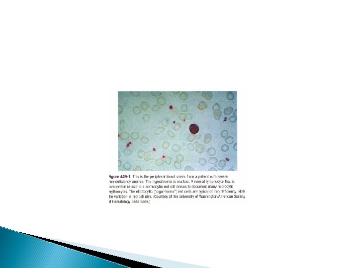
The blood smear reveals hypochromic, microcytic red cells with substantial variation in cell size. Elliptocytic or cigar-shaped red cells are often seen Detection of increased transferrin receptor and decreased reticulocyte hemoglobin concentration provides supporting diagnostic information when these studies are available.
White blood cell count (WBC) is normal, and thrombocytosis is often present. Thrombocytopenia is occasionally seen with very severe iron deficiency, potentially confusing the diagnosis with bone marrow failure disorders. Stool for occult blood should be checked to exclude blood loss as the cause of iron deficiency
In most instances, a complete blood count demonstrating a microcytic anemia with a high RDW, reduced RBC, normal WBC, and normal or elevated platelet count is sufficient for a presumptive diagnosis. Other laboratory such as reduced ferritin, reduced serum iron, and increased total ironbinding capacity, are not usually necessary unless severe anemia requires a more rapid diagnosis, other complicating clinical factors are present, or the anemia does not respond to iron therapy.
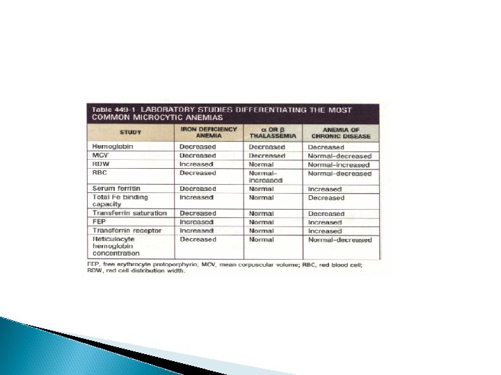
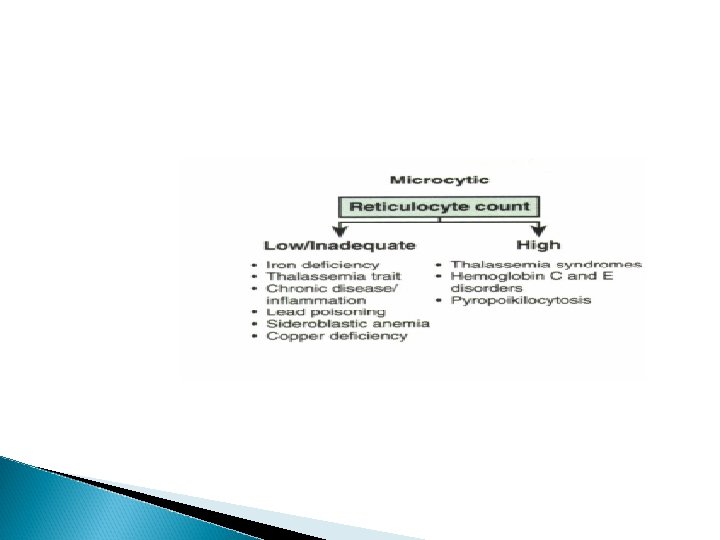
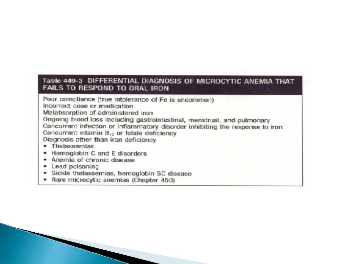
DIFFERENTIAL DIAGNOSIS The most common alternative causes of microcytic anemia are a or B -thalassemia and hemoglobinopathies, including hemoglobin E and C The thalassemia traits are most common and are associated with an elevated as opposed to decreased red blood cell count and a normal as opposed to elevated RDW
The anemia of chronic disease is usually normocytic but can be microcytic in a minority of cases Lead poisoning can cause microcytic anemia, but more often iron deficiency anemia causes pica, which then results in lead intoxication
PREVENTION Iron deficiency is best prevented to avoid both its systemic manifestations and the anemia. Breast-feeding should be encouraged, with the addition of iron-fortified cereals after 4 -6 mo of age. Infants who are not breast-fed should only receive iron-fortified formula (12 mg of iron per liter) for the first year, and thereafter bovine milk should be limited to <20 -24 oz daily
This approach encourages the ingestion of foods richer in iron and prevents blood loss due to bovine milk-induced enteropathy Routine screening using hemoglobin or hematocrit is done at 12 mo of age, or earlier if at 4 mo of age the child is assessed to be at high risk for iron deficiency.
TREATMENT The regular response of iron-deficiency anemia to adequate amounts of iron is a critical diagnostic and therapeutic feature Oral administration of simple ferrous salts (most often ferrous sulfate) provides inexpensive and effective therapy. There is no evidence that the addition of any trace metal, vitamin, or other hematinic substance significantly increases the response to simple ferrous salts.
Aside from the unpleasant taste of iron, intolerance to oral iron is uncommon in young children. In contrast, older children and adolescents sometimes have GI complaints.
The therapeutic dose should be calculated in terms of elemental iron. A daily total dose of 3 -6 mglkg of elemental iron in 3 divided doses is adequate, with the higher dose used in more severe cases. Ferrous sulfate is 20% elemental iron by weight and is ideally given between meals with juice, although this issue is usually not critical with a therapeutic dose.
Parenteral iron preparations are only used when malabsorption is present or when compliance is poor, because oral therapy is otherwise as fast, as effective, and much less expensive and less toxic. When necessary, parenteral iron sucrose and ferric gluconate complex have a lower risk of serious reactions than iron dextran
In addition to iron therapy, dietary counseling is usually necessary. Excessive intake of milk, particularly bovine milk, should be limited. Iron deficiency in adolescent girls secondary to abnormal uterine blood flow loss is treated with iron and hormone therapy
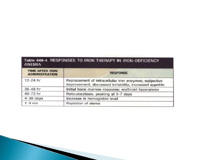
If the anemia is mild, the only additional study is to repeat the blood count approximately 4 wk after initiating therapy. At this point the hemoglobin has usually risen by at least 1 -2 GI d. L and has often normalized
If the anemia is more severe, earlier confirmation of the diagnosis can be made by the appearance of a reticulocytosis usually within 48 -96 hr of instituting treatment. The hemoglobin will then begin to increase 0. 1 -0. 4 Gl/ d. L per day depending on the severity of the anemia. Iron medication should be continued for 8 wk after blood values normalize to reestablish iron stores.
Good follow-up is essential to ensure a response to therapy. When the anemia responds poorly or not at all to iron therapy, there are multiple considerations, including diagnoses other than iron deficiency
Because a rapid hematologic response can be confidently predicted in typical iron deficiency, blood transfusion is rarely necessary. It should only be used when congestive heart failure is eminent or if the anemia is severe with evidence of substantial ongoing blood loss.