Lymphoma majid vafaie Lymphoma is the third most


 is a malignant process involving the lymphoreticular system that accounts for")


 cell, pathognomonic feature of HL, is a large cell (15")


 should be determined")


 can occur from rapid cell turnover, which is especially common in BL. TLS")

 should be")

- Slides: 63
Lymphoma majid vafaie
Lymphoma is the third most common cancer among U. S. children (age 14 yr or younger), with an annual incidence of 15 cases per 1 million children. It is the most common cancer in adolescents, accounting for >25% of newly diagnosed cancers in persons 15 -19 yr old.
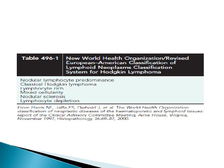
Hodgkin Lymphoma (HL) is a malignant process involving the lymphoreticular system that accounts for 6% of childhood cancers. In the United States, HL accounts for approximately 5% of cancers in persons 14 yr of age or younger it accounts for approximately 15% of cancers in adolescents (15 -19 yr of age)
EPIDEMIOLOGY The worldwide incidence of HL is approximately 2 -4 new cases/100, 000 population/yr there is a bimodal age distribution, with peaks at 15 -35 yr of age and again after 50 yr It is the most common cancer seen in adolescents and young adults, and the third most common in children younger than the age of 15 yr
In developing countries, the early peak tends to occur prior to adolescence. A male : female predominance is found among young children, but lessens with age. Infectious agents may be involved, such as human herpesvirus 6, cytomegalovirus, and (EBV).
Infection with EBV confers a 4 -fold higher risk of developing HL and may precede the diagnosis by years. EBV antigens have been demonstrated in HL tissues, particularly type II latent membrane proteins 1 and 2, although EBV status is not thought to be prognostic of outcom
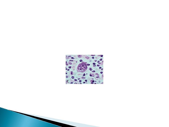
PATHOGENESIS The Reed-Sternberg (RS) cell, pathognomonic feature of HL, is a large cell (15 -45 μm in diameter) with multiple or multilobulated nuclei This cell type is considered the hallmark of HL, although similar cells are seen in infectious mononucleosis, NHL, and other conditions.
HL appears to arise in lymphoid tissue and spread to adjacent lymph node areas in a relatively orderly fashion. Hematogenous spread also occurs, leading to involvement of the liver, spleen, bone marrow, or brain, and is usually associated with systemic symptoms.
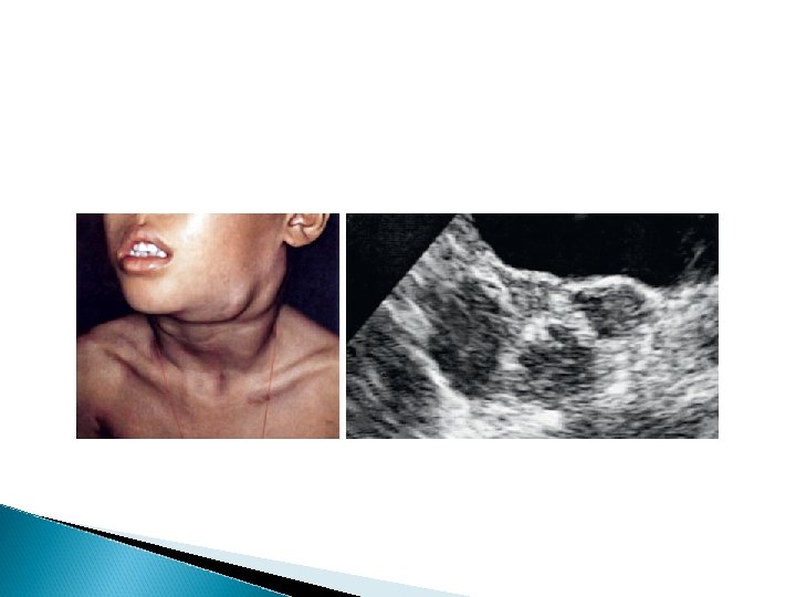
CLINICAL MANIFESTATIONS Patients commonly present with painless, nontender, firm, rubbery, cervical or supraclavicular lymphadenopathy and usually some degree of mediastinal involvement. Clinically detectable hepatosplenomegaly is rarely encountered.
Depending on the extent and location of nodal and extranodal disease, patients may present with symptoms and signs of airway obstruction (dyspnea, hypoxia, cough), pleural or pericardial effusion, hepatocellular dysfunction, Bonemarrow infiltration(anemia, neutropenia, or thrombocytopenia). Disease manifesting below the diaphragm is rare and occurs in approximately 3% of all cases.
Systemic symptoms Systemic symptoms, classified as B symptoms that are considered important in staging, are unexplained fever >38°C (100. 4°F), weight loss >10% total body weight over 6 mo, and drenching night sweats. Less common and not considered of prognostic significance are symptoms of pruritus, lethargy, anorexia, or pain that worsens after ingestion of alcohol. immune system abnormalities that often persist during and after therapy.
DIAGNOSIS Any patient with persistent, unexplained lymphadenopathy unassociated with an obvious underlying inflammatory or infectious process should undergo chest radiography to identify the presence of a large mediastinal mass before undergoing lymph node biopsy. Formal excisional biopsy is preferred over needle biopsy to ensure that adequate tissue is obtained, both for light microscopy and for appropriate immunohistochemical and molecular studies
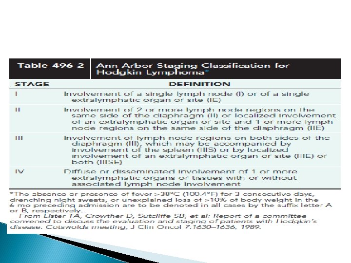
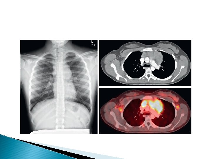
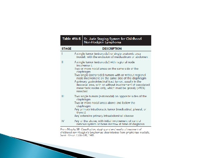
Once the diagnosis of HL is established, extent of disease (stage) should be determined to allow selection of appropriate therapy (Table 496 -2). Evaluation includes history, physical examination, and imaging studies, including chest radiograph; CT scans of the neck, chest, abdomen, and pelvis; and positron emission tomography (PET) scan
Staging of HD
Laboratory studies should include a cbc to identify abnormalities that might suggest marrow involvement; esr; serum ferritin, which is of some prognostic significance and, if abnormal at diagnosis, serves as a baseline to evaluate the effects of treatment.
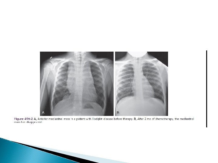
A cxr is particularly important for measuring the size of the mediastinal mass in relation to the maximal diameter of the thorax This determines “bulk”disease and becomes prognostically significant. Chest CT more clearly defines the extent of a mediastinal mass if present and identifies hilar nodes and pulmonary parenchymal involvement, which may not be evident on chest radiographs.
Bone marrow aspiration and biopsy should be performed to rule out advanced disease. Bone scans are performed in patients with bone pain and/or elevation of alkaline phosphatase. Gallium scan be particularly helpful in identifying areas of increased uptake, which can then be reevaluated at the end of treatment.
Fluorodeoxyglucose PET imaging has advantages over gallium scanning, as it is a 1 -day procedure with higher resolution, better dosimetry, less intestinal activity, and the potential to quantify disease. PET scans are being evaluated as a prognostic tool in HL, enabling therapy to be reduced in those predicted to have a good outcome.
Extralymphatic disease resulting from direct extension of an involved lymph node region is designated by category E.
A complete response : the complete resolution of disease on clinical examination and imaging studies or at least 70 -80% reduction of disease and a change from initial positivity to negativity on either gallium or PET scanning because residual fibrosis is common
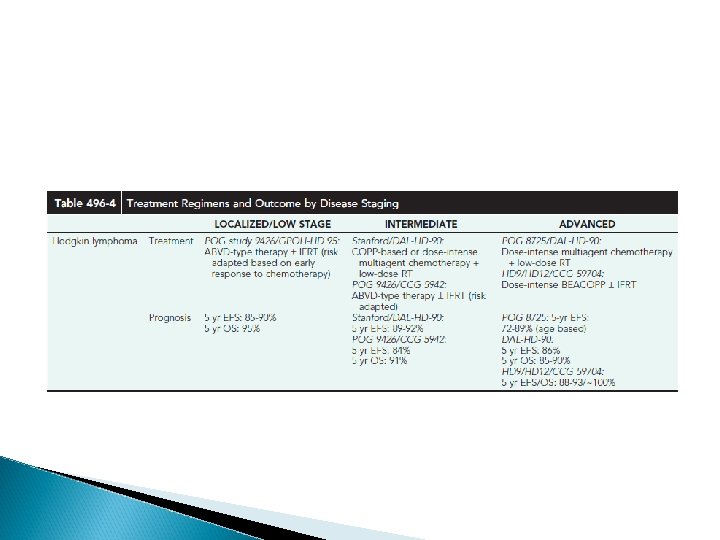
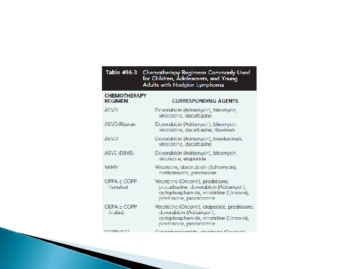
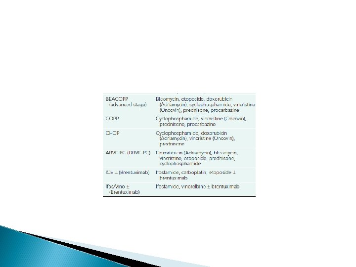
TREATMENT Chemotherapy and radiation therapy Treatment is risk adapted and involves the use of combined chemotherapy with or without low-dose involved-field radiation therapy based on response. Treatment is determined largely by disease stage, presence or absence of B symptoms, and the presence of bulky nodal disease.
Radiation therapy alone, once given at higher doses, initially resulted in prolonged remission and cure rates in patients with lowstage HL. However, this treatment also caused significant long-term morbidity in pediatric patients, including growth retardation, thyroid dysfunction, and cardiac and pulmonary toxicity.
Newer combinations of chemotherapy have reduced the risk of secondary cancers. The current Children’s Oncology Group trials are investigating whether radiation therapy can be eliminated
“Risk-adapted” protocols are based on both staging criteria and rapidity of response to initial chemotherapy. The aim is to reduce total drug doses and treatment duration and to eliminate radiation therapy if possible
Ongoing clinical trials report encouraging results with the use of anti-CD 20 antibody (rituximab), particularly in nodular lymphocytepredominant Hodgkin lymphoma where trials in relapsed disease have shown an overall response rate of 94%.
anti-CD 30 agents are being used that are targeted to the RS cells themselves, where CD 30 is abundantly expressed. Brentuximab vedotin is an antibody–drug conjugate that is now FDA approved to treat Hodgkin lymphoma It combines the chimeric anti-CD 30 antibody brentuximab linked to the antimitotic agent monomethyl auristatin E.
This agent shows impressive efficacy as single-agent therapy in refractory HL and is currently being tested as part of upfront therapy combined with chemotherapy in patients with newly diagnosed disease. EBV-specific cytotoxic T lymphocytes (CTLs) can also be generated from allogeneic donors for patients with advanced HL
RELAPSE Most relapses occur within the 1 st 3 yr after diagnosis, but relapses as late as 10 yr have been reported. Relapse cannot be predicted accurately with this disease.
Poor prognostic features include tumor bulk stage at diagnosis extralymphatic disease presence of B symptoms
Patients who achieve an initial chemosensitive response but relapse or progress less than 12 mo from diagnosis are candidates for myeloablative chemotherapy and autologous stem cell transplantation with or without the addition of radiation therapy.
For more difficult-to-treat refractory cases, agents such as Zevalin or Bexxar are being trialed, often in combination with stem cell transplantation strategies. Both are monoclonal anti-CD 20 antibodies to which a radioactive isotope is directly linked. Clinical trials show each to be more effective than rituximab in NHL patients
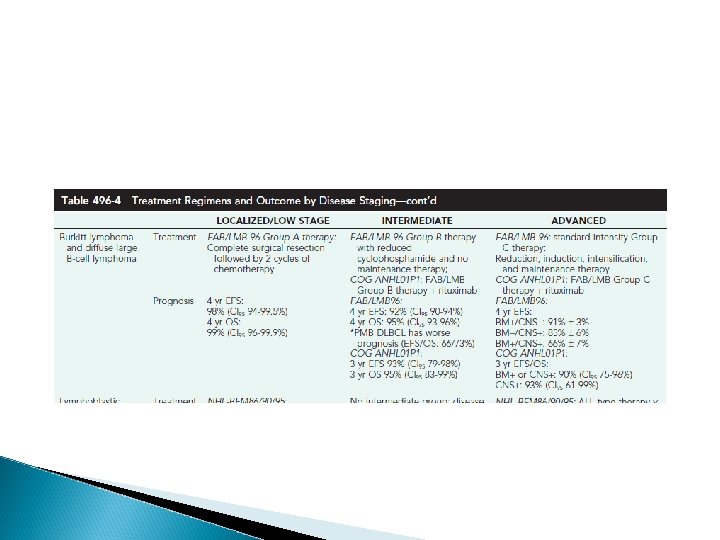
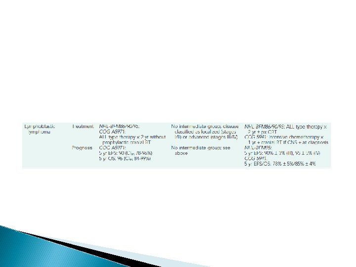
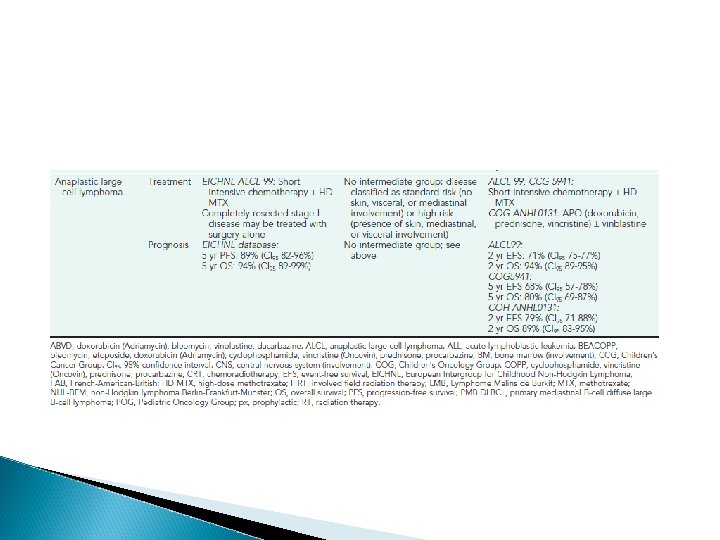
PROGNOSIS With the use of current therapeutic regimens, patients with favorable prognostic factors and early-stage disease have an event-free survival(EFS) of 85 -90% and an overall survival (OS) at 5 yr of >95%. Patients with advanced-stage disease have slightly lower EFS (80 -85%) and OS (90%), respectively, although OS has approached 100% with doseintensechemotherapy (Table 496 -4).
Prognosis after relapse depends on the time from completion of treatment to recurrence, site of relapse (nodal vs extranodal), and presence of B symptoms at relapse. Patients whose disease relapses >12 mo after chemotherapy alone or combinedmodality therapy have the best prognosis, and their relapses usually respond to additional standard therapy, resulting in a long-term survival of 60 -70%
A myeloablative autologous stem cell transplantation in patients with refractory disease or relapse within 12 mo of therapy results in a long-term survival rate of only 40 -50%. Allogeneic stem cell transplantation has shown promise in patients with poor risk features at relapse/progression.
Biopsy may also be indicated if there is an increase in size over baseline in 2 wk no decrease in size in 4 -6 wk no regression to “normal” in 8 -12 wk if new signs and symptoms develop Persistence of symptoms and lymphadenopathy greater than 2 wk and certain locations (supraclavicular, mediastinal, abdomen) also suggest malignancy
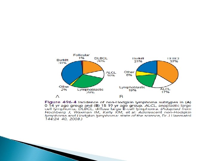
Non-Hodgkin Lymphoma NHL accounts for approximately 60% of lymphomas in children and is the second most common malignancy in patients age 15 -35 yr. The annual incidence of pediatric NHL in the United States is 750 -800 cases/yr. In contrast to adult NHL, which is typically indolent, pediatric NHL is usually high grade and aggressive. Although more than 70% of patients present with advanced disease, with survival rates of 90 -95% for localized disease and 70 -95% with advanced disease.
EPIDEMIOLOGY Although most children and adolescents with NHL present with de novo disease, a small number of patients have NHL secondary to specific etiologies, including inherited or acquired immune deficiencies(e. g. , severe combined immunodeficiency syndrome, Wiskott-Aldrich syndrome), viruses (e. g. , HIV, EBV), and as part of genetic syndromes (e. g. , ataxia-telangiectasia, Bloom syndrome). However most children in North America and Europe in whom NHL develops have no obvious genetic or environmental etiology
CLINICAL MANIFESTATIONS Approximately 70% of patients with NHL present with advanced disease (stage III or IV), including extranodal disease with bone marrow and central nervous system (CNS) involvement. B symptoms of fever, weight loss, and night sweats can be seen, particularly in ALCL, but are not prognostic. Capillary leak syndrome may be seen in ALK+ ALCLs.
LBL commonly manifests as an intrathoracic or mediastinal supradiaphragmatic mass and also has a pre-dilection for spreading to the bone marrow and CNS BL commonly manifests as abdominal (sporadic type) or head and neck (endemic type) tumor and can metastasize to the bone marrow or CNS
DLBCL commonly manifests as either an abdominal or mediastinal primary and, rarely, disseminates to the bone marrow or CNS. ALCL manifests either as a primary cutaneous manifestation (10%) or as systemic disease (90%) with dissemination to liver, spleen, lung, or mediastinum. Bone marrow or CNS disease is rare in ALCL
Site-specific manifestations of NHL include painless, rapid lymph node enlargement; cough or dyspnea with thoracic involvement; superior mediastinal syndrome; ascites, increased abdominal girth or intestinal obstruction with an abdominal mass; nasal congestion, earache, hearing loss, or tonsil enlargement with Waldeyer ring involvement; and localized bone pain.
NHL can present as a life-threatening oncologic emergency. These manifestations are important to recognize as they require intensive supportive care and, in some cases, alterative treatment. Sms can occur as a consequence of a large mediastinal mass causing obstruction of blood flow or respiratory airways. Spinal cord tumors can cause cord compression and acute paraplegias requiring emergent radiation therapy
(TLS) can occur from rapid cell turnover, which is especially common in BL. TLS can result in severe metabolic abnormalities including hyperuricemia, hyperphosphatemia, hyperkalemia, and hypocalcemia. This can rapidly lead to renal insufficiency/failure as well as cardiac abnormalities if not aggressively treated.
LABORATORY FINDINGS CBC; measurements of electrolytes, lactate dehydrogenase, uric acid, calcium, phosphorus, blood urea nitrogen, creatinine, bilirubin, alanine aminotransferase, and aspartate aminotransferase; bone marrow aspiration and biopsy; lumbar puncture with cerebrospinal fluid cytology, cell count and protein; chest radiographs; and neck, chest, abdominal, and pelvic CT scans (head CT for suspicion of CNS disease), and PET scan.
Tumor tissue (i. e. , biopsy, bone marrow, cerebrospinalfluid, or pleurocentesis/paracentesis fluid) should be tested by flow cytometry for immunophenotypic origin (T, B, or null) and cytogenetics (karyotype). Additional tests might include fluorescent in situ hybridization or quantitative reverse transcription PCR for specific genetic translocations, T- and B-cell gene rearrangement studies, and molecular profiling by oligonucleotide microarray.
TREATMENT Rituximab is a monoclonal antibody directed at CD 20 that improves outcomes in adult patients with B-NHL. As nearly all pediatric BLs and DLBCLs express CD 20, rituximab has been examined in pediatric B-NHL Nelarabine is a purine analog with significant T-lymphocyte toxicity; nelarabine has been investigated in T-LBL. Nelarabine is currently undergoing investigation in high-risk patients with T-ALL and T-LBL.