IRON METABOLISM IRON DEFICIENCY IRON OVERLOAD IRON 10

IRON METABOLISM IRON DEFICIENCY IRON OVERLOAD

IRON • 10 -15 mg/day in diet; 5 -10% absorbed – Absorption increased in iron deficiency, pregnancy, erythroid hyperplasia, hypoxia • Heme iron absorbed best • Fe 2+ much better than Fe 3+ – Some foods, drugs enhance and some inhibit absorption of ionic iron – Ability to regulate absorption limited • Absorption in proximal small intestine

IRON TRANSPORT AND STORAGE • Absorbed iron oxidized to Fe 3+ form • Bound tightly to transferrin in blood • Iron is transferred to cells and reduced to Fe 2+ form, then inserted into heme or stored • Storage iron (Fe 3+) bound to ferritin – Small amount of ferritin in blood (nanograms) correlates with body iron stores

Laboratory tests used to assess iron status • Serum iron: transferrin- bound iron being transported in the blood. • Total iron binding capacity (TIBC): the total amount of transferrin in blood. – Transferrin saturation = serum iron/TIBC (%) • Serum ferritin: Serum ferritin levels usually reflect body iron stores.

ASSESSMENT OF BODY IRON • Serum iron low in iron deficiency, inflammation • TIBC high in iron deficiency, normal or low in inflammation • Serum ferritin low in iron deficiency, increases in inflammation • Marrow iron stores (assessed by marrow biopsy) absent in iron deficiency

Marrow iron stores Marrow aspirate stained with Prussian Blue Absent iron stores Normal iron stores

IRON METABOLISM Ø Hepcidin is a peptide produced in the liver. Ø Hepcidin interacts with ferroportin to inhibit iron release from villus enterocytes and macrophages. Ø Hepcidin production is upregulated by high plasma iron levels or inflammation. Ø Low iron levels decrease hepcidin production, which in turn stimulates iron absorption and release into the blood. Ø The HFE gene modulates hepcidin production. Ø Mutations in HFE can cause diminished hepcidin release, and can eventually cause iron overload (hereditary hemochromatosis). N Engl J Med 2004; 350: 2383

Iron deficiency HFE mutation Hepcidin → Iron Ineffective erythropoiesis Liver disease
 Iron overload")
Inflammation Hepcidin → Iron Genetic conditions (rare) Iron overload
 J Clin Oncol 2010; 28:")
↑ Hepcidin → ↑ serum ferritin (more iron stored) J Clin Oncol 2010; 28: 2538 TIBC (mcg/d. L) Ferritin (ng/ml) Serum iron (mcg/d. L) Hepcidin levels in patients with anemia of inflammation ↑ Hepcidin → ↓ iron and TIBC (less iron transported)

Most of your iron is in your red cells 1 cc of red cells contains about 1 mg iron 1 cc of whole blood contains 0. 5 mg iron

IRON BALANCE • 1 -2 mg/day lost via desquamation, GI blood loss in adult • Normally we absorb about the same amount per day • Negative iron balance possible in early childhood • Menstruation, pregnancy, lactation promote negative balance • Positive balance (and eventual iron overload) can occur in inherited disorders (hemochromatosis), or as a result of repeated blood transfusions

• 20 mg of iron per day required for erythropoiesis • Most of this iron is recycled from old RBC after they are eaten by macrophages • 1 -2 mg of “new” iron absorbed from gut • 1 -2 mg of iron lost via sloughing of enterocytes N Engl J Med 2004; 350: 2383 • Excess iron stored – mainly in liver

Daily iron requirements vs age Dietary iron available ♀ Girls ♂ Boys Iron requirements may exceed dietary availability in infants and adolescent girls

Pregnancy depletes iron stores Normal iron stores in women = 200 -400 mg

IRON DEFICIENCY • Most common cause of anemia worldwide • Usually due to chronic blood loss – Exceptions: rapidly growing child, malabsorption • In young women this is usually due to menstrual blood loss and/or pregnancy • In anyone else: rule out GI blood loss – Esophageal disease, hiatal hernia, ulcer, inflammatory bowel disease, angiodysplasia, hemorrhoids, cancer

IRON DEFICIENCY ANEMIA • Microcytic, hypochromic (MCV may be normal in early or mild deficiency) • Reticulocyte count not increased • Aniso- and poikilocytosis in more severe cases • Serum ferritin usually low – Exception: inflammation or liver disease • Serum iron low, TIBC usually high

Iron deficiency Atrophic glossitis Pica “Spoon nails”

The evolution of iron deficiency anemia During transition from iron-deficient erythropoiesis to overt iron deficiency anemia, anemia is initially normocytic/normochromic and gradually becomes microcytic/hypochromic

IRON DEFICIENCY ANEMIA Treatment • Oral ferrous salts – Many patients have GI side effects – “Slow-release” forms often not well absorbed • Oral iron-polysaccharide complex • IV iron dextran or iron sucrose – If oral iron not absorbed or not tolerated – Slight risk of anaphylaxis • Should see increased hemoglobin within 2 -3 weeks

Other causes of microcytic anemia Decreased hemoglobin production due to: ØIron withheld from red cell precursors (increased hepcidin - anemia of inflammation) ØGlobin gene defects (thalassemias) Defects in heme synthetic pathway (sideroblastic anemias) • Inherited conditions • Heavy metal poisoning • Myelodysplasia (usually macrocytic/megaloblastic)

Differential diagnosis of microcytic anemia Variable Iron deficiency Thalassemia trait Inflammation MCV Low in proportion Low even in to anemia (may absence of be nl in early anemia stage) Normal or slightly low Serum iron Low Normal Low TIBC High Normal or low Serum ferritin Low Normal or high Marrow iron Absent Present Erythroblast iron Absent Present Absent Incr Hgb A 2 (β thal trait) Normal Hgb Normal electrophoresis

IRON OVERLOAD • Hereditary hemochromatosis – Autosomal recessive, HFE gene; genotype common but low penetrance • Other inherited disorders – Mutations in other genes that regulate iron metabolism – Africans, African-Americans • Chronic ineffective erythropoiesis – Thalassemia • Repeated transfusion – Toxicity after about 100 Units

HFE mutations disrupt signaling that normally increases hepcidin production in response to increased iron levels Hepcidin gene NEJM 2012; 366: 360
 •")
IRON OVERLOAD • Increased serum iron and high transferrin saturation (90%+ in hemochromatosis) • Very high serum ferritin (over 1000) • Increased liver and marrow iron – Quantitation of liver iron best indicator of severity • DNA test available for hereditary HC

IRON OVERLOAD Clinical consequences • • Cirrhosis, hepatocellular carcinoma Cardiomyopathy, heart failure Endocrine failure (esp diabetes) Arthropathy • Treatment of hereditary HC by phlebotomy prevents these problems and can reverse early tissue damage

HEMOCHROMATOSIS

NON-NUTRITIONAL HYPOPROLIFERATIVE ANEMIAS

ANEMIA OF INFLAMMATION AKA “anemia of chronic disease” • Most common cause of anemia in hospitalized patients • Hct rarely < 25 unless additional factors present • Causes: infection, autoimmune disorders, cancer • Mechanisms: – Inflammation → incr hepcidin expression → Impaired release of stored iron from macrophages – lower EPO production – Direct inhibition of red cell precursors by inflammatory cytokines – Shortened red cell survival • Benefit: decreased iron available to bacteria, etc

Inflammatory cytokines cause increased hepcidin production Hepcidin gene NEJM 2012; 366: 360

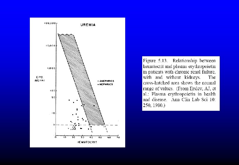
Erythropoietin levels are lower than expected for the degree of anemia in the presence of inflammation

ANEMIA OF INFLAMMATION Laboratory findings • Normocytic or mild microcytosis • Not many shift cells • Low serum iron, normal or low TIBC, normal or high serum ferritin • Relatively low EPO level for degree of anemia

LOW ERYTHROPOIETIN ANEMIA • Renal failure – May be compounded by blood loss during dialysis, inflammation, decreased rbc lifespan – Reversible with EPO injections • Endocrine disorders – Hypothyroidism, hypopituitarism • Protein-calorie malnutrition • Right-shifted hemoglobin O 2 dissociation curve
- Slides: 34