
Anemia under investigation Guided by Dr Sarita Patel
Anemia under investigation Guided by : Dr. Sarita Patel Dr. Sarita Mangukiya Dr. Manmeet Kochar Presented by : Group no. 2 Ankit bargujar-6 Antala harshvardhan-7 Badreshiya tarak-8 Bhalani shivani-9 Bhati virendra-10 29 -10 -2020
• 64 year female patient admitted in new civil hospital with following chief complain. • Chief Complaints(since 3 month) -Tremor in hands -Weakness in body -Body pain -Fever -Loss of apetite
• • 1. 2. 3. No significant family history Medical History Patient had typhoid 3 months ago before admitted Patient was admitted in Pooja hospital before 15 days for 1 week with same complain. In pooja hospital , 4 BT(blood transfusion) given. 29 -10 -2020
General Examination At the time of admission BP – 150/100 mmhg
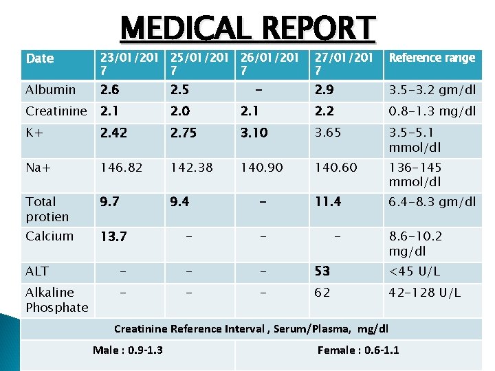
MEDICAL REPORT Date 23/01/201 25/01/201 26/01/201 7 7 7 27/01/201 7 Reference range Albumin 2. 6 2. 5 - 2. 9 3. 5 -3. 2 gm/dl Creatinine 2. 1 2. 0 2. 1 2. 2 0. 8 -1. 3 mg/dl K+ 2. 42 2. 75 3. 10 3. 65 3. 5 -5. 1 mmol/dl Na+ 146. 82 142. 38 140. 90 140. 60 136 -145 mmol/dl Total protien 9. 7 9. 4 - 11. 4 6. 4 -8. 3 gm/dl Calcium 13. 7 - - - ALT - - - 53 <45 U/L Alkaline Phosphate - - - 62 42 -128 U/L 8. 6 -10. 2 mg/dl Creatinine Reference Interval , Serum/Plasma, mg/dl Male : 0. 9 -1. 3 Female : 0. 6 -1. 1
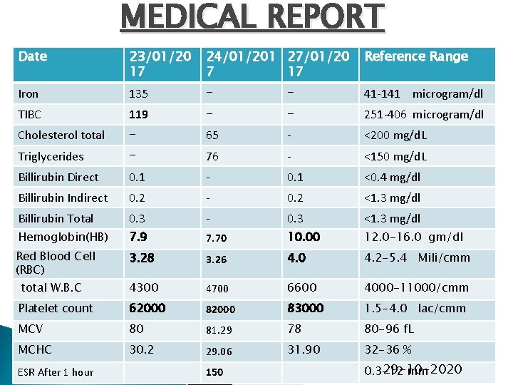
MEDICAL REPORT Date 23/01/20 17 24/01/201 27/01/20 7 17 Reference Range Iron 135 - - 41 -141 microgram/dl TIBC 119 - - 251 -406 microgram/dl Cholesterol total - 65 - <200 mg/d. L Triglycerides - 76 - <150 mg/d. L Billirubin Direct 0. 1 - 0. 1 <0. 4 mg/dl Billirubin Indirect 0. 2 - 0. 2 <1. 3 mg/dl Billirubin Total 0. 3 - 0. 3 <1. 3 mg/dl Hemoglobin(HB) 7. 9 7. 70 10. 00 12. 0 -16. 0 gm/dl Red Blood Cell (RBC) 3. 28 3. 26 4. 0 4. 2 -5. 4 Mili/cmm 4300 4700 6600 4000 -11000/cmm Platelet count 62000 83000 1. 5 -4. 0 lac/cmm MCV 80 81. 29 78 80 -96 f. L MCHC 30. 2 29. 06 31. 90 32 -36 % total W. B. C ESR After 1 hour 150 29 -10 -2020 0. 3 -12 mm
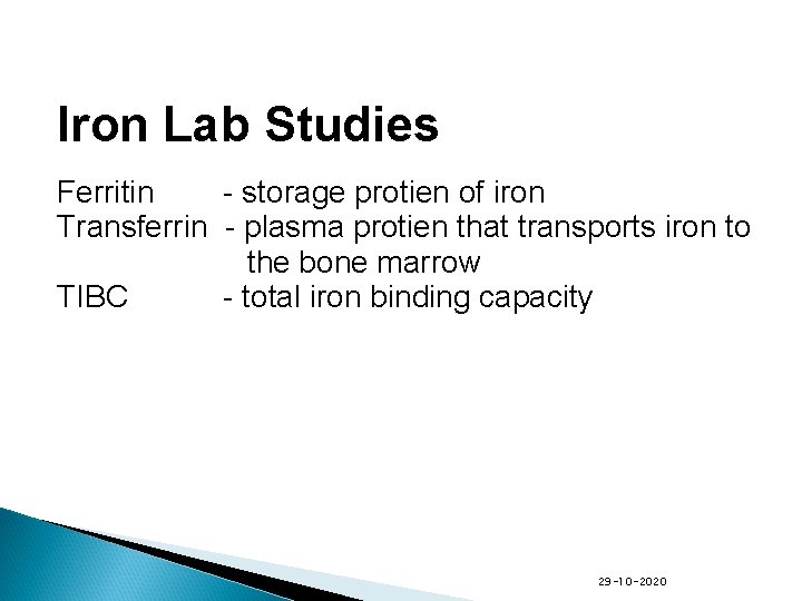
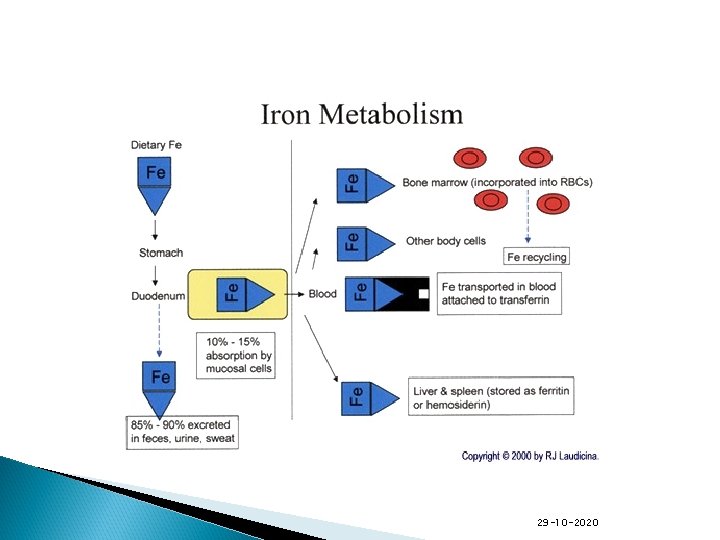
Iron Lab Studies Ferritin - storage protien of iron Transferrin - plasma protien that transports iron to the bone marrow TIBC - total iron binding capacity 29 -10 -2020
29 -10 -2020
29 -10 -2020
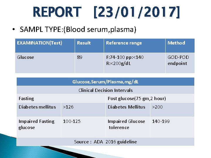
REPORT [23/01/2017] • SAMPL TYPE: (Blood serum, plasma) EXAMINATION(Test) Result Reference range Method Glucose 89 F: 74 -100 pp: <140 R: <200 g/d. L GOD-POD endpoint Glucose, Serum/Plasma, mg/d. L Clinical Decision Intervals Fasting Post glucose(75 gm, 2 hour) Diabetes mellitus >126 Diabetes Mellitus >200 Impaired Fasting glucose 100 -125 Impaired Glucose tolerence 140 -199 Source : ADA 2016 guideline
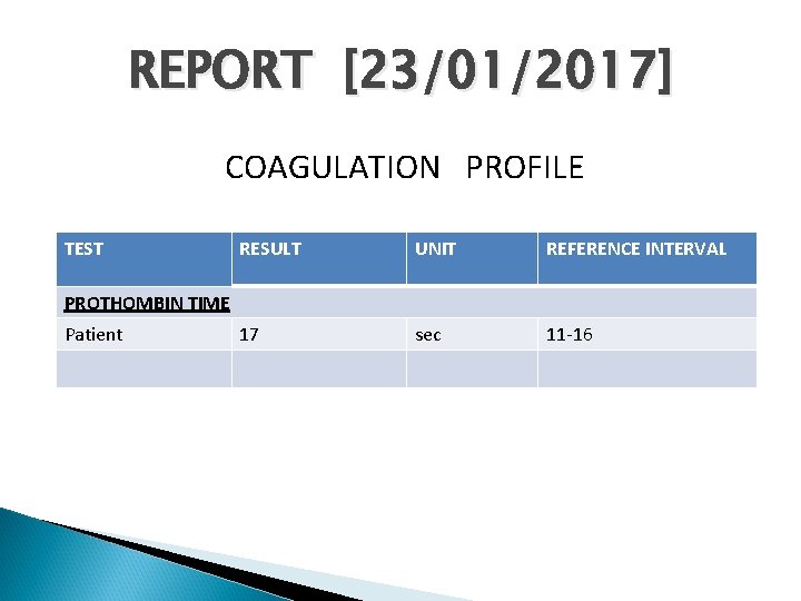
REPORT [23/01/2017] COAGULATION PROFILE TEST RESULT UNIT REFERENCE INTERVAL 17 sec 11 -16 PROTHOMBIN TIME Patient
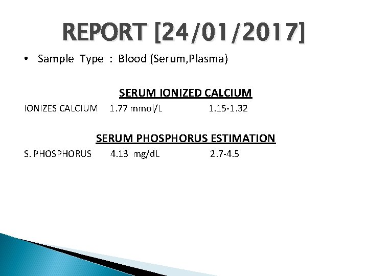
REPORT [24/01/2017] • Sample Type : Blood (Serum, Plasma) SERUM IONIZED CALCIUM IONIZES CALCIUM 1. 77 mmol/L 1. 15 -1. 32 SERUM PHOSPHORUS ESTIMATION S. PHOSPHORUS 4. 13 mg/d. L 2. 7 -4. 5
MCV-mean corpuscular volume Measure of average size of a single RBC Microcytic MCV<80 Macrocytic MCV>100 Normocytic MCV= 80 -100 29 -10 -2020
MCHC-mean corpuscular hemoglobin concentration -Measure of the Hg. B within a single RBC -Hypochronic-cell has a deficiency of Hg. B -Normochromic-value are normal 29 -10 -2020
TREATMENT v v BT Antibiotics Tab. Lasix(Furosemide) Antacid 29 -10 -2020
Role of lasix After BT , Lasix was given to reduce the fluid over-load. Role of antibiotics Because patient has c/o of fever. Role of antacids To reduce the gastric acid output 29 -10 -2020
Hypokalemia Drug induced- Diuratics(lasix)- inhibits sodium, potassium and chloride reabsorption from renal tubules. Insulin- intracellular shifting of potassium Inadequate potassium intake- it is uncommon Gastrointestinal loss- diarrhoea, vomitting kidney failure- rarely because mostly in kidney failure, loss the ability to remove potassium.
Symptoms of Hypokalemia Abnormal heart rhytham Muscle weakness Tremor Muscle cramps Constipation 29 -10 -2020
Hypercalcemia • Overactive parathyroid glands • • • hyperparathyrodism , secondary hyperparathyrodism(kideny failure) Cancer- Lung cancer and breast cancer, as well as some cancers of the blood, can increase your risk of hypercalcemia. Cancer cell may secret Parathyroid hormone related protein(PTHr. P) Spread of cancer (metastasis) to your bones also increases your risk of hypercalcemia. 29 -10 -2020
• • Immobility- People with cancer or other diseases that cause them to spend a great deal of time sitting or lying down may develop hypercalcemia. Over time, bones that don't bear weight release calcium into the blood Medications-such as lithium-may increase the release of parathyroid hormone. 29 -10 -2020
• • • Supplements Dehydration High bone turnover rates- Breakdown of bone • • Bone malignancy multiple myeloma 29 -10 -2020
Symptoms of hypercalcemia Loss of appetite. Fatigue, weakness, and muscle pain. Nausea and vomiting. Constipation and abdominal pain. Increased thirst and frequent urination. Confusion, disorientation 29 -10 -2020
Anemia -defined as a decrease in the total amount of red blood cells (RBCs) or hemoglobin in the blood. In Men <13 gm/dl In Women <12 gm/dl 29 -10 -2020
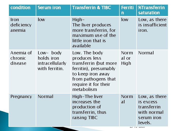
condition Serum iron Transferrin & TIBC Ferriti n %Transferrin saturation Iron deficiency anemia low High. The liver produces more transferrin, for maximum use of the little iron that is available low Low, as there is insufficient iron. Anemia of chronic disease Low- body holds iron intracellularly with ferritin. Low. The body produces less transferrin (but more ferritin), presumably to keep iron away from pathogens that require it for their metabolism Norm al or High Normal Pregnancy Normal High-The liver increases the production of transferrin, thus raising TIBC Norm al Low, as there is excess transferrin with normal serum iron levels. 29 -10 -2020
Classified based on • • • Cell size Diminished Production Accelerated loss 29 -10 -2020
Types Iron deficiency anemia. Vitamin deficiency anemia Aplastic anemia Anemias associated with bone marrow disease Hemolytic anemias 29 -10 -2020
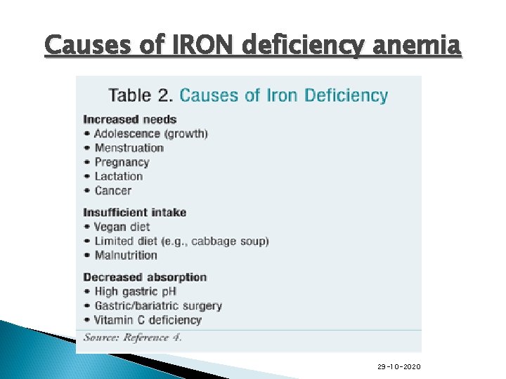
Causes of IRON deficiency anemia 29 -10 -2020
Iron Deficiency Anemia Signs and symptoms Fatigue Tachycardia/Palpitations Tachypnea on exertion Pallor 29 -10 -2020
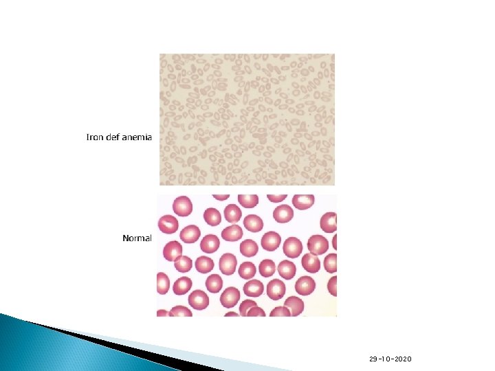
Iron Deficiency Anemia Laboratory findings Decreased serum ferritin Decreased serum iron Decreased MCV Increased total iron binding capacity(TIBC) 29 -10 -2020
29 -10 -2020
Anemia of Chronic Disease Essentials of Diagnosis Anemia (normochronic or microcytic) Normal or Increased ferritin Underlying Chronic Disease -chronic infection -chronic inflammation -Cancer -liver disease -Renal failure-decreased in EPO 29 -10 -2020
Anemia of Chronic Disease Signs and symptoms depends on causative condition Laboratory findings Hb mildly or moderately reduced MCV usually normal Serum ferritin normal or increased 29 -10 -2020
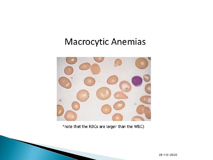
Macrocytic Anemias • • • Vitamin B 12 deficiency Folic acid deficiency There is a defect in DNA synthesis that affects the rapidly dividing cells in the bone marrow. 29 -10 -2020
29 -10 -2020
Causes of vitamin B 12 deficiency Dietary deficiency Decreased production pernicious anemia chronic atrophic gastritis Decreased B 12 absorption surgical resection of the ileum 29 -10 -2020
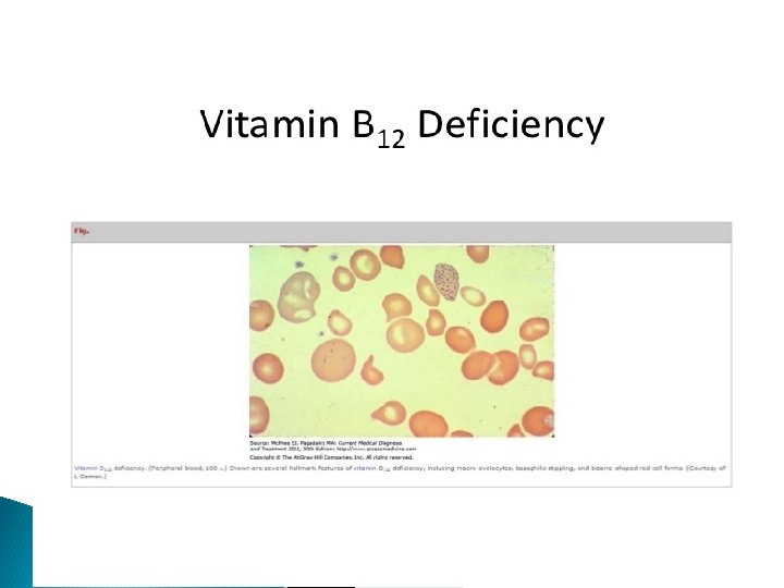
Vitamin B 12 Deficiency Signs and symptoms May be asymptomatic As anemia becomes more severe pallor glossitis Megaloblastic anemia (MCV>100) In sever deficiency- neurological symptoms present 29 -10 -2020
Vitamin B 12 deficiency Diagnosis -Macrocytic anemia -Hypersegmented neutrophils on PBS -Low Serum Vitamin B 12 levels 29 -10 -2020
29 -10 -2020
Folic acid dificiency Laboratory Findings macrocytic anemia hypersegmented netrophils on PBS normal serum vitamin B 12 levels reduced folate level 29 -10 -2020
Hemolytic anemias autoimmune or related to drugs, infection, lymphoproliferative disease, Rh or ABO incompatibility abnormal hemoglobin: sickle celldisease, thalassemia, metheoglobinemia membrane defect: hereditary spherocytosis, herediatry elliptocytosis, paroxymal noctural hemoglobinuria enzyme defect: G 6 PD deficirncy, pyruvate kinase deficiency 29 -10 -2020
Autoimmune hemolytic anemia Signs & Symptoms “ Rapid onset” anemia may be “life Threatening” fatigue jaudince splenomegaly 29 -10 -2020
Autoimmune hemolytic anemia Laboratory Hb low elevated reticulocyte level elevated Total & indirect billirubin 29 -10 -2020
Aplastic Anemia pancytopenia no abnormal cells seen on PBS hypocellular bone marrow 29 -10 -2020
Aplastic Anemia Laboratory Tests pancytopenia, although in early disease only one or two cell lines may be reduced Anemia may be severe Reticulocytes ALWAYS decreased Large RBC(macrocytosis) Neutrophils and platelets reduced in number, no immature or abnormal forms seen 29 -10 -2020
Causes of high total protien Chronic inflammatory conditions HIV/AIDS Bone marrow disorder Multiple myloma Amyloidosis Dehydration (which may make blood proteins appear falsely elevated 29 -10 -2020
Hypergammaglobulinemia Increase immunoglobulins leval may result from stimmulation of many clones of B cells(polyclonal hypergammaglobulinemia) or monoclonal proliferation(paraproteinemia). Polyclonal hypergammaglobulinemia: Stimulation of many clones of B cells produce a wide range of antibodies that appear as diffuse increase in gammaglobulinon electrophoresis. e. g. acute and chronic infections & autoimmune diseases Monoclonal hypergammaglobulinemia: Proliferation of a single immunoglobulin which appears as a discrete densely stained band(paraprotein or M band)on electrophoresis. Paraprotiens are characteristic of malignanat B-cell proliferation. Multiple myeloma is the commonest cause of paraproteinemia. 29 -10 -2020
About a : g ratio A high total protein level may be seen with chronic inflammation or infections such as viral hepatitis or HIV. Normal A: G Ratio <2 In Our patient A: G Ratio- 3. 9 (high) 29 -10 -2020
A low A/G ratio 1)overproduction of globulins, multiple myeloma or autoimmune diseases, 2)Underproduction of albumin, cirrhosis, kidney disease (nephrotic syndrome). . 29 -10 -2020
A high A/G ratio underproduction of immunoglobulins genetic deficiencies and in some leukemias 29 -10 -2020
ESR- causes and method to find ESR • • ESR-Erythrocyte sedimentation rate is the rate at Which RBC sediment in a period of one hour. It is common hematology test and is a non specific nature of inflammation. 29 -10 -2020
Methode of finding of ESR • • • To perform the test anticoagulated blood is traditionaly placed in an upright tube known as westergren tube. The fibrinogen is responsible for the ESR. Immunoglobulin are also responsible for the rouleaux formation. 29 -10 -2020
Factors affacting rate of ESR • • Factors incresing ESR: Inflammation Pregnancy Anemia Autoimmune disorders Infections Some kidney disease and cancers 29 -10 -2020
Factors affacting rate of ESR • • • Factor decreasing rate of ESR Polycythemia Hyperviscocity Leukemia Low plasma protien 29 -10 -2020
Creatinine is the waste product of muscle. • Kidney maintains the normal range of the creatinine in the body. • Poor clearence of creatinine by the kidney rises creatinine level in the body. Creatinine is high in our patient- May be due to some underlying kidney disease or may due to dehydration • 29 -10 -2020
Summary Our patient has low Hb, Low TIBC, decrease RBC and normal MCV and MCHC. Serum Iron is within normal range because of BT is given. Platelets are also decreased. Calcium is high, Total protein is high( globulin part is high) Potassium is low. 29 -10 -2020
Further investigation is required for diagnosis and management of this patient PTH level Urine Routine µscopic examination 24 hour Urine Protein estimation Stool Routine µscopic examination Serum Protein & Hb Electrophoresis X-ray skull and spine Bone marrow examination 29 -10 -2020
- Slides: 56