HYPOCHROMIC ANEMIA IRON METABOLISM OBJECTIVE Iron metabolism Iron

HYPOCHROMIC ANEMIA & IRON METABOLISM

OBJECTIVE • Iron metabolism • Iron distribution & transport • Dietary iron • Iron absorption • Iron requirements • Disorders of iron metabolism • Hypochromic anemia

• To accept & donate electron (Fe 2+ 3+ Fe ( • component of cytochromes, oxygen-binding molecules • cell growth, proliferation, differentiation • damage tissues H 2 O 2 Fe 2+ Fe 3+ OH

• Iron distribution & transport • transferrin, transferrin receptor • ferritin , hemosiderin (Fe 3+( • myoglobin, iron-containing enzymes • Dietary iron • Iron absorption • Iron requirements
 of")
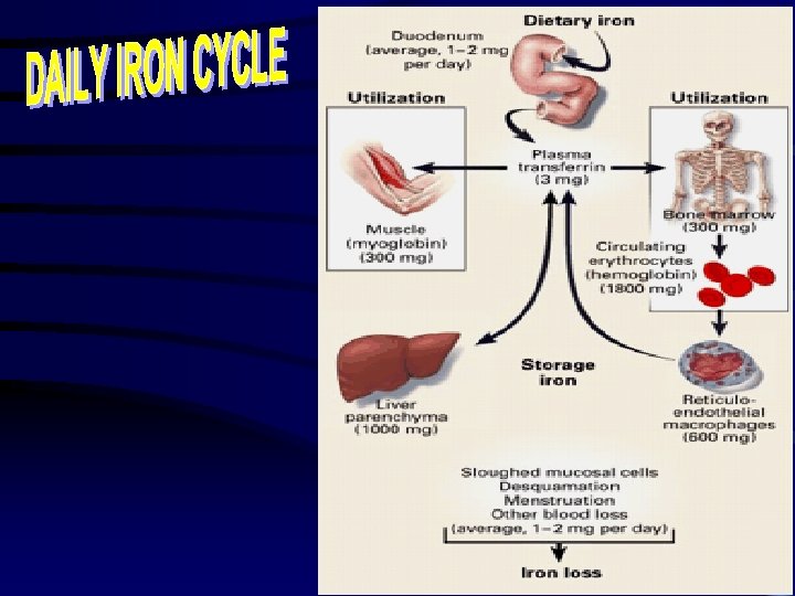
The distribution of body iron Amount of iron Male in average adult (g) of total Hb 2. 4 1. 7 65 ferritin & hemosiderin 1. 0 0. 3 30 Myoglobin 0. 15 0. 12 3. 5 Heme enzyme 0. 02 0. 15 0. 5 Transferrin-bound 0. 004 0. 003 0. 1 iron Female %

• Iron distribution & transport • Dietary iron • ferric hydroxides • ferric-protein complexs • heme-protein complexes • Iron absorption • Iron requirements

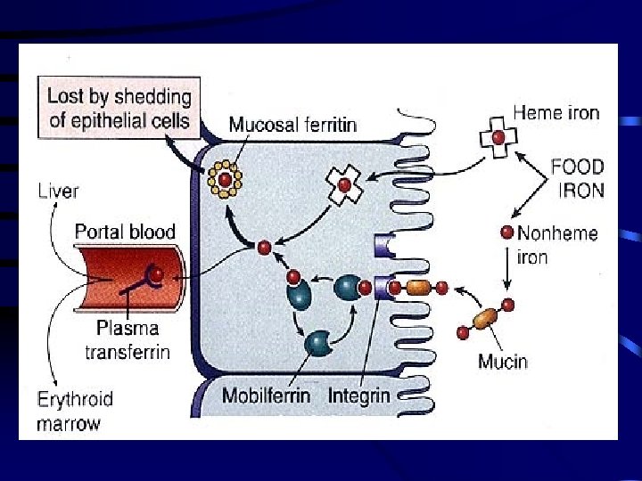
GI Absorption of Iron

INTRACELLULAR IRON TRANSPORT

• Iron distribution & transport • Dietary iron • Iron absorption • Iron requirements

Estimated daily iron requirements, Units are mg/day Adult men Postmenopausal female Menstruating female 0. 5 -1 1 -2 Pregnant female 1. 5 -3 Children 1. 1 Female (age 12 -15) 1. 6 -2. 6

Diseases of iron deficiency. 1 Iron-deficiency anemia (IDA(. 2 Anemia of chronic disease (ACD( Diseases of iron overload

What is iron-deficiency anemia? It is the lack of iron in the blood, which is necessary to make hemoglobin.

Symptoms Fatigue - Sometimes out of proportion to anemia Atrophic glossitis Pica Koilonychia (Nail spooning) Esophageal Web

Causes of Iron Deficiency • Chronic blood loss • Uterine • GI tract • Increased demands • Prematurity • Growth • Pregnancy • Malabsorption • gastrectomy • Poor diet

Iron Deficiency Anemia (IDA( • Most common cause of anemia • Microcytic hypochromic anemia • MCV, MCHC are reduced • blood film : small red cells (microcytic( : pale red cells (hypochromic(

Laboratory findings. 1 Red cell indices & blood film. 2 Bone marrow iron. 3 Serum iron & iron binding capacity. 4 Serum transferrin receptor (s. Tf. R(. 5 Serum ferritin. 6 Zinc protoporphyrin

TIBC Normal Iron def. ACD Iron overload Serum iron & iron binding capacity

• Chronic inflammatory diseases • Infections • Non-infectious • Malignant diseases • release of iron from macrophage to plasma • red cell life span • response to EPO • release IL-1 & TNF

• Increased iron absorption • Increased iron uptake • Repeated red cell transfusions

• Iron-deficiency anemia (IDA( • Anemia of chronic disease (ACD( • Sideroblastic anemia • Thalassemia • Lead poisoning

The Cause of Hypochromic Anemia Iron Protoporphyrin • Iron deficiency • Chronic inflammation or malignant • Sideroblastic anemia Heme + Globin • Thalassemia Hemoglobin

• A defect in heme synthesis • Hereditary & Acquired • mitochondrial defects • pyridoxal-6 -phosphate • mutation in the d-aminolevulinic acid synthase ) ring sideroblasts in BM( • myelodysplasia syndrome • Hypochromic & microcytic red cells

• Inhibits both heme & globin synthesis • Interferes with the breakdown of RNA by inhibiting pyrimidine 5’nucleotidase • accumulation of denatured RNA in red cells ) basophilic stippling( • Hypochromic anemia • Ring sideroblasts (BM( • Free erythrocyte protoporphyrin is raised

Differential diagnosis of hypochromic anemia IDA ACD Thalassemia Siderblastic anemia Serum iron N TIBC N serum ferritin BM iron stores+ N/ + + N - Erythroblast iron - - + Hb N N Hb. A 2 electrophoresis N ring forms N

Hemochromatosis A genetically determined form of iron overload that results in progressive hepatic, pancreatic, cardiac, and other organ damage

Hemochromatosis • It is one of the most common genetic disorders in the U. S. • Present in heterozygous (one gene) form in 12% of nonblacks and 30% of blacks • Present in homozygous form (2 gene) in 1 in 200 nonblacks and 1 in 100 blacks • Homozygotes will die of iron overload unless they give blood frequently • Homozygotes absorb three times more iron from food than other people • Even heterozygotes may be at risk for iron overload, increasing risk of heart disease

Hemochromatosis: Risk Factors • Higher risk in people of northern European descent • Men tend to manifest symptoms earlier because they have no way to dispose of excess iron (menstruation, pregnancy, lactation) • Men may develop symptoms in their 30 s but may not be diagnosed until their 50 s • Women often develop symptoms after menopause

Hemochromatosis: Symptoms • • Joint pain Fatigue Lack of energy Abdominal pain Loss of sex drive Heart problems Abnormal pigmentation of the skin, making it look gray or bronze

Hemochromatosis: if untreated, may result in • • Arthritis Liver disease: cirrhosis, cancer, liver failure Damage to the pancreas, leading to diabetes Heart abnormalities, including arrhythmias and heart failure • Impotence or early menopause • Thyroid or adrenal problems

Hemochromatosis: Diagnosis and Treatment • Testing: serum ferritin and transferrin saturation can reveal excess stores of iron; followed by HFE (genetic) test and possible liver biopsy • Treatment: regular phlebotomy to remove excess iron • Avoidance of iron supplements and sources of iron in the diet, especially heme iron • Awareness of iron cooking vessels

- Slides: 33