IN THE NAME OF GOD PRINCIPLES OF DXA






- Slides: 98
IN THE NAME OF GOD
PRINCIPLES OF DXA SCAN INTERPRETATION Dr Mozhgan Karimifar Assistant Prof. of Endocrinology Isfahan University of Medical Sciences
Skeletal Anatomy in Densitometry
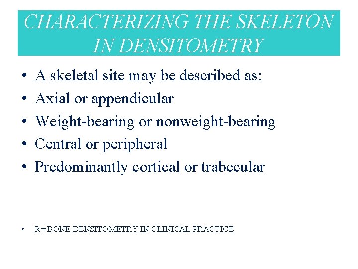
CHARACTERIZING THE SKELETON IN DENSITOMETRY • • • A skeletal site may be described as: Axial or appendicular Weight-bearing or nonweight-bearing Central or peripheral Predominantly cortical or trabecular • R= BONE DENSITOMETRY IN CLINICAL PRACTICE
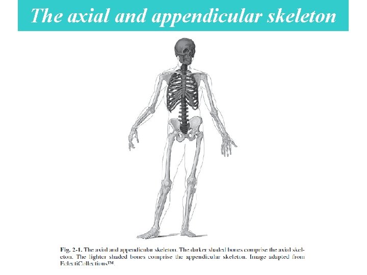
The axial and appendicular skeleton
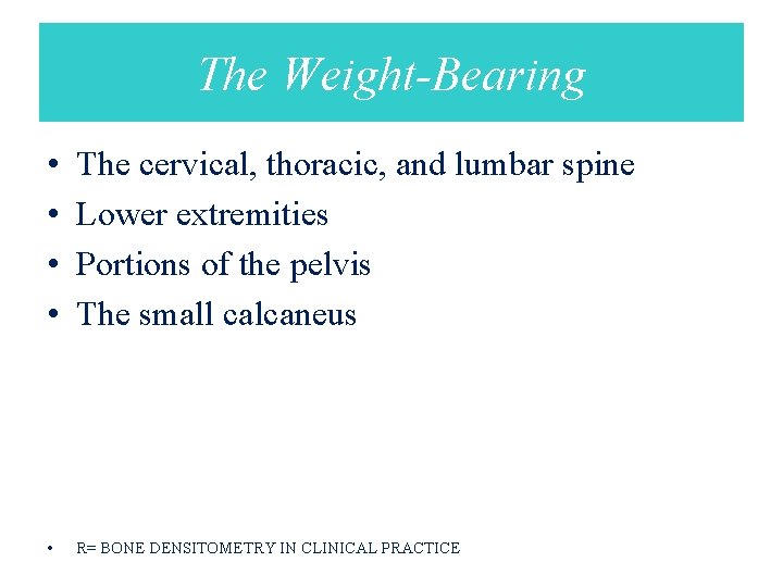
The Weight-Bearing • • The cervical, thoracic, and lumbar spine Lower extremities Portions of the pelvis The small calcaneus • R= BONE DENSITOMETRY IN CLINICAL PRACTICE
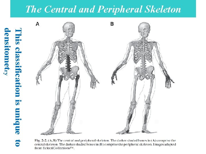
The Central and Peripheral Skeleton This classification is unique to densitometry
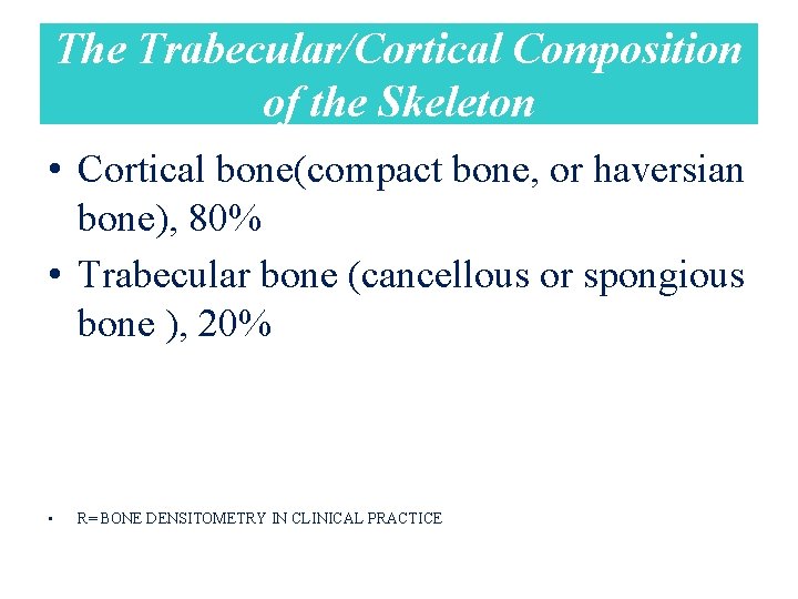
The Trabecular/Cortical Composition of the Skeleton • Cortical bone(compact bone, or haversian bone), 80% • Trabecular bone (cancellous or spongious bone ), 20% • R= BONE DENSITOMETRY IN CLINICAL PRACTICE
Skeletal metabolism • Cortical bone = Trabecular bone • R= BONE DENSITOMETRY IN CLINICAL PRACTICE
The greatest magnitude of change (BMD) • Axial trabecular bone > Appendicular trabecular bone • R= BONE DENSITOMETRY IN CLINICAL PRACTICE
Followed over time for changes in the BMD • from a disease process or therapeutic intervention: • Trabecular bone > Cortical bone • R= BONE DENSITOMETRY IN CLINICAL PRACTICE
Hyperparathyroidism • May cause demineralization at predominantly cortical sites, like the femoral neck or 33% radial site. • R= BONE DENSITOMETRY IN CLINICAL PRACTICE
Cushing’s disease • may preferentially destroy the trabecular bone of the axial skeleton. • R= BONE DENSITOMETRY IN CLINICAL PRACTICE
Acromegaly • hypogonadism may cause a profound decrease in the trabecular bone of the spine • while excessive growth hormone causes an increase in the density of the cortical bone of the appendicular skeleton. • R= BONE DENSITOMETRY IN CLINICAL PRACTICE
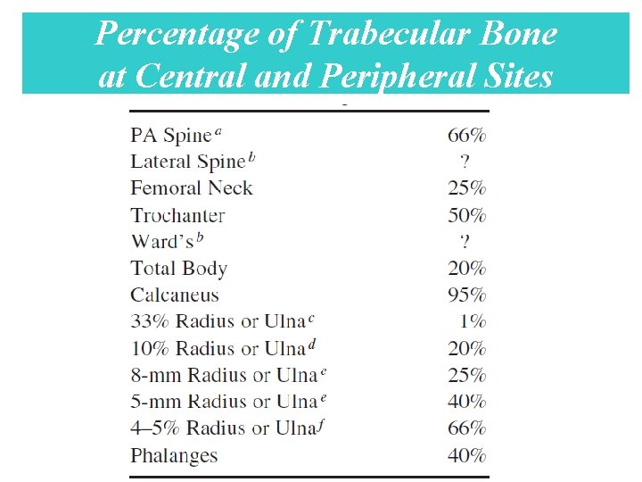
Percentage of Trabecular Bone at Central and Peripheral Sites
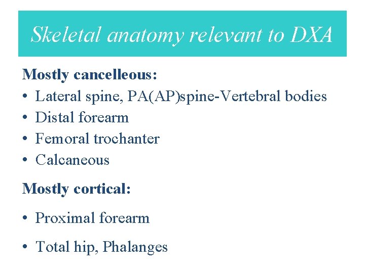
Skeletal anatomy relevant to DXA Mostly cancelleous: • Lateral spine, PA(AP)spine-Vertebral bodies • Distal forearm • Femoral trochanter • Calcaneous Mostly cortical: • Proximal forearm • Total hip, Phalanges
Dual-energy x-ray absorptiometry BMD
BMD • DEXA is the technique of choice in the assessment of bone mineral density (BMD, the average concentration of mineral in a defined section of bone. • Dual-Energy X-Ray Absorptiometry in the Diagnosis of Osteoporosis: A Practical Guide(AJR: 196, April 2011)
DEXA • • • Quick method Accurate (exact measurement of BMD) Precise (reproducible) Flexible (different regions can be scanned) Low radiation dose • Dual-Energy X-Ray Absorptiometry in the Diagnosis of Osteoporosis: A Practical Guide(AJR: 196, April 2011)
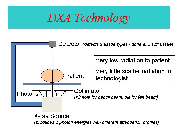
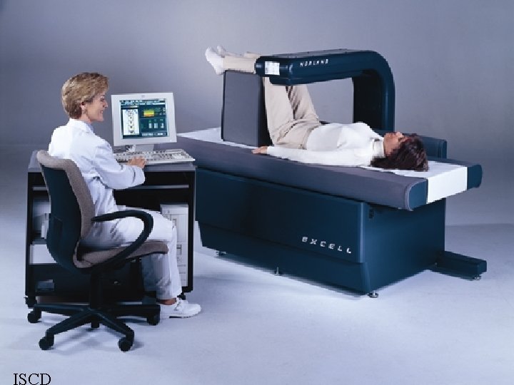
DXA Technology • A DEXA scanner consists of a low-dose x-ray tube with two energies for separating mineral and soft-tissue components and a highresolution multi detector array. • Dual-Energy X-Ray Absorptiometry in the Diagnosis of Osteoporosis: A Practical Guide(AJR: 196, April 2011)
DXA Technology Detector (detects 2 tissue types - bone and soft tissue) Very low radiation to patient. Patient Photons Very little scatter radiation to technologist Collimator (pinhole for pencil beam, slit for fan beam) X-ray Source (produces 2 photon energies with different attenuation profiles)
Areas Scanned • Lumbar spine • Proximal femur • Forearm – Two regions should be measured so that if one is unevaluable, the forearm can be imaged. • Dual-Energy X-Ray Absorptiometry in the Diagnosis of Osteoporosis: A Practical Guide(AJR: 196, April 2011)
For children (younger than 20 years) • Only the lumbar spine • Variability in femoral maturation results in lack of reproducibility in the hip region • so the reference database is available only for the spine • Dual-Energy X-Ray Absorptiometry in the Diagnosis of Osteoporosis: A Practical Guide(AJR: 196, April 2011)
Analysis of BMD • • • Five steps: 1 - Identification character 2 - Good scan criterion 3 - Region of Interest (ROI) 4 - Area 5 - Interpretation
Identification character • • • Name Age BW Height Reference population
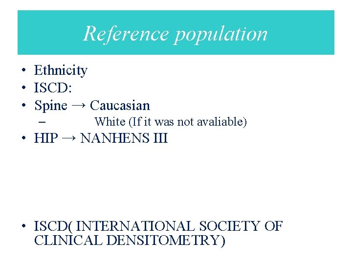
Reference population • Ethnicity • ISCD: • Spine → Caucasian – White (If it was not avaliable) • HIP → NANHENS ΙΙΙ • ISCD( INTERNATIONAL SOCIETY OF CLINICAL DENSITOMETRY)
Good scan criterion • 1 - Appropriate patient positioning • 2 - Artifact • 3 - Scan Range – Upper – Lower – Lateral
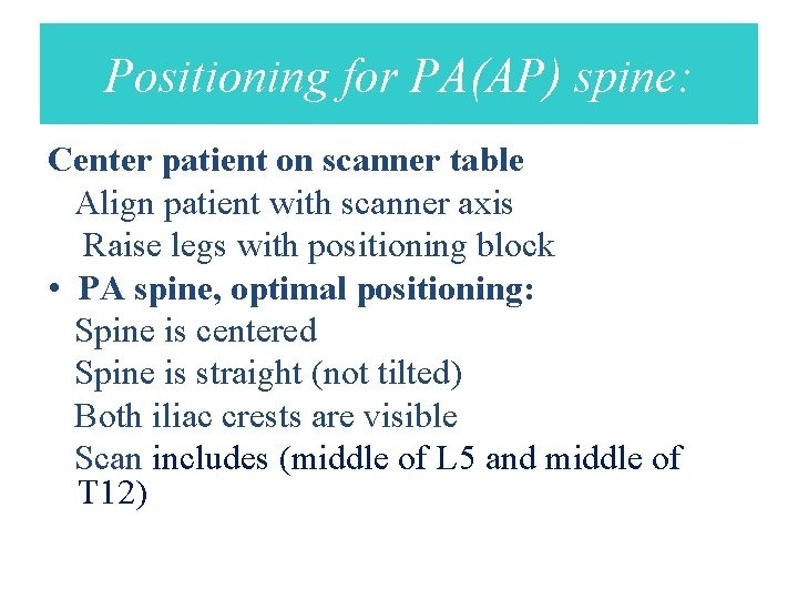
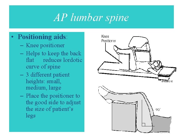
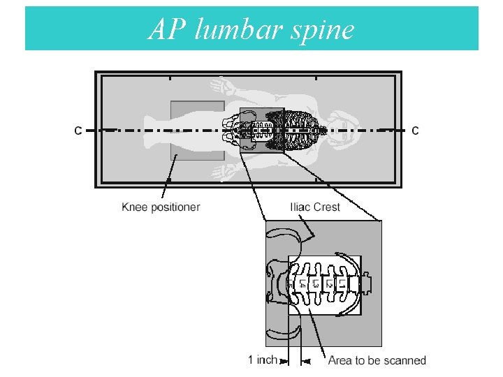
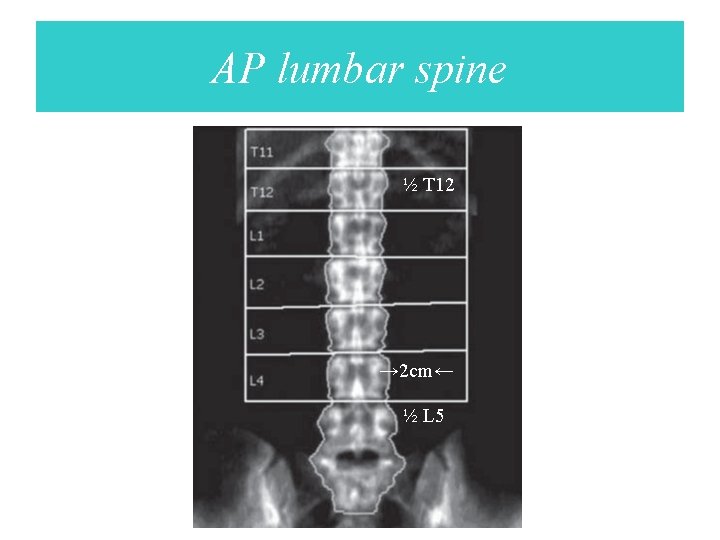
Positioning for PA(AP) spine: Center patient on scanner table Align patient with scanner axis Raise legs with positioning block • PA spine, optimal positioning: Spine is centered Spine is straight (not tilted) Both iliac crests are visible Scan includes (middle of L 5 and middle of T 12)
AP lumbar spine • Positioning aids: – Knee positioner – Helps to keep the back flat reduces lordotic curve of spine – 3 different patient heights: small, medium, large – Place the positioner to the good side to adjust the size of patient’s legs
AP lumbar spine
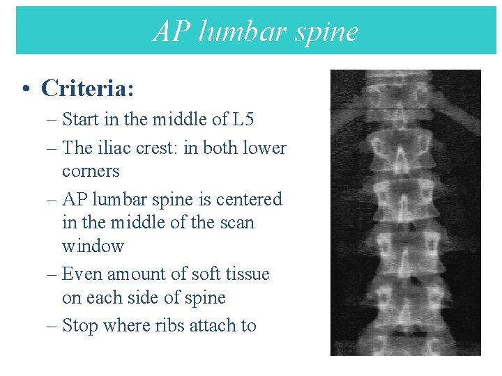
AP lumbar spine AP Lumbar spine • Criteria: – Start in the middle of L 5 – The iliac crest: in both lower corners – AP lumbar spine is centered in the middle of the scan window – Even amount of soft tissue on each side of spine – Stop where ribs attach to T 12
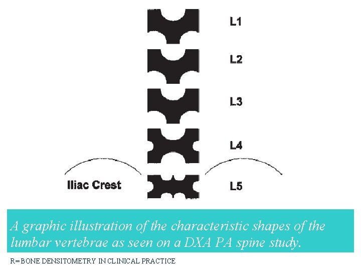
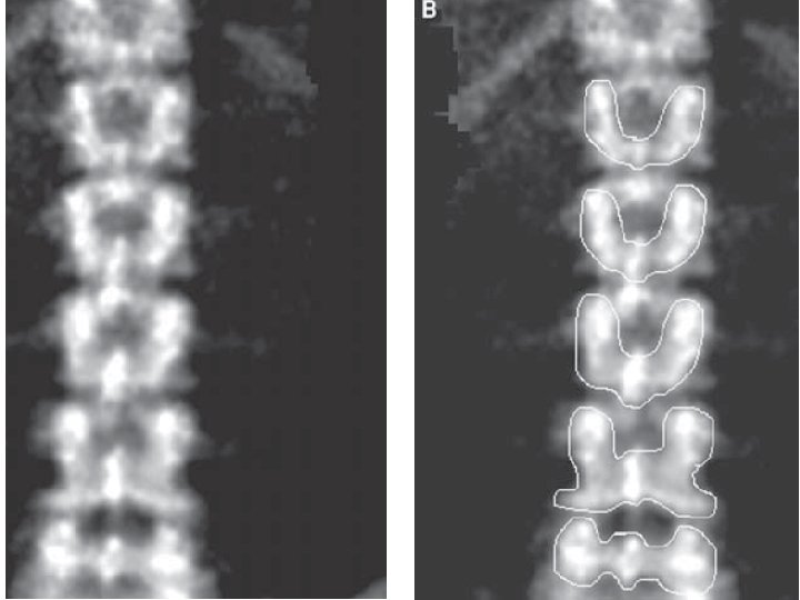
A graphic illustration of the characteristic shapes of the lumbar vertebrae as seen on a DXA PA spine study. R= BONE DENSITOMETRY IN CLINICAL PRACTICE
AP lumbar spine ½ T 12 → 2 cm← ½ L 5
Lumbar spine • Posteroanterior images of the lumbar spine include vertebral bodies L 1–L 4. • Dual-Energy X-Ray Absorptiometry in the Diagnosis of Osteoporosis: A Practical Guide
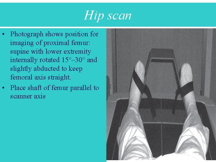
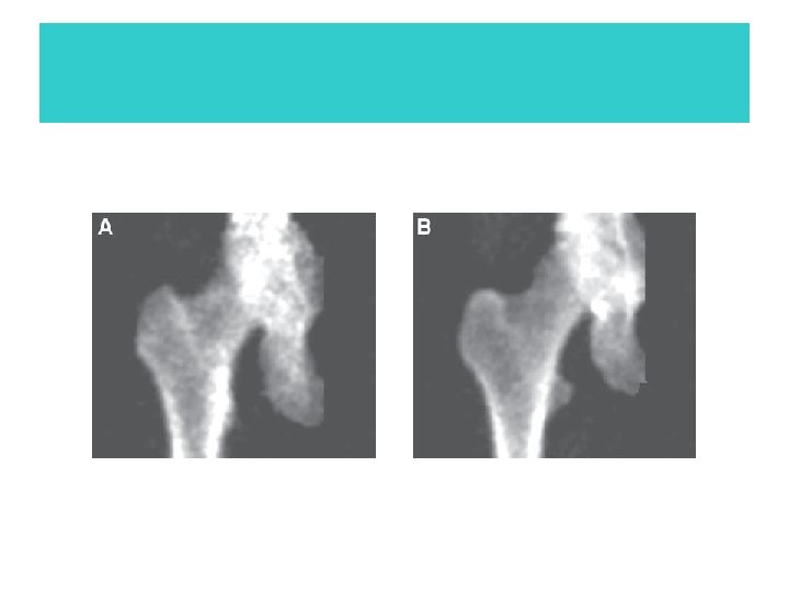
Hip scan • Photograph shows position for imaging of proximal femur: supine with lower extremity internally rotated 15°– 30° and slightly abducted to keep femoral axis straight. • Place shaft of femur parallel to scanner axis
Proper rotation of hip • The size of the lesser trochanter is the best indication of the degree of rotation • BMD values →degree of rotation of proximal femur and the position of the femoral neck ROI • Internal or external rotation →↑ BMD • BONE DENSITOMETRY IN CLINICAL PRACTICE APPLICATION AND INTERPRETATION SECOND EDITION
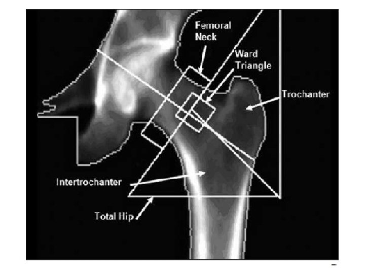
Proximal femur • Either hip • The lowest-level data on the femoral neck and total hip are used for diagnosis. • Total hip is the most reproducible measurement of the hip.
Forearm • is used in three conditions: • when the hip and spine cannot be measured the data interpreted • in examinations of patients with hyperparathyroidism • Those weight exceeds the limit for the table • Dual-Energy X-Ray Absorptiometry in the Diagnosis of Osteoporosis: A Practical Guide
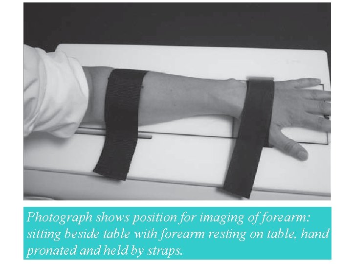
Photograph shows position for imaging of forearm: sitting beside table with forearm resting on table, hand pronated and held by straps.
Forearm scan • Optimal positioning: Forearm is centered Radius and ulna straight Distal cortex of radius and ulna visible Check for artifacts
Primary hyperparathyroidism • • The areas imaged are total bone third of the radius the ultradistal radius, and the ulna
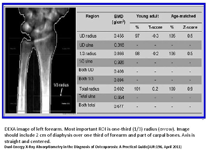
Primary hyperparathyroidism • The most useful measurements are • the ultradistal portion of the radius as an indicator of trabecular bone loss and • the distal one third of the radius (distal radial diaphysis, excluding the ultradistal portion) as an indicator of cortical bone mineral loss.
Primary hyperparathyroidism • • According to the International Society for Clinical Densitometry, the distal one third (33% radius) of the nondominant forearm is the region of choice in the assessment of osteoporosis.
DEXA image of left forearm. Most important ROI is one-third (1/3) radius (arrow). Image should include 2 cm of diaphysis over one third of forearm and part of carpal bones. Axis is straight and centered. Dual-Energy X-Ray Absorptiometry in the Diagnosis of Osteoporosis: A Practical Guide(AJR: 196, April 2011)
Vertebral Anatomy
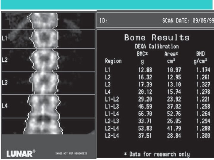
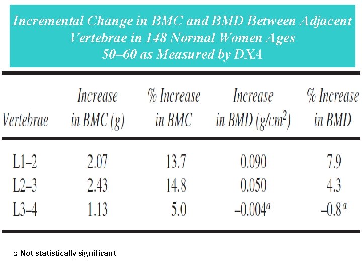
• L 1 frequently has the lowest BMC and BMD • the BMC increased between L 1–L 2, L 2– 3, and L 3– 4 although the increase • between L 3– 4 was roughly half that seen at the other levels • The largest increase in BMD occurred between L 1– 2.
Incremental Change in BMC and BMD Between Adjacent Vertebrae in 148 Normal Women Ages 50– 60 as Measured by DXA a Not statistically significant
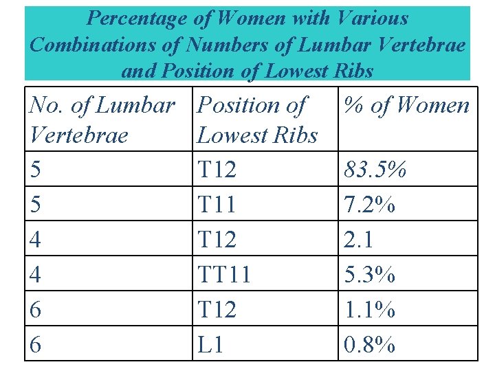
• The majority of individuals have five lumbar vertebrae with the lowest set of ribs on T 12
Percentage of Women with Various Combinations of Numbers of Lumbar Vertebrae and Position of Lowest Ribs No. of Lumbar Vertebrae 5 5 4 4 6 6 Position of Lowest Ribs T 12 T 11 T 12 TT 11 T 12 L 1 % of Women 83. 5% 7. 2% 2. 1 5. 3% 1. 1% 0. 8%
Artifacts in PA or AP Spine Densitometry
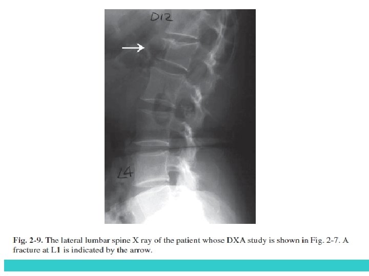
VERTEBRAL FRACTURES • The BMD of a fractured vertebra will be increased • Vertebral fractures in osteoporosis frequently occur in the: • T 7–T 9 • T 12–L 2
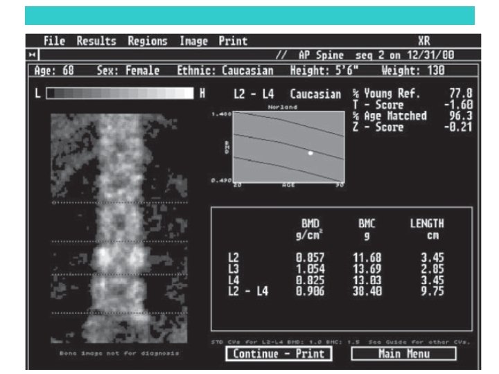
VERTEBRAL FRACTURES • FX lumbar vertebra can be excluded • ↓ the maximum number of contiguous vertebrae • For reasons of statistical accuracy and precision, the BMD for three or four contiguous vertebrae is preferred over two-vertebrae averages or the BMD of a single vertebra.
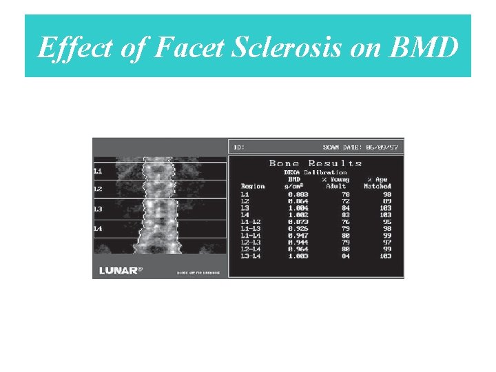
DEGENERATIVE CHANGES AND DYSTROPHIC CALCIFICATION • ↑ BMD: • Osteophytes and facet scleros • Aortic calcification
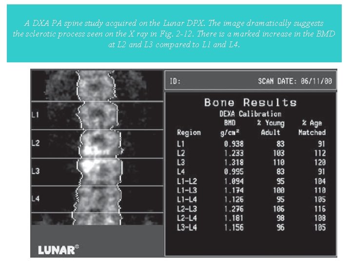
A DXA PA spine study acquired on the Lunar DPX. The image dramatically suggests the sclerotic process seen on the X ray in Fig. 2 -12. There is a marked increase in the BMD at L 2 and L 3 compared to L 1 and L 4.
Effect of Aortic Calcification on BMD. • Although it did not significantly increase • BMD, vascular calcification was seen in 24. 3% of the 144 postmenopausal women • studied by Rand et al. • The percentage with any degree of aortic calcification • was extremely low under age 60 but increased dramatically in women aged 60 and • older.
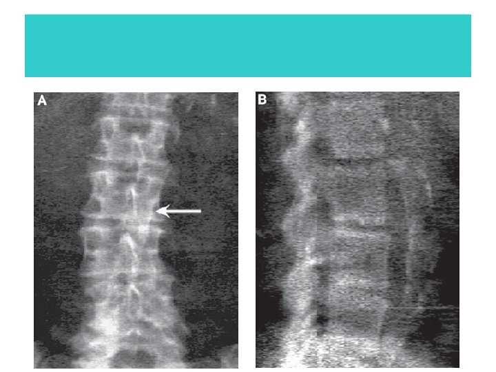
Effect of Facet Sclerosis on BMD
THE VERTEBRAL ROTATION AND PA LUMBAR SPINE BONE DENSITY • Rotation of the vertebral bodies is often a component of idiopathic scoliosis, although • it is not frequently seen in adult-onset degenerative scoliosis. • In clinical practice then, rotation of the spine for any reason, should be expected to cause • an apparent decrease in bone density when measured with DXA.
increases in the BMD • • • pancreatic calcifications Renal stones gall stones contrast agents ingested calcium tablets in addition to osteophytes • aortic calcification • fractures.
THE PROXIMAL FEMUR IN DENSITOMETRY • • the femoral neck Ward’s area the trochanteric region and the shaft
• In positioning the patient for a proximal femur study, internally rotating the femur • 15 to 20° will bring the femoral neck parallel to the plane of the scan table. • If the femoral neck rotation is increased or decreased from this position, the femoral neck BMD value will increase.
Effect of Leg Dominance on BMD in the Proximal Femur • In general, there does not seem to be a significant difference in the BMD in the regions of the proximal femur between the right and left legs of normal individuals
Effect of Scoliosis, Osteoarthritis, Osteophytes, Surgery, and Fracture on BMD in the Proximal Femur • Osteoarthritic change in the hip joint→↑BMD
the preferred site to evaluate in patients with osteoarthritis of the hip • The trochanteric region(not apparently affected)
In patients with scoliosis • • however, lower bone densities have been reported on the side of the convexity. If a “worst-case” measurement is desired, the bone density in the proximal femur should be measured in the femur on the side of the • convexity.
THE FOREARM IN DENSITOMETRY
The forearm is used in three conditions: • The hip and spine cannot be measured or the data interpreted • Patients with hyperparathyroidism • Whose weight exceeds the limit for the table • Dual-Energy X-Ray Absorptiometry in the Diagnosis of Osteoporosis: A Practical Guide(AJR: 196, April 2011)
Primary hyperparathyroidism • Recommendations include measurement at the three sites (hip, spine, and radius) for diagnosis and for follow-up after surgical and medical treatment.
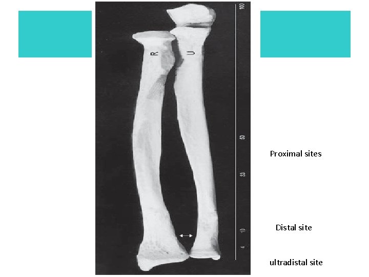
• Commonly measured sites are the 33% or 1/3 rd site 3, the • 50%, and 10% sites, the 5 -mm and 8 -mm sites, and the ultradistal site. The sites designated • by a percentage are named based on the location of the site in relation to the overall • length of the ulna.
Proximal sites Distal site ultradistal site
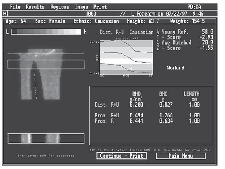
Norland
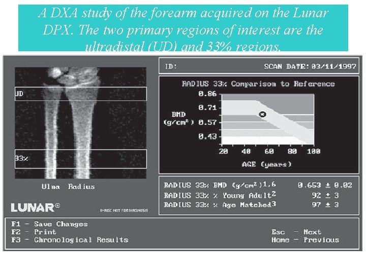
A DXA study of the forearm acquired on the Lunar DPX. The two primary regions of interest are the ultradistal (UD) and 33% regions.
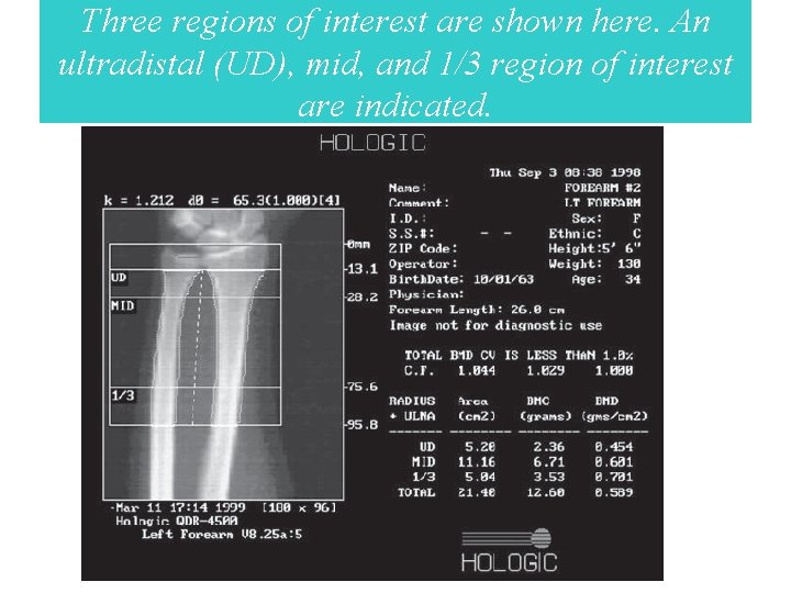
Three regions of interest are shown here. An ultradistal (UD), mid, and 1/3 region of interest are indicated.
Effect of Arm Dominance on Forearm BMD • Unlike the proximal femur, arm dominance has a pronounced effect on the bone density • in the forearm. In healthy individuals, the BMC at the 33% radial site differs by 6 to 9% • between the dominant and nondominant arms. • A difference of 3% has been reported • at the 8 -mm site
• Because • of these recognized differences, the nondominant arm has traditionally been studied when • the bone content or density is quantified for the purposes of diagnosis or fracture risk • assessment.
• Some manufacturers supply databases for the dominant arm that can • be used for comparisons if the dominant arm is to be studied. The operator’s manual for the • densitometry device should be consulted to determine which arm was used to create the • database(s) provided by the manufacturer.
The effect of movement during a forearm scan • The presence of a prior fracture → ↑BMD • Movement artifact appeared to slightly decrease the measured BMD
THE STANDARDIZED BMD
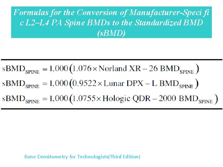
Conversion Formulas for L 2 -L 4 BMDs of the PA Spine Between DXA Devices Bone Densitometry for Technologists(Third Edition)
Formulas for the Conversion of Manufacturer-Speci fi c L 2–L 4 PA Spine BMDs to the Standardized BMD (s. BMD) Bone Densitometry for Technologists(Third Edition)
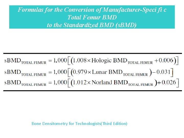
Formulas for the Conversion of Manufacturer-Speci fi c Total Femur BMD to the Standardized BMD (s. BMD) Bone Densitometry for Technologists(Third Edition)
The Utility of the s. BMD • The s. BMD is attractive as a means of comparing a BMD value obtained on one manufacturer’s device with a BMD value obtained on another. • This is useful in large population studies and clinical trials in which devices from several manufacturers must be used. • Bone Densitometry for Technologists(Third Edition)
Interpretation of BMD • BMD g/cm 2 • T-score, which represents the SD by which the BMD differs from the mean BMD of a young adult reference population of the same ethnicity and sex • Dual-Energy X-Ray Absorptiometry in the Diagnosis of Osteoporosis: A Practical Guide(AJR: 196, April 2011)
Interpretation of BMD • Z-score, which is the SD by which the BMD differs from the mean BMD of a healthy population of the same ethnicity, sex, and age as the person undergoing DEXA. Dual-Energy X-Ray Absorptiometry in the Diagnosis of Osteoporosis: A Practical Guide(AJR: 196, April 2011)
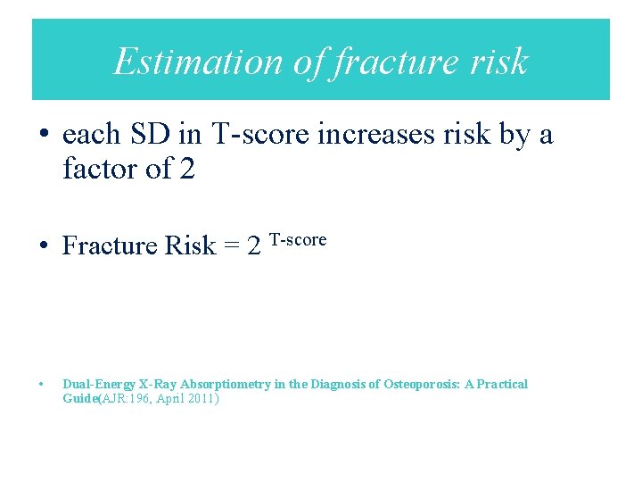
Estimation of fracture risk • each SD in T-score increases risk by a factor of 2 • Fracture Risk = 2 T-score • Dual-Energy X-Ray Absorptiometry in the Diagnosis of Osteoporosis: A Practical Guide(AJR: 196, April 2011)
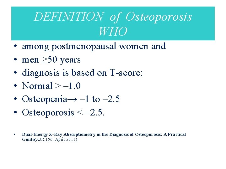
DEFINITION of Osteoporosis WHO • • • among postmenopausal women and men ≥ 50 years diagnosis is based on T-score: Normal > – 1. 0 Osteopenia→ – 1 to – 2. 5 Osteoporosis < – 2. 5. • Dual-Energy X-Ray Absorptiometry in the Diagnosis of Osteoporosis: A Practical Guide(AJR: 196, April 2011)
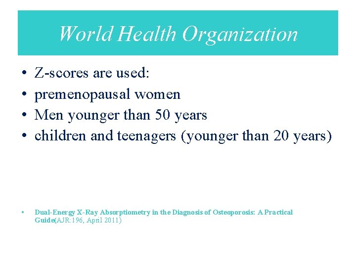
World Health Organization • • Z-scores are used: premenopausal women Men younger than 50 years children and teenagers (younger than 20 years) • Dual-Energy X-Ray Absorptiometry in the Diagnosis of Osteoporosis: A Practical Guide(AJR: 196, April 2011)
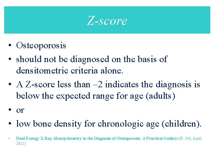
Z-score • Osteoporosis • should not be diagnosed on the basis of densitometric criteria alone. • A Z-score less than – 2 indicates the diagnosis is below the expected range for age (adults) • or • low bone density for chronologic age (children). • Dual-Energy X-Ray Absorptiometry in the Diagnosis of Osteoporosis: A Practical Guide(AJR: 196, April 2011)
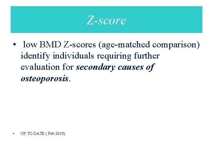
Z-score • low BMD Z-scores (age-matched comparison) identify individuals requiring further evaluation for secondary causes of osteoporosis. • UP TO DATE ( Feb 2019)