HYPERTENSIVE DISORDER OF PREGNANCY AN OVER VIEW Prof















- Slides: 129
HYPERTENSIVE DISORDER OF PREGNANCY- AN OVER VIEW Prof. S. P. Jaiswar Department of Obst. & Gynae. K. G. Medical University, Lucknow
INTRODUCTION Ø Commonest medical disorders diagnosed by obstetrician Ø Spectrum of severity ranges mild to severe Ø There is controversy and lack of clarity about mild to moderate HDP
INTRODUCTION Ø Despite of years of research in this field still there is lack of consensus about- § Definition § Classification § Level of blood pressure at which antihypertensive therapy needs to be started
INTRODUCTION Responsible for a high proportion of - hospital admission - labour inductions - fetal morbidity and mortality (20 -25%) - major cause - maternal mortality (15 -20%) Incidence: 8 -10%
INTRODUCTION Hypertension in pregnancy complicates 5% of all pregnancies & 11% of first pregnancies ä Leading cause of Maternal Mortality ä Account for 40, 000 maternal deaths annually ä In India 30% maternal deaths due to pre- eclampsia & eclampsia (Chhabra et al) ä
Measuring Blood Pressure ä ä ä ä At every visit Woman should be sitting with back & arm support without crossing legs for 5 min BP is lowest in lateral recumbent position, & the BP of the superior arm in this position is 10 -12 mm Hg lower than the inferior arm Remove all tight clothing Always measure in right arm Instrument at level of heart Diastolic BP at Korotkoff V
DEFINITION According to Working Group (2000) of High Blood Pressure Education Programme hypertension is diagnosed when: Blood pressure >140/90 mm of Hg - >30 mm of Hg or more in SBP. - >15 mm of Hg or more in DBP. This rise of BP is observed at interval of 4 -6 hrs.
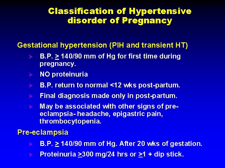
Classification of Hypertensive disorder of Pregnancy Gestational hypertension (PIH and transient HT) Ø B. P. > 140/90 mm of Hg for first time during pregnancy. Ø NO proteinuria Ø B. P. return to normal <12 wks post-partum. Ø Final diagnosis made only in post-partum. Ø May be associated with other signs of preeclampsia- headache, epigastric pain, thrombocytopenia. Pre-eclampsia Ø B. P. > 140/90 mm of Hg. After 20 wks of gestation. Ø Proteinuria >300 mg/24 hrs or >1 + dip stick.
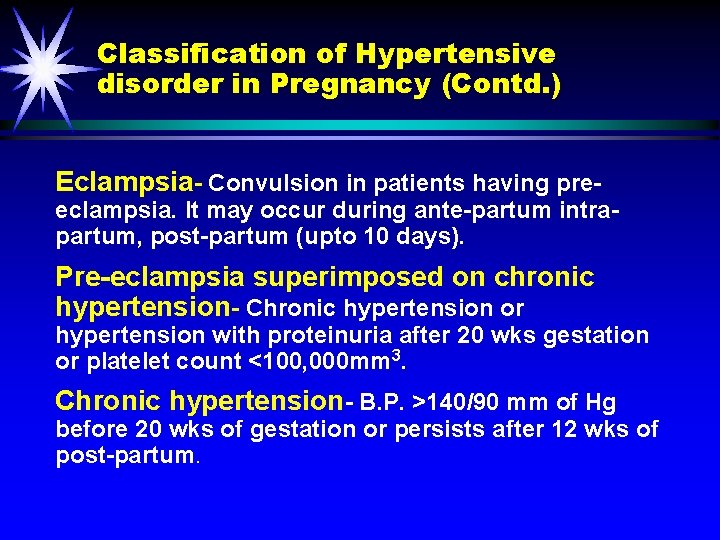
Classification of Hypertensive disorder in Pregnancy (Contd. ) Eclampsia- Convulsion in patients having pre- eclampsia. It may occur during ante-partum intrapartum, post-partum (upto 10 days). Pre-eclampsia superimposed on chronic hypertension- Chronic hypertension or hypertension with proteinuria after 20 wks gestation or platelet count <100, 000 mm 3. Chronic hypertension- B. P. >140/90 mm of Hg before 20 wks of gestation or persists after 12 wks of post-partum.
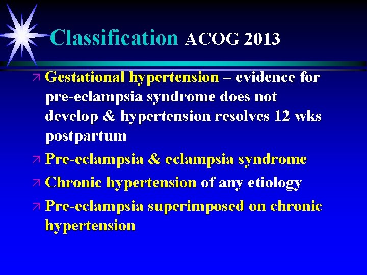
Classification ACOG 2013 ä Gestational hypertension – evidence for pre-eclampsia syndrome does not develop & hypertension resolves 12 wks postpartum ä Pre-eclampsia & eclampsia syndrome ä Chronic hypertension of any etiology ä Pre-eclampsia superimposed on chronic hypertension
Pre-eclampsia ä ä ä ä Hypertension (≥ 140/90 mm. Hg) on 2 occasions 4 hours apart after 20 wks gestation) + Proteinuria − ≥ 300 mg/24 hr or − Protein ceratinine ratio ≥ 0. 3 or − Dipstick 1+ persistent OR Thrombocytopenia – Platelets < 1, 000/µl Renal insufficiency – creatinine >1. 1 mg% or doubling of baseline (no prior renal disease) Liver involvement – Serum transaminase levels (AST or ALT) twice normal) Pulmonary edema Cerebral Symptoms – Headache, Visual, disturbances, convulsions
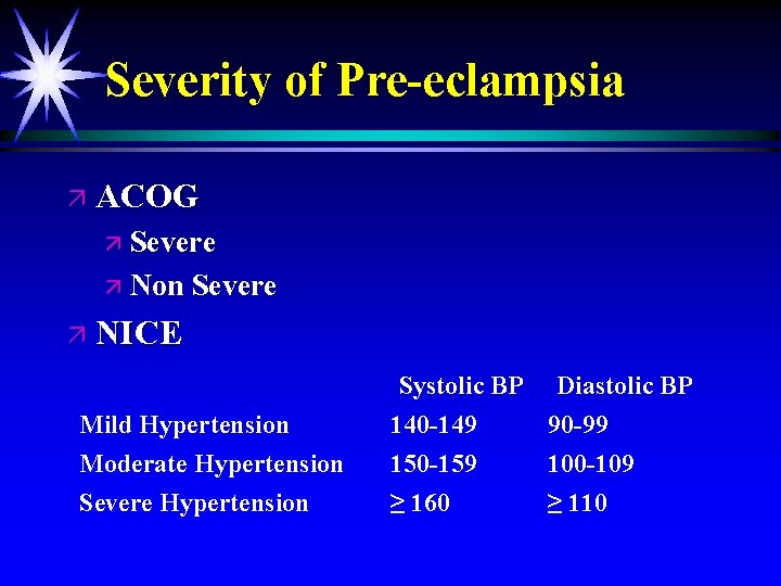
Severity of Pre-eclampsia ä ACOG ä Severe ä Non Severe ä NICE Mild Hypertension Moderate Hypertension Severe Hypertension Systolic BP Diastolic BP 140 -149 90 -99 150 -159 100 -109 ≥ 160 ≥ 110
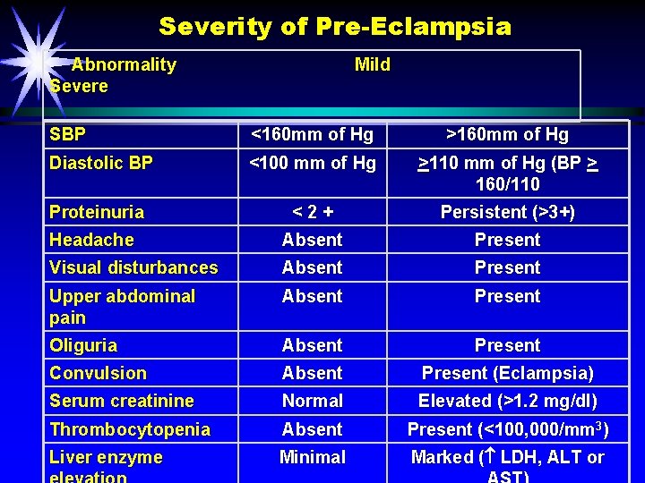
Severity of Pre-Eclampsia Abnormality Mild Severe SBP <160 mm of Hg >160 mm of Hg Diastolic BP <100 mm of Hg >110 mm of Hg (BP > 160/110 Proteinuria < 2 + Persistent (>3+) Headache Absent Present Visual disturbances Absent Present Upper abdominal pain Absent Present Oliguria Absent Present Convulsion Absent Present (Eclampsia) Serum creatinine Normal Elevated (>1. 2 mg/dl) Thrombocytopenia Absent Present (<100, 000/mm 3) Liver enzyme Minimal Marked ( LDH, ALT or
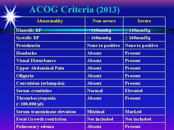
ACOG Criteria (2013) Abnormality Non-severe Severe Diastolic BP < 110 mmhg ≥ 110 mm. Hg Systolic BP < 160 mmhg ≥ 160 mm. Hg Proteinuria None to positive Headache Absent Present Visual Disturbance Absent Present Upper Abdominal Pain Absent Present Oliguria Absent Present Convulsion (eclampsia) Absent Present Serum creatinine Normal Elevated Thrombocytopenia (<100, 000/µl) Absent Present Serum transminase elevation Minimal Marked Fetal Growth restriction Not included Pulmonary edema Absent Present
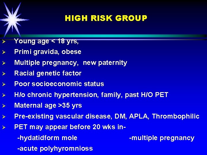
HIGH RISK GROUP Ø Young age < 18 yrs, Ø Primi gravida, obese Ø Multiple pregnancy, new paternity Ø Racial genetic factor Ø Poor socioeconomic status Ø H/o chronic hypertension, family, past H/O PET Ø Maternal age >35 yrs Ø Pre-existing vascular disease, DM, APLA, Thrombophilic Ø PET may appear before 20 wks in- -hydatidform mole -multiple pregnancy -acute polyhyromnioss
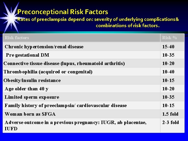
Preconceptional Risk Factors Rates of preeclampsia depend on: severity of underlying complications& combinations of risk factors. Risk factors Risk % Chronic hypertension/renal disease 15 -40 Pre gestational DM 10 -35 Connective tissue disease (lupus, rheumatoid arthritis) 10 -20 Thrombophilia (acquired or congenital) 10 -40 Obesity/insulin resistance 10 -15 Age older than 40 y 10 -20 Limited sperm exposure 10 -35 Family history of preeclampsia/ cardiovascular disease 10 -15 Woman born as SFGA 1. 5 fold Adverse outcome in a previous pregnancy: IUGR, ab placentae, IUFD 2 -3 fold
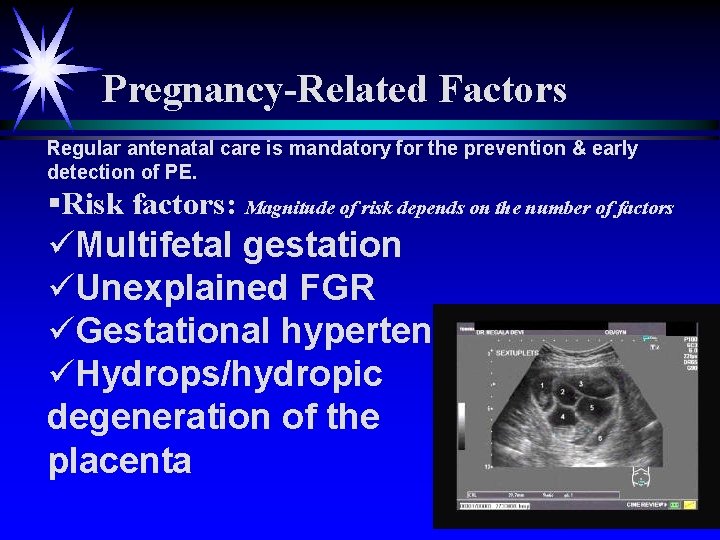
Pregnancy-Related Factors Regular antenatal care is mandatory for the prevention & early detection of PE. §Risk factors: Magnitude of risk depends on the number of factors üMultifetal gestation üUnexplained FGR üGestational hypertension üHydrops/hydropic degeneration of the placenta
ä Incidence of Pre-eclamsia- 5 -15% ä Primi-gravida- 3 -10% ä Multi-gravida- 5%
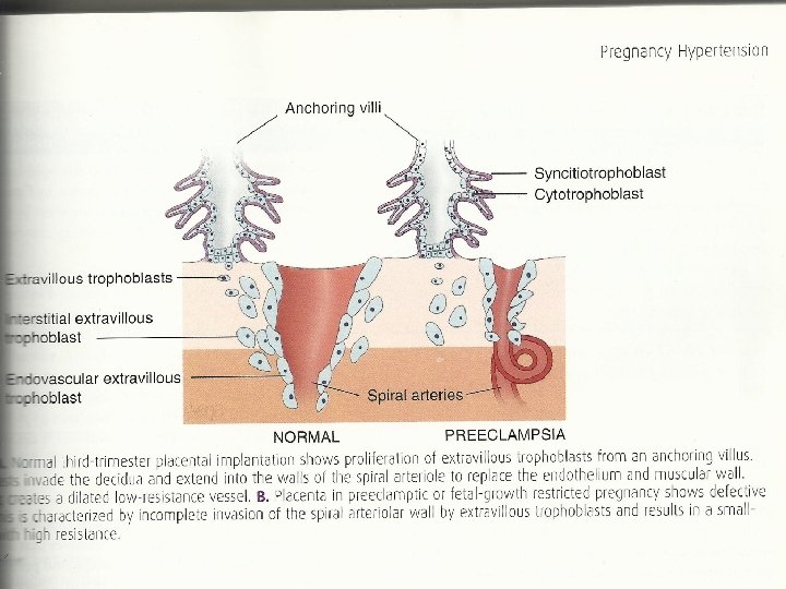
PATHOPHYSIOLOGY OF PRE-ECLAMSIA Ø Ø The exact pathophysiology is not clear. Pre-eclampsia is a disorder of endothelial function with vasospasm. Histopathology of placenta showsØ Diffuse placental thrombosis Ø Inflammatory placental decidual vasculopathy Ø Abnormal trophoblastic invasion of end arteries. Altered maternal immune response to fetal/ placental tissue.
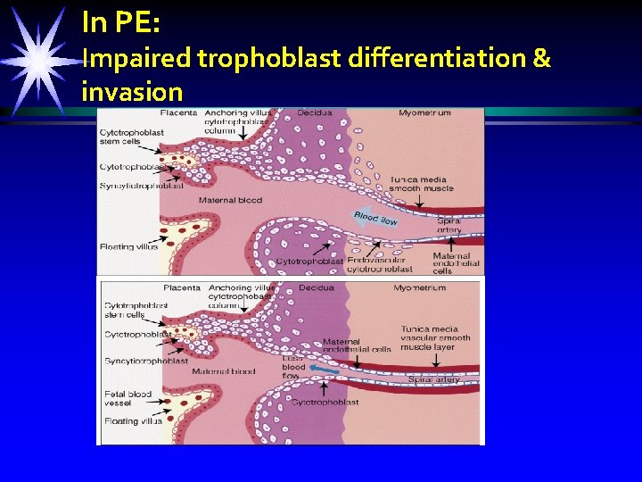
Pathogenesis: • unknown (Barton& Sibai, 2008). ØImpaired trophoblast differentiation& invasion ØPlacental & endothelial dysfunction ØImmune maladaptation to paternal ages. ØExaggerated systemic inflammatory response.
In PE: Impaired trophoblast differentiation & invasion
PATHOPHYSIOLOGY OF PREECLAMSIA ä Increase vascular sensitivity to angiotensin II. Ø Imbalance between prostacyclin and thromboxane A 2 ä ä ä Increase oxygen free radicals Abnormal calcium metabolism Vit E, C, Antioxidant and micronutrient deficiency Genetic predisposition Inhibition of Nitric Oxide synthesis- increases main arterial pressure
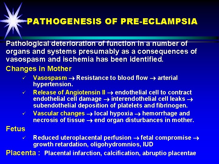
PATHOGENESIS OF PRE-ECLAMPSIA Pathological deterioration of function in a number of organs and systems presumably as a consequences of vasospasm and ischemia has been identified. Changes in Mother ü ü ü Vasospasm Resistance to blood flow arterial hypertension. Release of Angiotensin II endothelial cell to contract endothelial cell damage interendothelial cell leaks subendothelial deposition of platelets and fibrinogen. Vascular changes local hypoxia hemorrhage and necrosis of tissue end organ disturbances in mother. Fetus Reduced uteroplacental perfusion fetal compromise growth retardation, oligohydromnios, IUD Placenta : Placental infarction, calcification, abruptio placentae ü
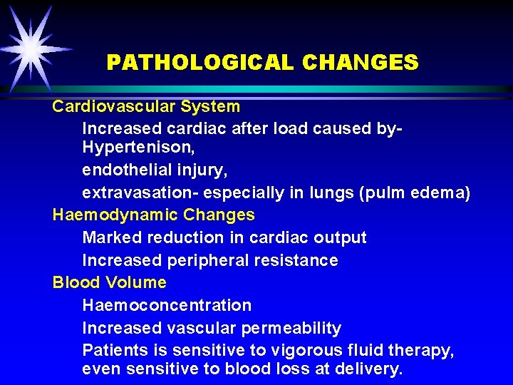
PATHOLOGICAL CHANGES Cardiovascular System Increased cardiac after load caused by. Hypertenison, endothelial injury, extravasation- especially in lungs (pulm edema) Haemodynamic Changes Marked reduction in cardiac output Increased peripheral resistance Blood Volume Haemoconcentration Increased vascular permeability Patients is sensitive to vigorous fluid therapy, even sensitive to blood loss at delivery.
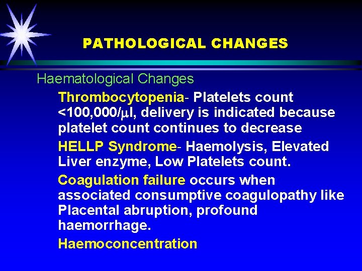
PATHOLOGICAL CHANGES Haematological Changes Thrombocytopenia- Platelets count <100, 000/ l, delivery is indicated because platelet count continues to decrease HELLP Syndrome- Haemolysis, Elevated Liver enzyme, Low Platelets count. Coagulation failure occurs when associated consumptive coagulopathy like Placental abruption, profound haemorrhage. Haemoconcentration
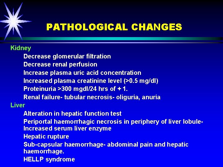
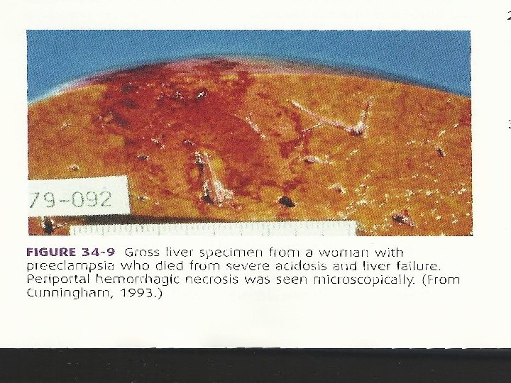
PATHOLOGICAL CHANGES Kidney Decrease glomerular filtration Decrease renal perfusion Increase plasma uric acid concentration Increased plasma creatinine level (>0. 5 mg/dl) Proteinuria >300 mgdl/24 hrs of + 1. Renal failure- tubular necrosis- oliguria, anuria Liver Alteration in hepatic function test Periportal haemorrhagic necrosis in periphery of liver lobule- Increased serum liver enzyme Hepatic rupture Sub-capsular haemorrhage- abdominal pain and hepatic haemorrhage. HELLP syndrome
ä HELLP syndrome- characterized by H- haemolysis EL- elevated liver enzyme LP- low platelet count It occurs in 20% of women with sever PET associated with placental abruption, renal failure, sub capsular hematoma Increase fetal and maternal mortality.
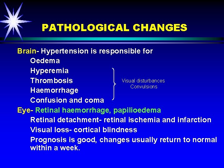
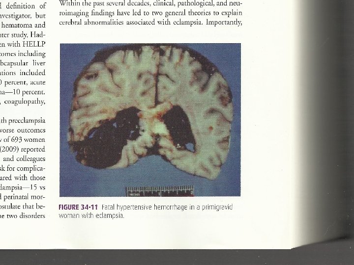
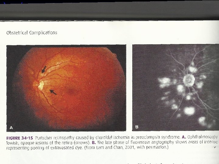
PATHOLOGICAL CHANGES Brain- Hypertension is responsible for Oedema Hyperemia Visual disturbances Thrombosis Convulsions Haemorrhage Confusion and coma Eye- Retinal haemorrhage, papilioedema Retinal detachment- retinal ischemia and infarction Visual loss- cortical blindness Prognosis is good, changes usually return to normal within a week.
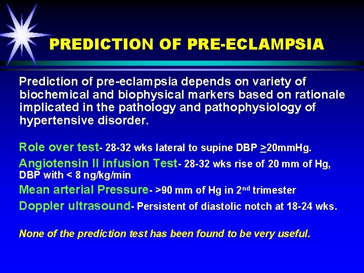
PREDICTION OF PRE-ECLAMPSIA Prediction of pre-eclampsia depends on variety of biochemical and biophysical markers based on rationale implicated in the pathology and pathophysiology of hypertensive disorder. Role over test- 28 -32 wks lateral to supine DBP >20 mm. Hg. Angiotensin II infusion Test- 28 -32 wks rise of 20 mm of Hg, DBP with < 8 ng/kg/min Mean arterial Pressure- >90 mm of Hg in 2 nd trimester Doppler ultrasound- Persistent of diastolic notch at 18 -24 wks. None of the prediction test has been found to be very useful.
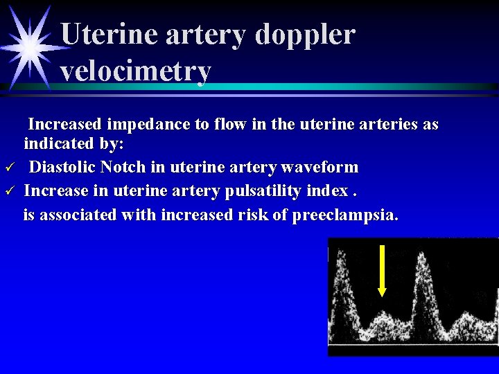
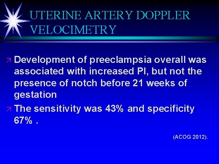
Uterine artery doppler velocimetry ü ü Increased impedance to flow in the uterine arteries as indicated by: Diastolic Notch in uterine artery waveform Increase in uterine artery pulsatility index. is associated with increased risk of preeclampsia.
UTERINE ARTERY DOPPLER VELOCIMETRY ä Development of preeclampsia overall was associated with increased PI, but not the presence of notch before 21 weeks of gestation ä The sensitivity was 43% and specificity 67%. (ACOG 2012).
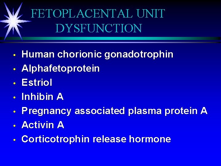
FETOPLACENTAL UNIT DYSFUNCTION • • Human chorionic gonadotrophin Alphafetoprotein Estriol Inhibin A Pregnancy associated plasma protein A Activin A Corticotrophin release hormone
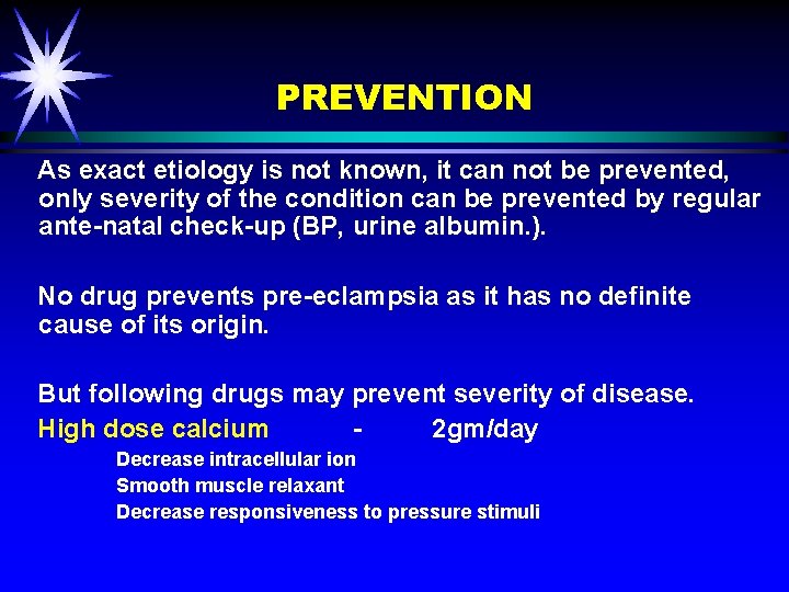
PREVENTION As exact etiology is not known, it can not be prevented, only severity of the condition can be prevented by regular ante-natal check-up (BP, urine albumin. ). No drug prevents pre-eclampsia as it has no definite cause of its origin. But following drugs may prevent severity of disease. High dose calcium 2 gm/day Decrease intracellular ion Smooth muscle relaxant Decrease responsiveness to pressure stimuli
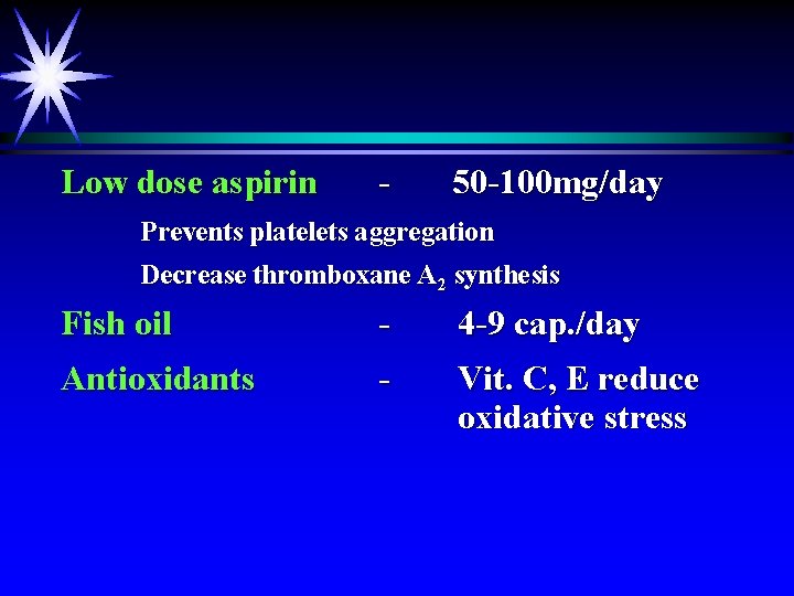
Low dose aspirin - 50 -100 mg/day Prevents platelets aggregation Decrease thromboxane A 2 synthesis Fish oil - 4 -9 cap. /day Antioxidants - Vit. C, E reduce oxidative stress
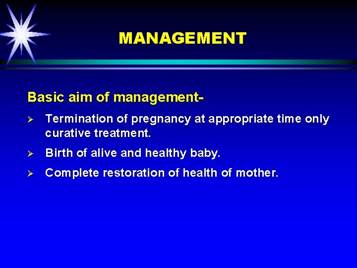
MANAGEMENT Basic aim of managementØ Termination of pregnancy at appropriate time only curative treatment. Ø Birth of alive and healthy baby. Ø Complete restoration of health of mother.
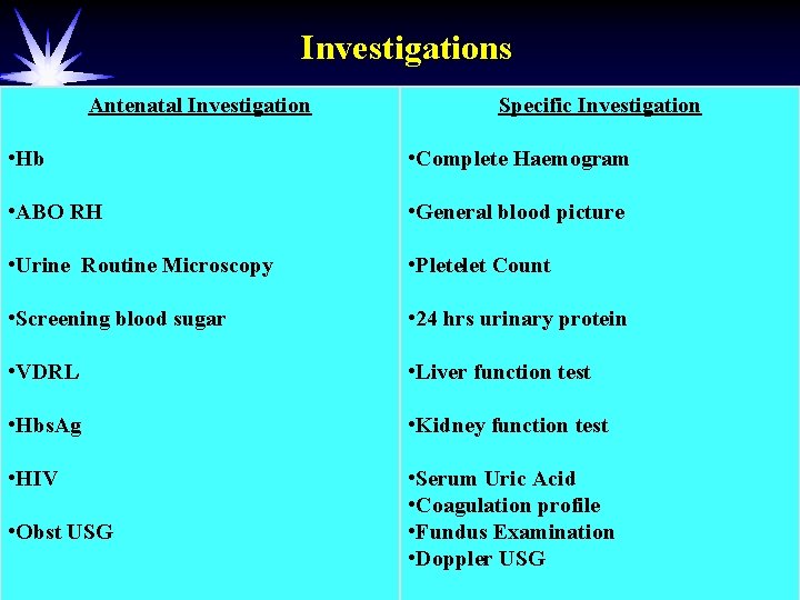
Investigations Antenatal Investigation Specific Investigation • Hb • Complete Haemogram • ABO RH • General blood picture • Urine Routine Microscopy • Pletelet Count • Screening blood sugar • 24 hrs urinary protein • VDRL • Liver function test • Hbs. Ag • Kidney function test • HIV • Serum Uric Acid • Coagulation profile • Fundus Examination • Doppler USG • Obst USG
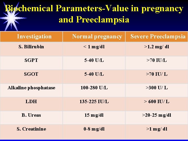
Biochemical Parameters-Value in pregnancy and Preeclampsia Investigation Normal pregnancy Severe Preeclampsia S. Bilirubin < 1 mg/dl >1. 2 mg/ dl SGPT 5 -40 U/L >70 IU/L SGOT 5 -40 U/L >70 IU/ L Alkaline phosphatase 100 -280 U/L >300 U/ L LDH 135 -225 IU/L > 600 IU/ L B. Ureas 15 mg/dl >20 -25 mg/dl S. Creatinine 0 -8 mg/dl >1 mg/ dl
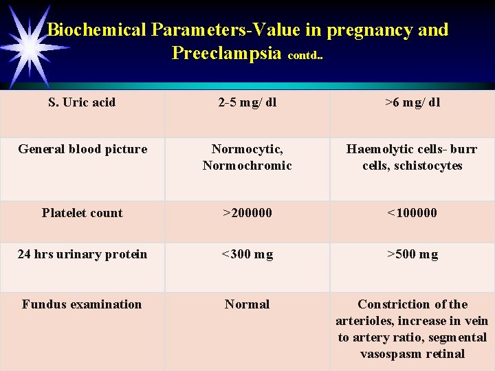
Biochemical Parameters-Value in pregnancy and Preeclampsia contd. . S. Uric acid 2 -5 mg/ dl >6 mg/ dl General blood picture Normocytic, Normochromic Haemolytic cells- burr cells, schistocytes Platelet count >200000 <100000 24 hrs urinary protein <300 mg >500 mg Fundus examination Normal Constriction of the arterioles, increase in vein to artery ratio, segmental vasospasm retinal
MANAGEMENT For early detectionØ Increase antenatal visit especially 3 rd trimester Ø Blood pressure >140/90 mm of Hg- to be admitted to the hospital for evaluation of severity. Ø Mild disease may often manage as out patient. Ø Hospitalization- hypertension, proteinuria Persistent or worsening of Patient develops
MANAGEMENT In Hospital Ø For systemic evaluation detailed history of headache, visual disturbance, epigastric pain, rapid weight gain. Ø Daily weight recording Ø Urine for protein- every 2 nd day Ø Blood pressure recording 4 hrly in sitting position Ø Estimation of S. creatinine, hematocrit, platelets count, S. liver enzyme Ø Evaluation of fetal size, ammniotic fluid- clinically, USG.
MANAGEMENT Care to be taken Ø Reduced activity is beneficial Ø Absolute bed rest is not necessary Ø Adequate protein and calories should be given Ø Sodium and fluid intake should be adequate Ø Do not give diuretics to correct oliguria or anuria Ø Do not give diazepam to stop convulsion- apnea, aspiration
Maternal Surveillance in mild Preeclampsiaä ä ä Measure BP-4 times per day Measure body weight- weekly Daily urine dipstick evaluation of protein in urine Proteinuria in a 24 hr specimen every week CBC with platelet count, LDH, AST, ALT twice per week Inquire in each contact about fetal movements, development of scotoma, headache, epigastric pain.
Maternal Surveillance in sever Preeclampsiaä ä ä ä Measure BP 4 times per day Measure body weight every day Daily input and output Daily urine dipstick evaluation of urine protein Proteinuria in 12 hr specimen CBC with platelet count, LDH, AST, ALT, KFT every other day or more frequent Inquire in each contact about fetal movements, development of scotoma, headache, epigastric pain.
Fetal Surveillance in mild Preeclampsiaä Daily fetal heart rate monitoring ä Daily fetal movement count ä Modified biophysical profile every week ä Fetal biometry every 3 weeks ä Umbilical and cerebral doppler every week
Fetal surveillance Baseline USG for AFI, fetal weight, Doppler ä DFMC ä Weekly NST ä ä NICE guidelines recommend 2 weekly Doppler for mild PE ä No large prospective studies comparing frequency of monitoring
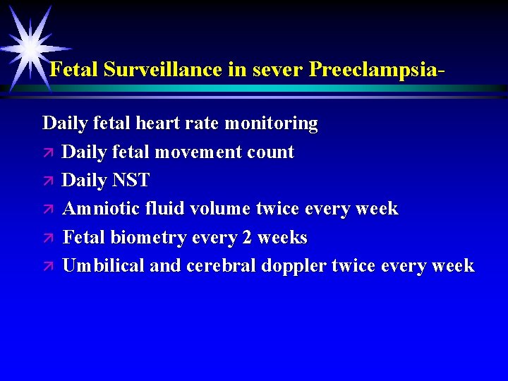
Fetal Surveillance in sever Preeclampsia. Daily fetal heart rate monitoring ä Daily fetal movement count ä Daily NST ä Amniotic fluid volume twice every week ä Fetal biometry every 2 weeks ä Umbilical and cerebral doppler twice every week
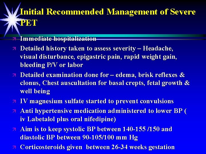
Initial Recommended Management of Severe PET ä ä ä ä Immediate hospitalization Detailed history taken to assess severity – Headache, visual disturbance, epigastric pain, rapid weight gain, bleeding P/V or labor Detailed examination done for – edema, brisk reflexes & clonus, Chest auscultation for basal crepts, fetal growth & well being IV magnesium sulfate started to prevent convulsions Anti hypertensive medication administered to lower BP ( iv Labetalol plus oral nifedipine) Aim is to keep systolic BP between 140 -155 /150 and diastolic BP between 90 -105/100 mm Hg Corticosteroids given between 26 -34 weeks gestation
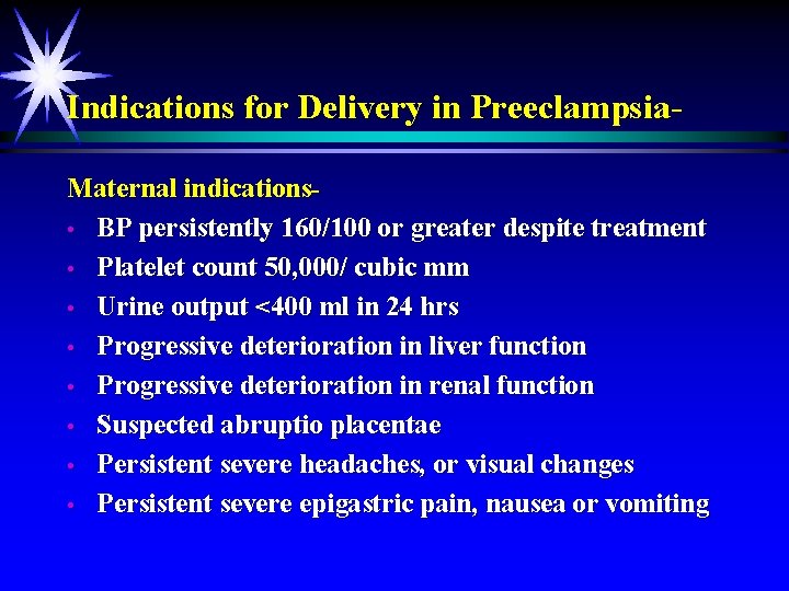
Indications for Delivery in Preeclampsia. Maternal indications • BP persistently 160/100 or greater despite treatment • Platelet count 50, 000/ cubic mm • Urine output <400 ml in 24 hrs • Progressive deterioration in liver function • Progressive deterioration in renal function • Suspected abruptio placentae • Persistent severe headaches, or visual changes • Persistent severe epigastric pain, nausea or vomiting
Fetal indicationä Severe growth restriction ä Nonreassuring fetal cardiotocography ä Oligohydramnios
There is general agreement that all such patients should be delivered if the disease develops after 34 weeks gestation There is disagreement about management of patients with severe PET before 34 weeks gestation Prompt delivery is indicated ä ä ä Imminent eclampsia Multi-organ dysfunction Severe IUGR < 5 th percentile Suspected abruption placentae Non reassuring fetal testing ä ä Some consider delivery as the definite treatment regardless of gestational age Others recommend prolonging pregnancy for fetal lung maturity till 34 weeks
Although delivery is beneficial to mother , it must be weighed against the risk for prematurity ä ä It was believed that premature infants of severe PET have accelerated lung and neurological maturation as a result of stress in utero Had lower rates of morbidity and mortality with similar gestation age of normal infants ä This has been revealed wrong ä They have higher rates of admission in neonatal intensive care unit Sibai 2007
Timing and mode of Delivery Continue pregnancy till 37 weeks ä Expectant management vs Induction of labor (Hypitat trial 2010) ä Reduced rates of severe HT (induction group). ä No difference in CS delivery and neonatal outcome. ä Induction prevents complications however little they are ä
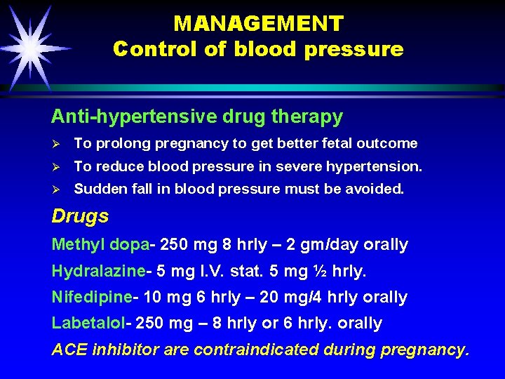
MANAGEMENT Control of blood pressure Anti-hypertensive drug therapy Ø To prolong pregnancy to get better fetal outcome Ø To reduce blood pressure in severe hypertension. Ø Sudden fall in blood pressure must be avoided. Drugs Methyl dopa- 250 mg 8 hrly – 2 gm/day orally Hydralazine- 5 mg I. V. stat. 5 mg ½ hrly. Nifedipine- 10 mg 6 hrly – 20 mg/4 hrly orally Labetalol- 250 mg – 8 hrly or 6 hrly. orally ACE inhibitor are contraindicated during pregnancy.
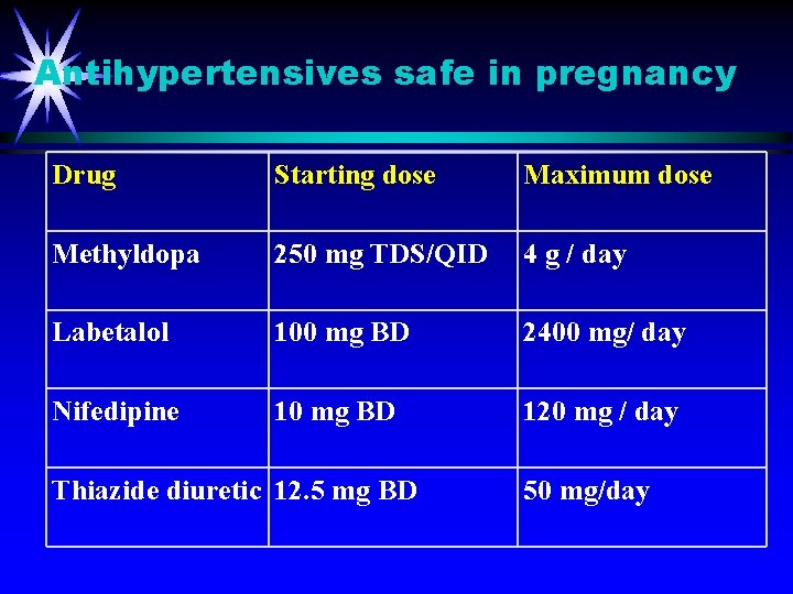
Antihypertensives safe in pregnancy Drug Starting dose Maximum dose Methyldopa 250 mg TDS/QID 4 g / day Labetalol 100 mg BD 2400 mg/ day Nifedipine 10 mg BD 120 mg / day Thiazide diuretic 12. 5 mg BD 50 mg/day
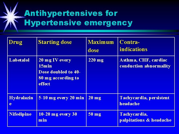
Antihypertensives for Hypertensive emergency Drug Starting dose Maximum Contraindications dose Labetalol 20 mg IV every 15 min Dose doubled to 4080 mg according to effect 220 mg Hydralazin e 5 -10 mg every 20 min 20 mg Tachycardia, persistent headache Nifedipine 10 -20 mg every 30 min Tachycardia, palpitations & headache 50 mg Asthma, CHF, cardiac conduction abnormality
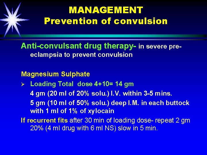
MANAGEMENT Prevention of convulsion Anti-convulsant drug therapy- in severe preeclampsia to prevent convulsion Magnesium Sulphate Ø Loading Total dose 4+10= 14 gm (20 ml of 20% solu. ) I. V. within 3 -5 mins. 5 gm (10 ml of 50% solu. ) deep I. M. in each buttock with 1 ml of 1% of xylocain If recurrent fits after 30 min of loading dose- repeat 2 gm 20% (4 ml drug with 6 ml NS) slow in 5 min.
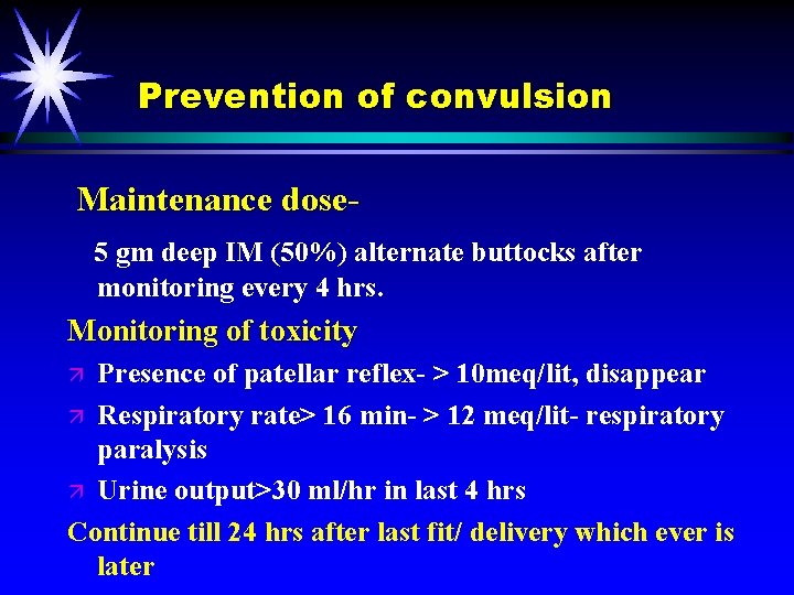
Prevention of convulsion Maintenance dose 5 gm deep IM (50%) alternate buttocks after monitoring every 4 hrs. Monitoring of toxicity Presence of patellar reflex- > 10 meq/lit, disappear ä Respiratory rate> 16 min- > 12 meq/lit- respiratory paralysis ä Urine output>30 ml/hr in last 4 hrs Continue till 24 hrs after last fit/ delivery which ever is later ä
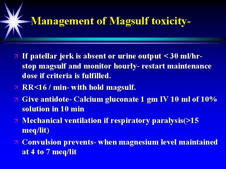
Management of Magsulf toxicityä ä ä If patellar jerk is absent or urine output < 30 ml/hrstop magsulf and monitor hourly- restart maintenance dose if criteria is fulfilled. RR<16 / min- with hold magsulf. Give antidote- Calcium gluconate 1 gm IV 10 ml of 10% solution in 10 min Mechanical ventilation if respiratory paralysis(>15 meq/lit) Convulsion prevents- when magnesium level maintained at 4 to 7 meq/lit
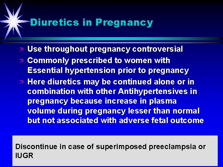
Diuretics in Pregnancy ä ä ä Use throughout pregnancy controversial Commonly prescribed to women with Essential hypertension prior to pregnancy Here diuretics may be continued alone or in combination with other Antihypertensives in pregnancy because increase in plasma volume during pregnancy lesser than normal but not associated with adverse fetal outcome Discontinue in case of superimposed preeclampsia or IUGR
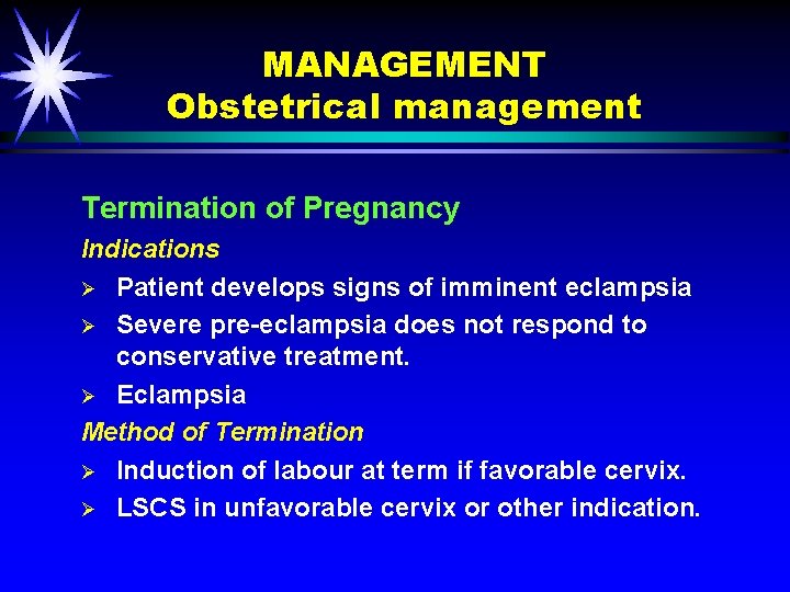
MANAGEMENT Obstetrical management Termination of Pregnancy Indications Ø Patient develops signs of imminent eclampsia Ø Severe pre-eclampsia does not respond to conservative treatment. Ø Eclampsia Method of Termination Ø Induction of labour at term if favorable cervix. Ø LSCS in unfavorable cervix or other indication.
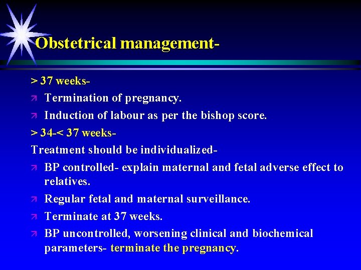
Obstetrical management> 37 weeksä Termination of pregnancy. ä Induction of labour as per the bishop score. > 34 -< 37 weeks. Treatment should be individualizedä BP controlled- explain maternal and fetal adverse effect to relatives. ä Regular fetal and maternal surveillance. ä Terminate at 37 weeks. ä BP uncontrolled, worsening clinical and biochemical parameters- terminate the pregnancy.
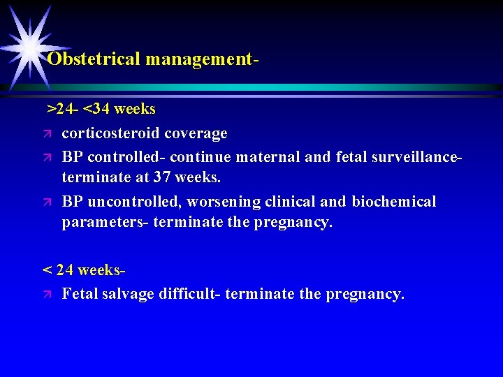
Obstetrical management>24 - <34 weeks ä corticosteroid coverage ä BP controlled- continue maternal and fetal surveillanceterminate at 37 weeks. ä BP uncontrolled, worsening clinical and biochemical parameters- terminate the pregnancy. < 24 weeksä Fetal salvage difficult- terminate the pregnancy.
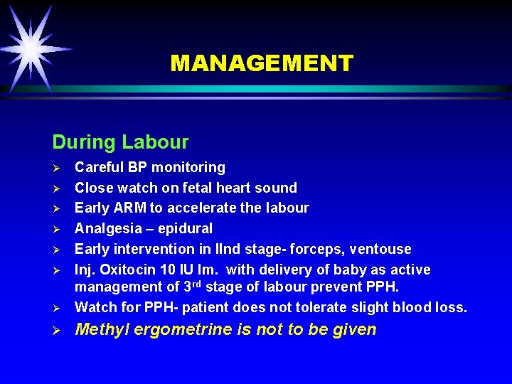
MANAGEMENT During Labour Ø Careful BP monitoring Close watch on fetal heart sound Early ARM to accelerate the labour Analgesia – epidural Early intervention in IInd stage- forceps, ventouse Inj. Oxitocin 10 IU Im. with delivery of baby as active management of 3 rd stage of labour prevent PPH. Watch for PPH- patient does not tolerate slight blood loss. Ø Methyl ergometrine is not to be given Ø Ø Ø
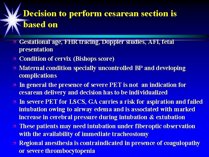
Decision to perform cesarean section is based on ä ä ä ä Gestational age, FHR tracing, Doppler studies, AFI, fetal presentation Condition of cervix (Bishops score) Maternal condition specially uncontrolled BP and developing complications In general the presence of severe PET is not an indication for cesarean delivery and decision has to be individualized In severe PET for LSCS, GA carries a risk for aspiration and failed intubation owing to airway edema and is associated with marked increase in cerebral pressure during intubation & extubation These patients may need intubation under fiberoptic observation with the availability of immediate tracheostomy Regional anesthesia is contraindicated in presence of coagulopathy or severe thrombocytopenia
MANAGEMENT Post-natal Period Ø Dose of antihypertensive is usually reduced Ø Methyl dopa is discontinued- causes post-partum depression Ø ACE inhibitor may be started- if required Ø Patient must be counseled for recurrence in future pregnancy or develop hypertension in later life.
Antihypertensives in Postpartum period ä ä ä Should be started if SP > 150 mm Hg & DP > 100 mm Hg Discontinued if BP remains below hypertensive level for atleast 48 hours Causes of exacerbation of Hypertension in Postpartum period: - IV fluids given in labour - IV Oxytocin in labour - Mobilization of extracellular fluid to intravascular compartment
Antihypertensives in Postpartum period & Breast feeding ä All antihypertensives are found in breast milk but plasma: milk ratio of drugs is different ä Long term effects of these drugs on breastfeeding infants not studied
Choice of Antihypertensives in Postpartum period • Labetalol & Nifedipine drug of choice • Labetalol/ Hydralazine (IV)- in Hypertensive emergency ä ä Diuretics- only indicated in Pulmonary edema, decrease milk production ACE inhibitors - adverse effect on neonatal renal function Methyldopa- Postpartum depression Amlodipine- also a calcium channel blocker but not used due to limited studies in literature supporting use
Eclampsia Dr. S. P. Jaiswar Professor Dept. of Obst. & Gynae KGMU Lucknow
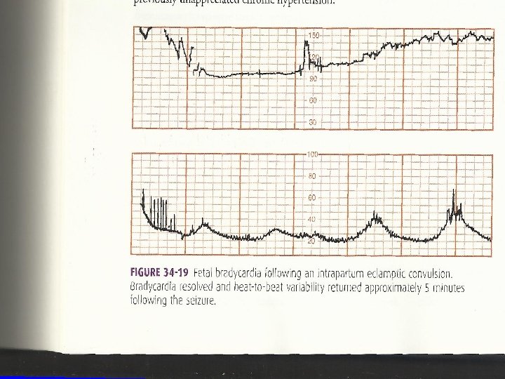
Introduction ä Convulsive state of pre-eclampsia is called eclampsia. ä Pre-eclampsia is complicated by generalized tonic and clonic convulsions and coma.
Incidence ä ä 1 in 500 to 1 in 30 More common in primi-gravida Five time more common in twins 50% occur in late pregnancy According to time of onset of convulsions ä Ante- partum- 50% ä Intra- partum- 30% Intercurrent ä Post- partum- 20%
Cause of Convulsions ä Cerebral irritation- due to cerebral hypoxia - due to spasm of cerebral vessels ä Cerebral oedema- perivascular oedema ä Cerebral dysarrythmia - Following anoxia or oedema
Cerebral Pathologyä Cortical and sub- cortical oedema ä Infarction ä Haemorrhage Hypoxia, ischaemia or oedema
Clinical features. Sometimes patient shows premonitory symptoms before developing convulsions Divided into four stagesä Pre monitory Stage ä Tonic Stage ä Clonic Stage ä Stage of Coma
Premonitory Stageä Patient become unconscious ä Twitching of muscles of face, tongue, limbs ä Eyeballs roll or turned to one side, become fixed ä Its last for 30 sec.
Tonic Stageä Whole body goes into a tonic spasm ä Respiration ceases, tongue protrudes out ä Cyanosis appears, eyeballs become fixed ä This stage last for 30 sec
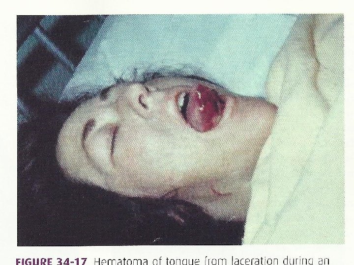
Clonic Stageä Voluntary muscles undergo alternative contraction and relaxation ä Twitching start from the face then involve one side of extremities and ultimately the whole body ä Biting of tongue ä Breathing is laboured and blood stained frothy secretions ä Cyanosis gradually disappear ä Lasts for 1 -4 min.
Stage of Coma- Following fits patient passes in stage of coma Status eclampticus. Multiple recurring fits at varying intervals
Differential Diagnosisä Epilepsy ä Hysteria ä Encephalitis and Meningitis ä Puerperal cerebral thrombosis ä Poisoning ä Cerebral malaria ä Intra- cranial tumors
Prognosis Immediate Mortality-2 -30% maternal mortality Causes of Maternal deathä Cardiac Failure ä Pulm oedema ä Aspiration septic pneumonitis ä Cerebral hemorrhage ä Acute renal failure ä Cardiopulmonary arrest ä Adult respiratory distress syndrome ä Post partum shock ä Puerperal sepsis
Fetal Complicationsä Peri natal mortality- 30 -50% ä Pre maturity- spontaneous, induced ä Intra uterine asphyxia- Infarction, retroplacental, haemorrhage spasm ä Effect of drugs ä Trauma
Management- Preventionä To control blood pressure ä Prophylactic magsulf therapy ä Patient to be referred to tertiary centre
Hospital Managementä ä ä ä ä Shout for help Maintain airway, breathing and circulation Oxygen administration 8 -10 L/min Arrest convulsions Ventilator support Prevention of injury Hemodynamic stabilization Delivery within 6 -8 hr, maximum 12 hrs Prevention of complications ä Post partum care
Specific Management. Anti-convulsions- ä Magnesium sulphate is the drug of choice Pritchard regimen (Intramuscular) 4 gm I/V, over 3 -5 min(20 ml of 20% solution) 10 gm I/M deep(5 gm in each buttock-50% solution) Maintenance dose- 5 gm I. M. 4 hrly ä Intravenous- Zuspan or Sibai ä 4 -6 gm IV over 15 -20 min 1 -2 gm/hr IV infusion ä ä
INTRAMUSCULAR DOSAGE SCHEDULE ä ä Magnesium sulphate 8 ml (50% weight by volume = 4 gm) dissolved in 12 ml of saline ( total=20 ml) for 20 % w/v intravenous dose Magnesium sulphate 10 ml (5 gm) injected deep intramuscular in each buttock. (50% w/v dose) Addition of 1 ml of 2% lidocaine reduces pain. LOADING DOSE : 4 gm of magnesium sulphate as 20% solution intravenously at rate of 1 gm/min + 10 gm of 50% magnesium sulphate solution , one half (5 gm) injected deeply in upper outer quadrant of each buttock.
INTRAMUSCULAR DOSAGE SCHEDULE MAINTAINENCE DOSE ä Every four hour give 5 gm of 50% w/v solution (10 ml= 5 gm) injected deep intramuscular in upper outer quadrant of alternate buttocks ä BUT only after ensuring that : 1. Patellar reflex is present 2. Respiration not depressed 3. Urine output in previous 4 hour exceeded 100 ml. ä
ä In case of recurrent fits half loading dose : 2 gm more intravenously as 20% solution is given. ä 4 ml of 50% w/v (2 gm of magnesium sulphate) dissolved in 6 ml of saline to make 2 gm of 20% w/v solution( 2 gm in 10 ml)
ADVERSE EFFECTS OF MAGSULPH ä ä ä Fits controlled at blood levels of 4 -7 meq/l 10 meq/l: patellar reflexes disappear >= 10 meq/l : respiratory depression >=12 meq/l : respiratory paralysis and respiratory arrest Neonatal respiratory depression only if severe hypermagnesemia at delivery Neonatal compromise usually not problematic
Antidote ä Calcium gluconate or calcium chloride 1 gm intravenously and witholding further magnesium sulphate reverses mild to moderate respiratory depression ä For severe cases prompt tracheal intubation and mechanical ventilation required.
Magnesium Sulphate- Preferred Control fits without depression in mother and foetus Reduce of recurrent convulsions Significantly reduce maternal death (3%) Reduce perinatal mortality Anti hypertensive drugs- BP >160/105 mm of Hg (NHBPEP-2000) Diuretics-In pulmonary oedema(20 -40 mg IV)
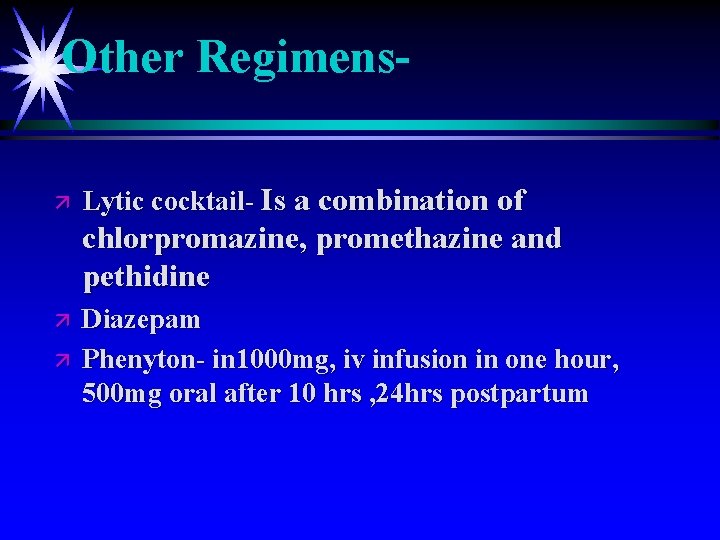
Other Regimensä Lytic cocktail- Is a combination of chlorpromazine, promethazine and pethidine ä ä Diazepam Phenyton- in 1000 mg, iv infusion in one hour, 500 mg oral after 10 hrs , 24 hrs postpartum
Other Regimensä Lytic cocktail- Is a combination of chlorpromazine, promethazine and pethidine ä ä Diazepam Phenyton- in 1000 mg, iv infusion in one hour, 500 mg oral after 10 hrs , 24 hrs postpartum
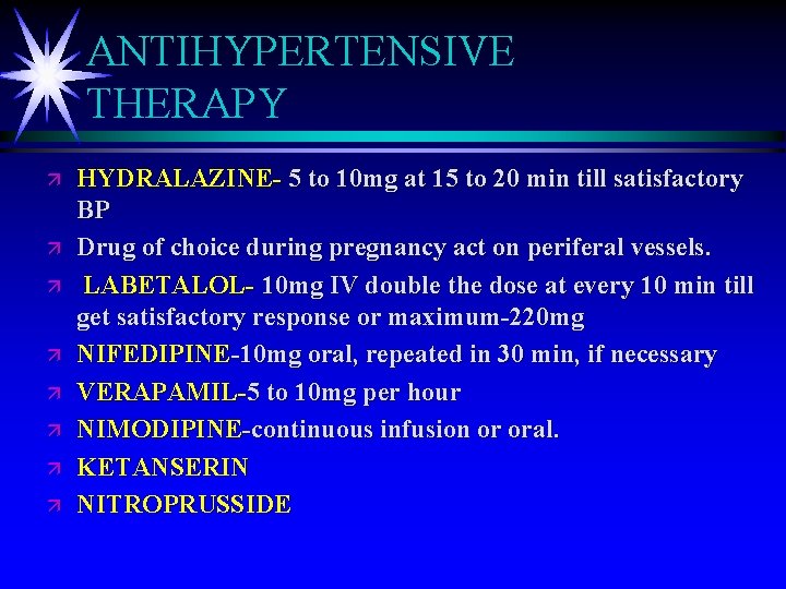
ANTIHYPERTENSIVE THERAPY ä ä ä ä HYDRALAZINE- 5 to 10 mg at 15 to 20 min till satisfactory BP Drug of choice during pregnancy act on periferal vessels. LABETALOL- 10 mg IV double the dose at every 10 min till get satisfactory response or maximum-220 mg NIFEDIPINE-10 mg oral, repeated in 30 min, if necessary VERAPAMIL-5 to 10 mg per hour NIMODIPINE-continuous infusion or oral. KETANSERIN NITROPRUSSIDE
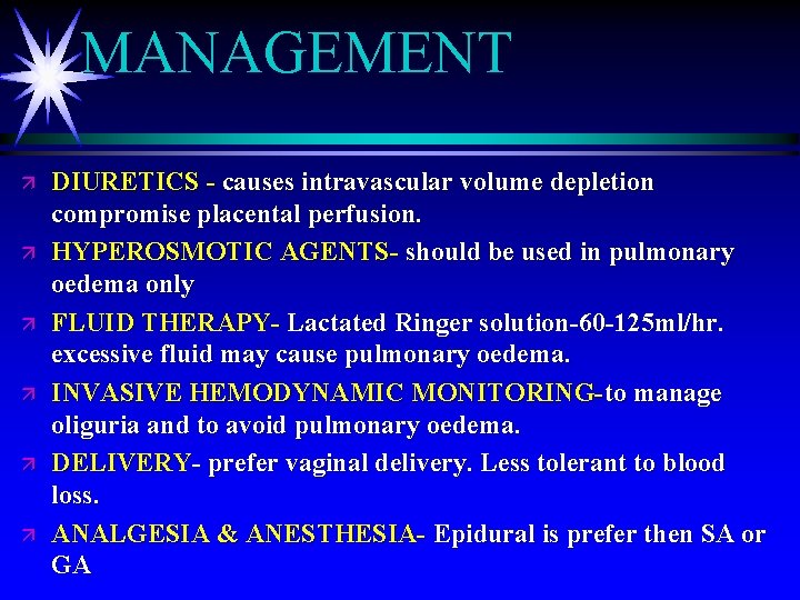
MANAGEMENT ä ä ä DIURETICS - causes intravascular volume depletion compromise placental perfusion. HYPEROSMOTIC AGENTS- should be used in pulmonary oedema only FLUID THERAPY- Lactated Ringer solution-60 -125 ml/hr. excessive fluid may cause pulmonary oedema. INVASIVE HEMODYNAMIC MONITORING-to manage oliguria and to avoid pulmonary oedema. DELIVERY- prefer vaginal delivery. Less tolerant to blood loss. ANALGESIA & ANESTHESIA- Epidural is prefer then SA or GA
Management of complicationsä Pulmonary oedema ä Heart failure ä Anuria ä Hyperpyrexia ä Psychosis
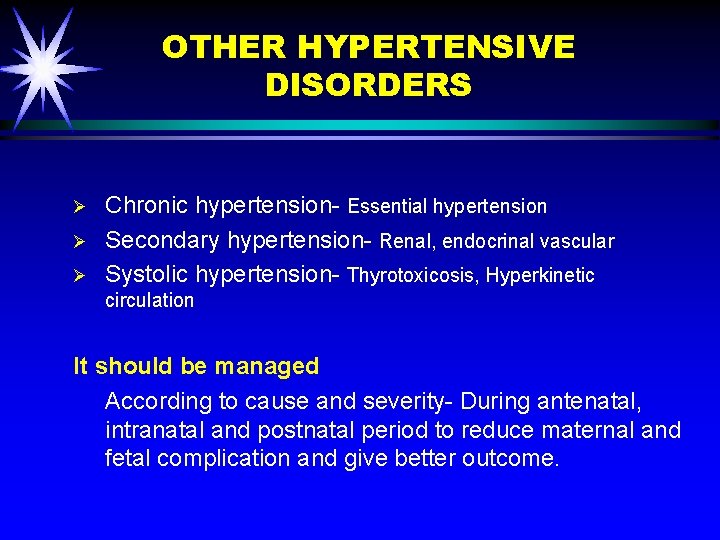
OTHER HYPERTENSIVE DISORDERS Ø Ø Ø Chronic hypertension- Essential hypertension Secondary hypertension- Renal, endocrinal vascular Systolic hypertension- Thyrotoxicosis, Hyperkinetic circulation It should be managed According to cause and severity- During antenatal, intranatal and postnatal period to reduce maternal and fetal complication and give better outcome.
CONCLUSION Severe manifestation and complication can be decreased by. Early detection Ø Meticulous antenatal care Ø Timely intervention. To reduce maternal and fetal morbidity and mortality and to get better outcome. Ø
Hypertensive Disorders Of Pregnancy Questionaire
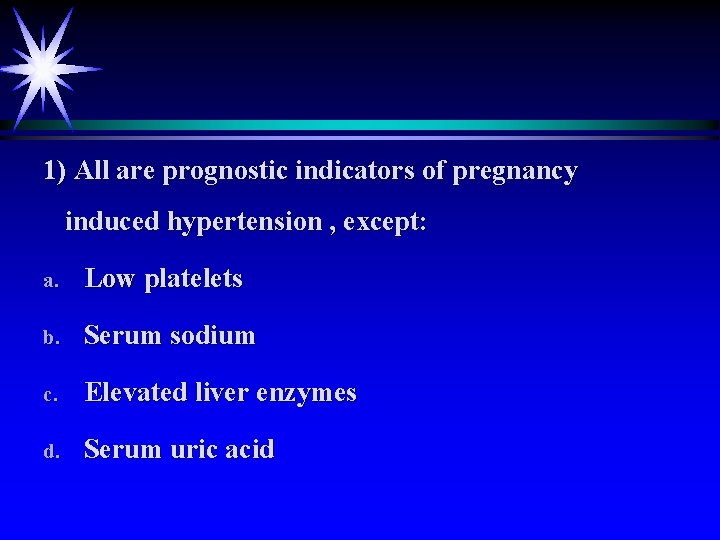
1) All are prognostic indicators of pregnancy induced hypertension , except: a. Low platelets b. Serum sodium c. Elevated liver enzymes d. Serum uric acid
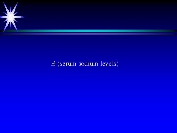
B (serum sodium levels)
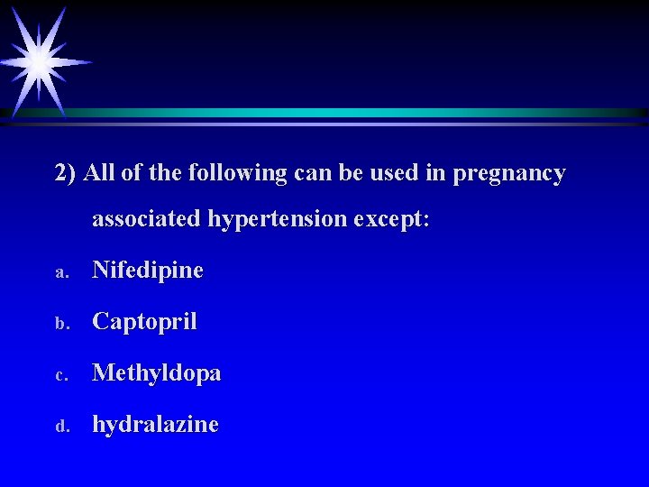
2) All of the following can be used in pregnancy associated hypertension except: a. Nifedipine b. Captopril c. Methyldopa d. hydralazine
B (captopril)
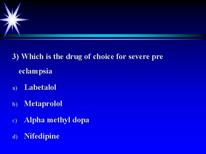
3) Which is the drug of choice for severe pre eclampsia a) Labetalol b) Metaprolol c) Alpha methyl dopa d) Nifedipine
A ( labetalol)
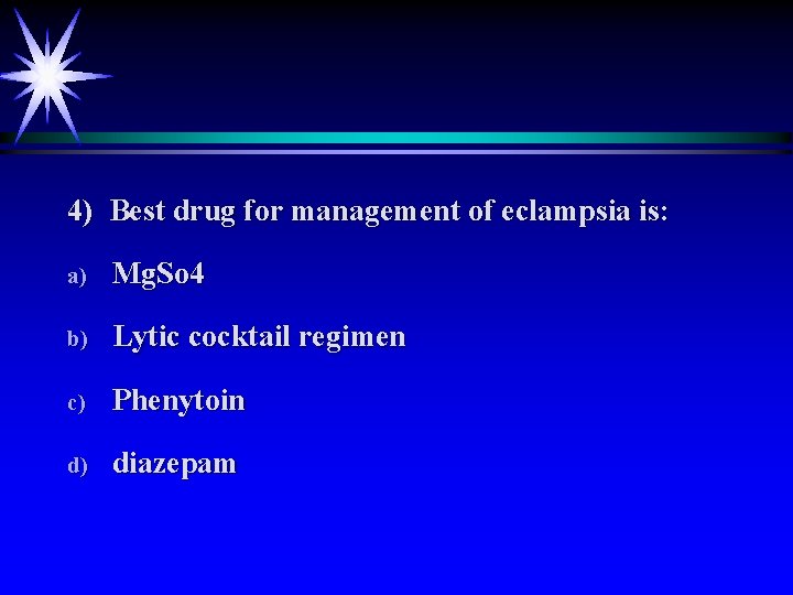
4) Best drug for management of eclampsia is: a) Mg. So 4 b) Lytic cocktail regimen c) Phenytoin d) diazepam
A (Mg. SO 4)
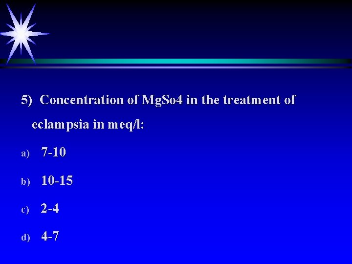
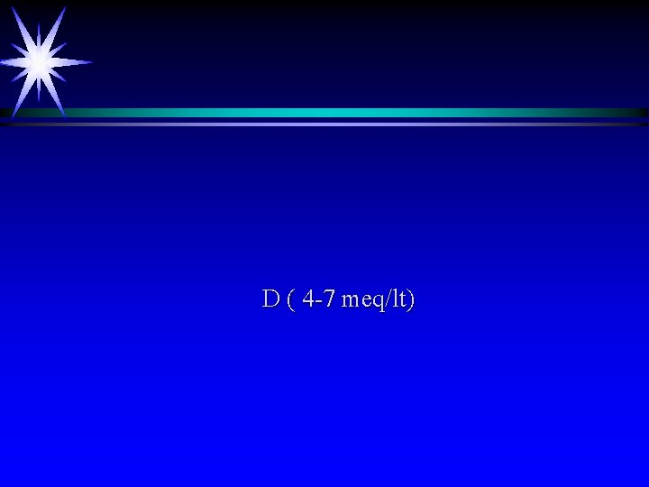
5) Concentration of Mg. So 4 in the treatment of eclampsia in meq/l: a) 7 -10 b) 10 -15 c) 2 -4 d) 4 -7
D ( 4 -7 meq/lt)
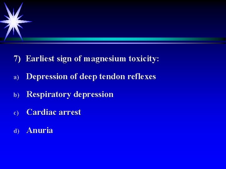
7) Earliest sign of magnesium toxicity: a) Depression of deep tendon reflexes b) Respiratory depression c) Cardiac arrest d) Anuria
A (depression of deep tendon reflexes)
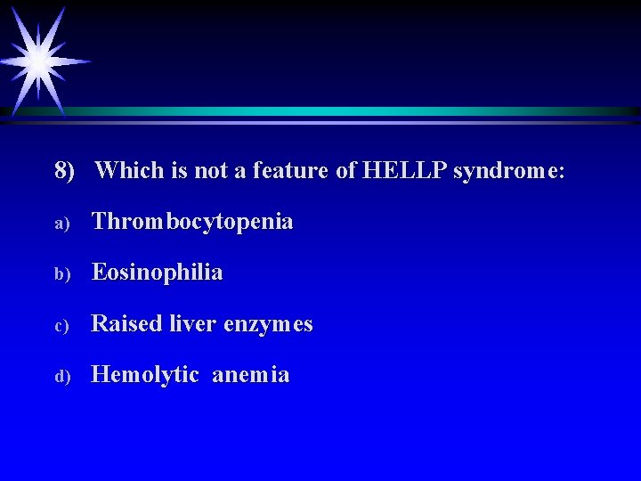
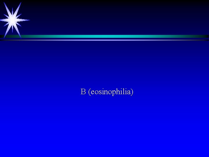
8) Which is not a feature of HELLP syndrome: a) Thrombocytopenia b) Eosinophilia c) Raised liver enzymes d) Hemolytic anemia
B (eosinophilia)
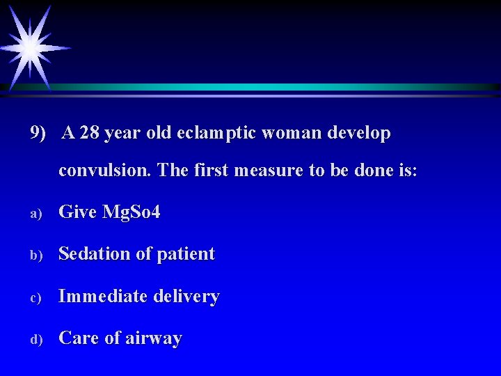
9) A 28 year old eclamptic woman develop convulsion. The first measure to be done is: a) Give Mg. So 4 b) Sedation of patient c) Immediate delivery d) Care of airway
D ( care of airway)
10) In severe pregnancy induced hypertension BP is a) 160/110 mm Hg b) 150/100 mm Hg c) 260/100 mm Hg d) 120/80 mm Hg
A ( 160/110 mm Hg)
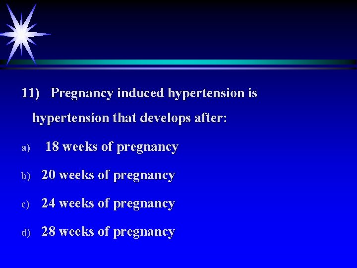
11) Pregnancy induced hypertension is hypertension that develops after: a) 18 weeks of pregnancy b) 20 weeks of pregnancy c) 24 weeks of pregnancy d) 28 weeks of pregnancy
B ( 20 weeks of pregnancy)
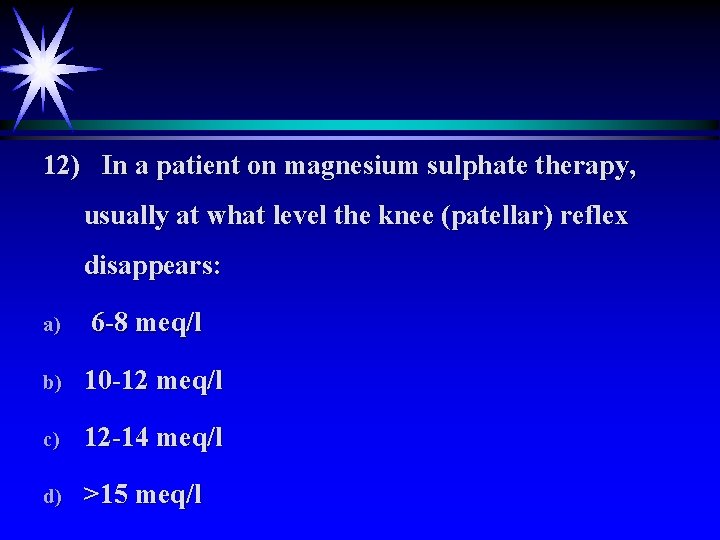
12) In a patient on magnesium sulphate therapy, usually at what level the knee (patellar) reflex disappears: a) 6 -8 meq/l b) 10 -12 meq/l c) 12 -14 meq/l d) >15 meq/l
B ( 10 -12 meq/lt)
THANK YOU