Pregnancy Development and Lactation Chapter 18 Species Development


")


- Slides: 64
Pregnancy, Development and Lactation Chapter 18
Species Development • Species can only be perpetuated if pregnancy, development of offspring and lactation are appropriately carried out.
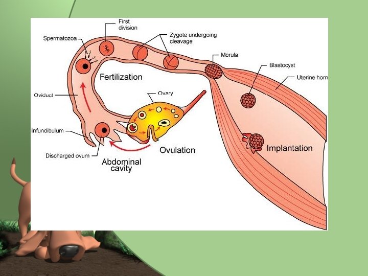
Fertilization • Copulation- the act of breeding allowed by the female during estrous (heat) period. – Usually in a mounted position • Intromission- the insertion of the penis into the vagina. • Ejaculation- when the semen is deposited in the upper portion of the vagina – Horse and pig deposit semen directly into uterus through open cervix.
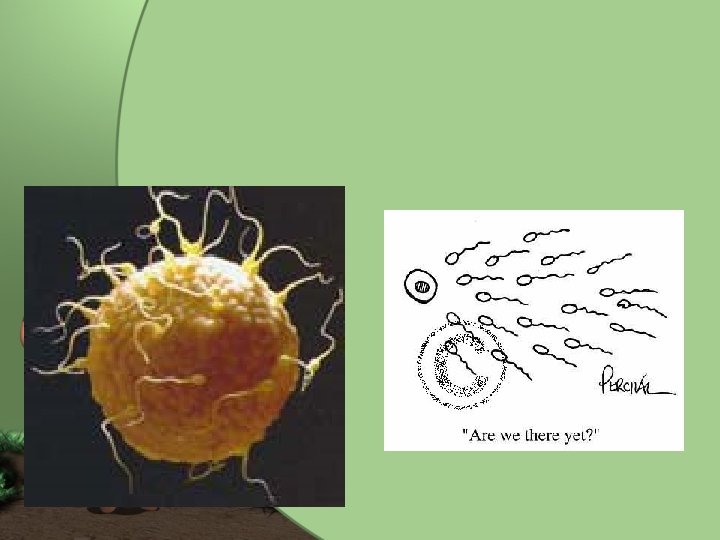
Transport of Spermatozoa • Start actively swimming as soon as deposited trying to make their way to oviducts. – Would take over an hour under own power – Are helped out by uterine contractions • Copulation causes posterior pituitary gland to release oxytocin which causes smooth muscle of reproductive tract to contract helping spermatozoa to the ovum.
Capacitation • Series of changes that spermatozoa undergo in the female reproductive tract to increase chances of fertilization. – Changes of ion movement through cell membranes – Increase in cell’s metabolic rates – Increase in rate of use of simple sugars for energy production. – Allows acrosome enzymes to be released.
Fertilization of the Ovum • Spermatozoa are programmed to seek out something large and round attempt to penetrate it. – Some try to fertilize non-ovum things. • Once ovum is found, many spermatozoa may swarm around it and start tunneling through the layers. – Process aided by enzymes of acrosome. • Once one spermatozoa penetrates ovum, change in membrane prevents any other sperm from entering.
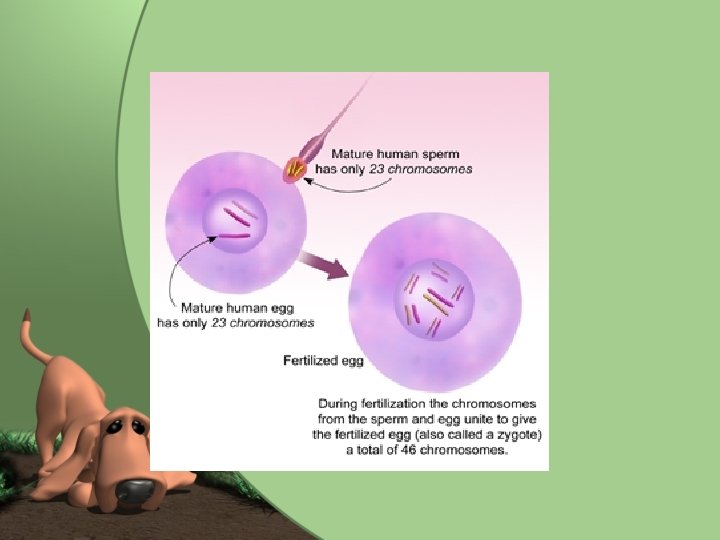
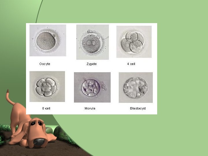
The Zygote • Once ovum is fertilized, it becomes a zygote. • Male pronucleus- the nucleus of the male spermatozoan immediately after fertilization. • Female pronucleus- the nucleus of the ovum immediately after fertilization. – Each pronucleus carries haploid number – Join together to get diploid number • This joining establishes genetic information for offspring.
Cleavage • The rapid division of the zygote once single nucleus has been established. • Cell divides rapidly but overall size remains the same because are dividing so quickly do not have time to grow. • Once zygote is a solid mass of cells, is in morula stage. • During this time, zygote is moving down ovum to uterus – Propelled by cilia and muscular contractions
Blastocyst • Cells of morula stage continued to divide and hollow cavity is formed in center of zygotic cell. • Once bump of cells on one side is developed, this is now the blastocyst.
Implantation • The means by which the blastocyst makes itself a home by attaching itself to the lining of the uterus (endometrium). • Once blastocyst comes to rest beside uterine lining, enzymes produced by blastocyst dissolve away some lining and implants itself into this pit in the lining. • Placenta begins to form as soon as implantation occurs. – Way of transporting oxygen and nutrients to blastocyst.
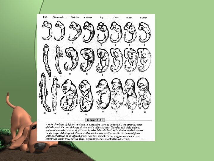
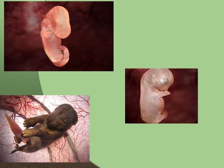
Terminology • Embryo-what developing offspring is referred to during early part of pregnancy. • Fetus- What developing offspring is referred to during later part of pregnancy.
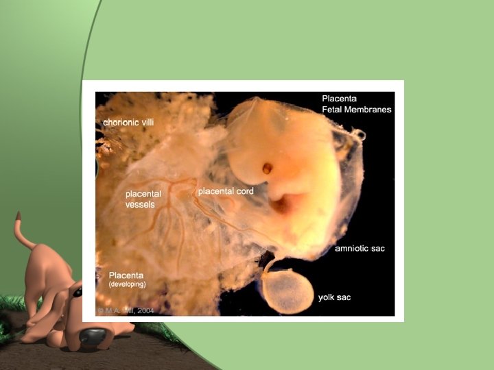
The Placenta • Life-support system of the developing fetus. • Fetus is a parasite on mother during pregnancy. • Grows along with fetus to enable for appropriate transfer of waste products, etc.
Structure of the Placenta • Multi-layered, fluid-filled membranous sac. • Develops around embryo and is connected to it by umbilical cord. – Smaller connections between outermost layer of placenta and lining of uterus. • This is where exchange of nutrients and wastes takes place. • Fetal and maternal blood does not mix but runs in close proximity to one another.
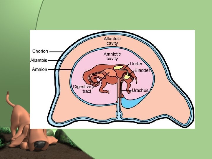
Structure of Placenta Continued… • 3 layers of placenta – 1. Amnion • Forms amniotic sac directly around fetus – 2. Allantois • Outside amniotic sac and forms allantoic sac. – 3. Chorion • Outside of allantoic sac and attaches to uterine lining. • Linked to fetus by umbilical cord.
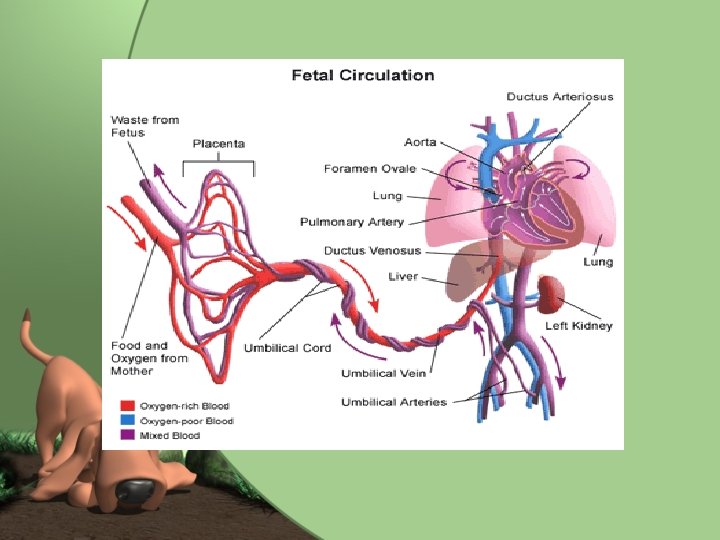
The umbilical cord • Link between the fetus and nutrient and waste exchange structures of the placenta. • Cordlike in structure – Contains umbilical arteries (2) and umbilical vein. • Umbilical arteries carry unoxygenated, waste filled blood from fetus to placenta • Umbilical vein carries nutrient and oxygen rich blood back from placenta to fetus. – Contains drainage tube from fetus’ urinary bladder (urachus) • Tube runs from the cranial tip of fetus’ bladder through umbilical cord to allantoic sac. • Fetus does not produce urine but do produce fluid that must be eliminated.
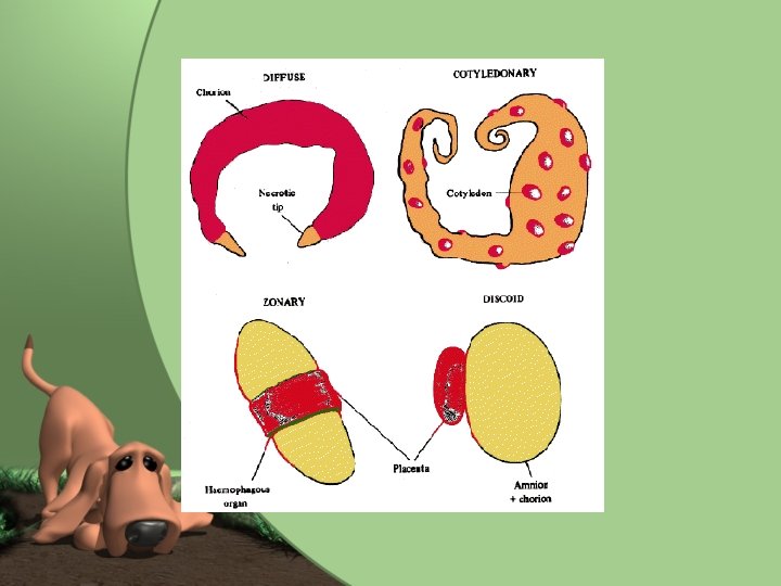
Attachment to the Uterus • Where chorion attaches to lining of the uterus. • Type of attachement varies among species and is one of four types: – 1. Diffuse Attachment – 2. Cotyledonary Attachment – 3. Zonary Attachment – 4. Discoid Attachment.
Diffuse Attachment • Means that attachment sites are spread diffusely over the whole surface of the placenta and the whole lining of the uterus. • No small, limited areas of attachment. • Found in pigs and horses • Detaches easily from uterine lining and is passed after the delivery of the newborn.
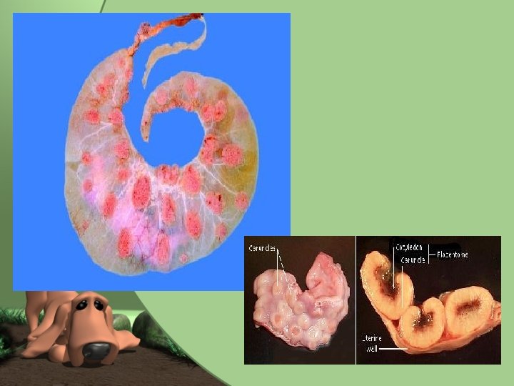
Cotyledonary Attachment • Most complicated type and is somewhat opposite of diffuse attachment. • Areas of attachment are small, separate, and numerous. • Placentome- attachment sites. • Cotyledon- area on surface of the placenta • Caruncle-area on surface of uterus (mushroom-like). – Cotyledon and caruncle interdigitate with one another. • Each placentome must separate completely for placenta to pass after birth. – If not completely passed, may be retained and can cause other problems.
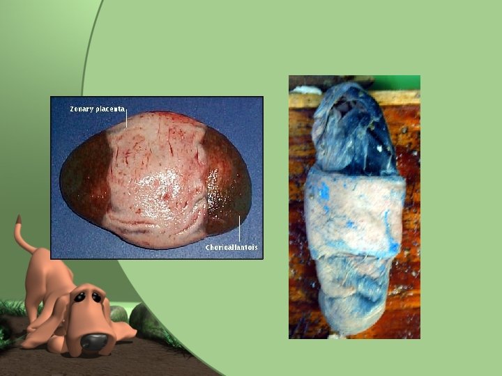
Zonary Attachment • Placenta attaches to the uterus in beltshaped area that encircles the placenta. • Found in dogs and cats • Detaches easily after delivery.
Discoid Attachment • Area of attachment between placenta and uterus is a single-disk shaped area. • Found in humans and other primates
Pregnancy • Also called gestation period • Time from implantation to delivery of the newborn. • Judge time by time since fertlization. • Is divided into three segments called trimesters – 1 st- period of the embryo implanting and organizing and placental development – 2 nd- embryo now called fetus and is fetal development period. Parts are taking shape and differentiating. – 3 rd- fetal growth. All parts grow dramatically
Gestation Periods Species • Cat • Dog • Cow • Elephant • Ferret • Goat, Sheep • Hamster • Horse • Human • Pig • Rabbit Approx Gestation Period 2 mo (56 -69 days) 2 mo (59 -68 days) 9 mo (271 -291 days) 21 mo (615 -650 days) 6 w (42 days) 5 mo (143 -155 days) 3 w (19 -20 days) 11 mo (321 -346 days) 9 mo (280 days) 3 mo, 3 w, 3 d (110 -116 days) 1 mo (30 -32 days)
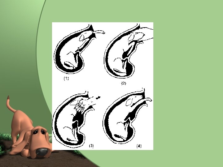
Parturition • • Birth process Lungs of newborn become functional Goes from parasite to independent being. Parturition is triggered by size and weight of uterus and changing hormone levels. – Progesterone of dam declines – Progesterone has kept myometrium from contracting – Increased levels of glucocorticoid hormones stimulate rise in estrogen levels – These increase sensitivity to oxytocin, released from posterior pituitary gland. • Oxytocin stimulates contractions which starts labor process
3 Stages of Labor • 1. Uterine Contractions – Presses fetus against uterus – Causes cervix to gradually dilate – Dam may appear restless in this stage. • 2. Delivery of the Newborn – Combination of uterine and abdominal muscle contractions – “Water” or amniotic and allantoic sacs rupture. • 3. Delivery of the placenta – Placenta separates from wall of the uterus and is expelled by uterine contractions. – Dam often eats the placenta.
Labor Continued • Multiparous animals: the second and third stages of parturition intermix with one another. – Newborn and placenta are delivered alternatively. – Next newborn will not be delivered typically until previous placenta has been expelled.
Parturition: normal presentation
Dystocia • “difficult birth” • Most common cause is that fetus is too large to pass or is in wrong orientation for delivery. • May have to Repell the fetus or deliver through Cesarean section. • If fetus is dead, may have to be removed in segments- called Embryotomy.
Parturition: abnormal presentations
Delivery
Placental Delivery
Whelping/ Parturition Normal Presentation of two sacs
Involution of the Uterus • After parturition is complete, uterus gradually returns to nonpregnant size. • Process is called involution. • At placental attachment sites, endometrium sloughs into uterus and areas heal over. • Myometrium contractions continue slowly, pushing contents through birth canal. • Will pass from bright red blood, to dead tissue over course of weeks to about a month.
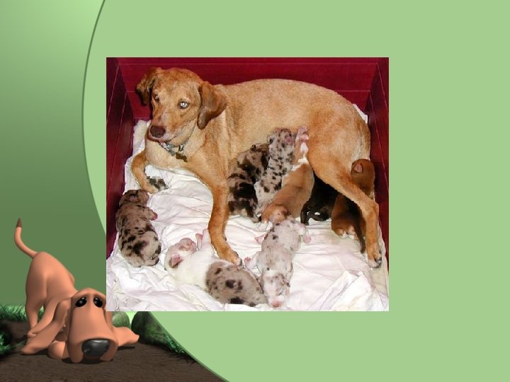
Mammary Glands and Lactation • Play an important role during neonatal period. • Mammary glands are specialized skin glands. • Produce colostrum and milk which are crucial to early life. • Present in both males and females – Females secrete appropriate hormones for them to become functional
Species Differences • Number, size and location varies from animal to animal. • Cattle, sheep and goats only have one opening per teat • Dogs have up to 20 openings per nipple.
Number of Mammary Glands for Common animals SPECIES Cats Horses NUMBER OF GLANDS 10 2 Dogs Humans 10 2 Cattle Pigs 4 14
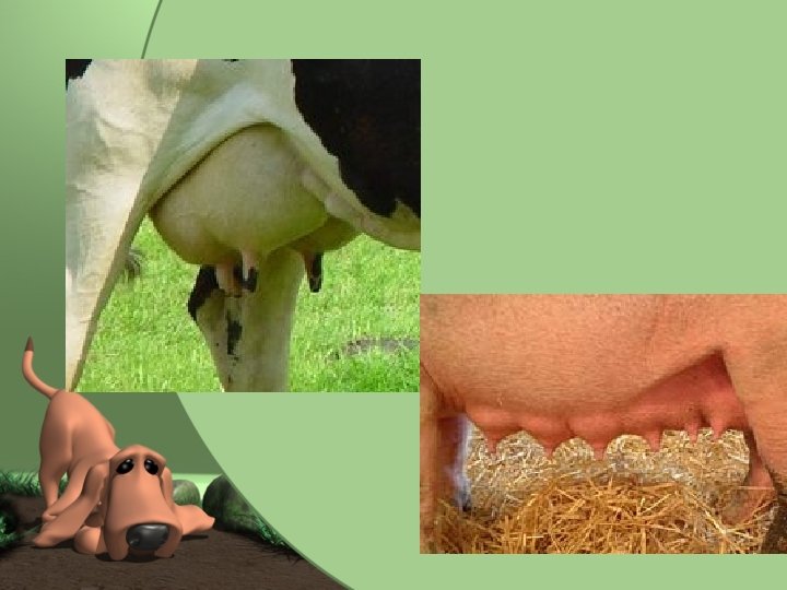
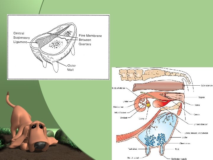
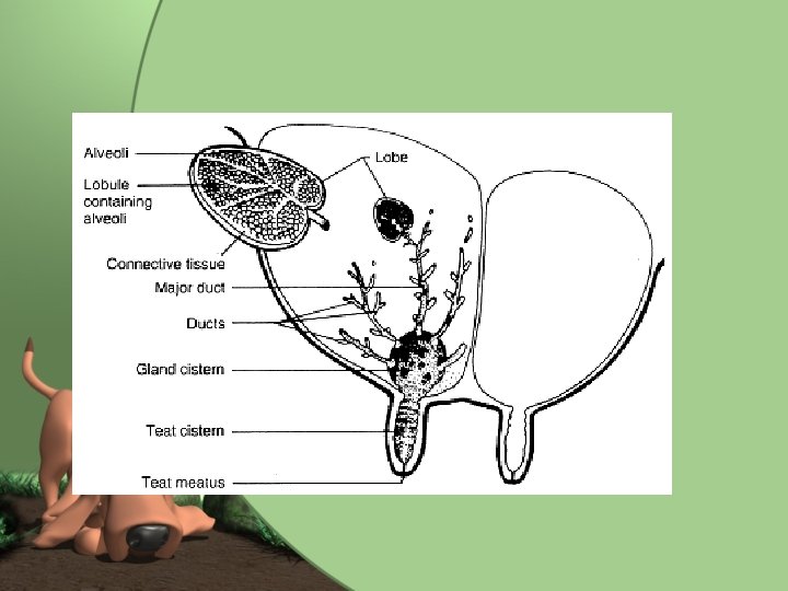
Udder of the Cow • Udder is term used for mammary glands. • Exaggerated in size, but composition is similar to other animal’s mammary glands • Four mammary glands (quarters) • Quarters are completely separate units from each other • Each quarter has its own milk-secreting systems and ducts leading down to separate teats • Suspended by strong suspensory ligaments that allow it to stretch. – Acts as a shock absorber.
Udder Continued • Mastitis- infection of the mammary gland – Since are separate, is unlikely to spread from one quarter to another. – Can spread through bloodstream.
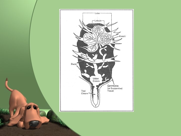
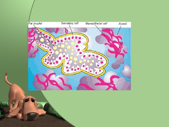
Alveoli and Duct System • Alveoli- milk-secreting units of the mammary gland • Alveolar duct- alveoli secrete milk into tube – Similar in make up to Alveoli found in lungs. • Ducts empty into large space called gland sinus which is continuous with the teat sinus which is where milk is extracted by suckling young. • Tip of teat has streak canal- passageway from teat sinus to outside. – Surrounded by elastic fibers and ringlike sphincter muscle that keeps it closed to prevent leakage.
Mammary Gland Development • Mammary glands develop in response to hormones produced at puberty – Prolactin and Growth Hormone directly encourage mammary gland development – Estrogen and progesterone encourage mammary alveoli and duct systems to develop • Influenced by FSH and LH on ovaries – Certain drugs may inhibit normal mammary gland development
Lactation • Process of milk production • Begins at end of pregnancy and is obvious at time of parturition. • Prolactin and Growth Hormone from anterior pituitary gland hormones from adrenal cortex are involved with the starting of lactation.
Colostrum • “First milk” or “premilk”. • Contains large amounts of proteins, lipids, and amino acids than milk and high levels of essential vitamins. – Antibodies for defense • Supplies important nutrients and defenses that newborn can not receive elsewhere. – If does not receive within first few hours, body can no longer process appropriately. • Has laxative effect to clear meconium ( first feces) from newborn’s intestinal tract. • Involved with passive immunity from dam to newborn • Those without appropriate colostrum tend to be weaker and do not grow as rapidly. • Why we wait for vaccines.
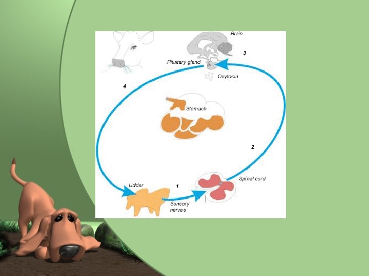
Maintenance and Lactation • Lactation continues as long as mammary gland is emptied regularly. • Physical stimulation of nipple. • This sends nerve impulses to brain which continues stimulation of appropriate hormones for milk production. • When nursing stops, signal stops. – Will lead to involution of the mammary gland or “drying up”.
Milk Letdown • Immediate effect of nursing or milking • Milk accumulates high up in mammary gland in alveoli and small ducts and does not move down until Milk letdown occurs. • Oxytocin causes myoepithelial cells around the alveoli and small ducts to contract – Squeezes milk down the large ducts and sinuses. – Can take from few seconds to minute for milk to flow freely.