Flagellates v Intestinal flagellates v Genital flagellates v

 (人內滴蟲) 恩特若姆那斯鞭毛蟲")


 Common infection in both tropic and sub-tropic area")

 2 oval nuclei; 4")
 4")
 Vomiting 嘔吐")


 hydrochloride(鹽酸奎那克林) 5 mg/kg/day in divided doses thrice daily for")


 resulting in a frothy and creamy white discharge")
 trophozoites Semen or prostatic fluid examination (man) Artificial")


內臟利什曼原蟲症 India, Bangladesh, scattered areas in")


 Single mitochondrian Extends along the whole length of the parasite Reveals")


內臟利什曼原蟲症: Kala-azar黑熱病 Incubation period is 4 to 10 months Early")
皮下利什曼原蟲症 Starts as a painless papule(丘疹) on exposed parts of")


 Pathogens:Trypanosoma rhodesiense 羅德西亞錐蟲 Trypanosoma gambiense 甘比亞錐蟲 (morphologically indistinguishable) Vector:biting flies;")


 0. 2 g intravenously injection")
 cells and protein levels higher than")

 Geographical distribution South America Central America Vectors Reduviid bugs: Triatoma,")


 Flagellum鞭毛 –roma An")


- Slides: 84
Flagellates 鞭毛蟲 v Intestinal flagellates v Genital flagellates v Haemoflagellates
Intestinal and genital flagellates Intestinal flagellates 腸道鞭毛蟲 • Enteromonas hominis; (non pathogenic) (人內滴蟲) 恩特若姆那斯鞭毛蟲 • Retortamonas intestinalis; 腸旋滴蟲 (non pathogenic) • Chilomastix mesnili脣形鞭毛蟲 • Giardia lamlbia梨形鞭毛蟲 • Trichomonas hominis (large intestine) 腸道鞭毛滴蟲 • Trichomonas tenax 口腔毛滴蟲(mouth) • Pentatrichomonas hominis 人毛滴蟲
Intestinal and genital flagellates Genital flagellates 生殖道鞭毛蟲 * Trichomonas vaginalis 陰道鞭毛蟲
Chilomastix mesnili 麥氏唇形鞭毛蟲 Non-pathogenic Large intestine Life cycle Trophozoite: long cytostome 3 anterior flagella 1 cytostomal flagellum Cyst: single nucleus lemon-shape p. 30 fig. 21
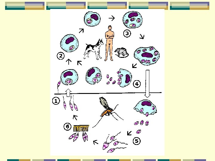
Giarida lamblis 梨形鞭毛蟲 (Syn. G. duodenalis) Common infection in both tropic and sub-tropic area Small intestine infection Cystic infection, 10 cysts may produce infection Infection via contaminated food and water Excystation occurs in upper small intestine
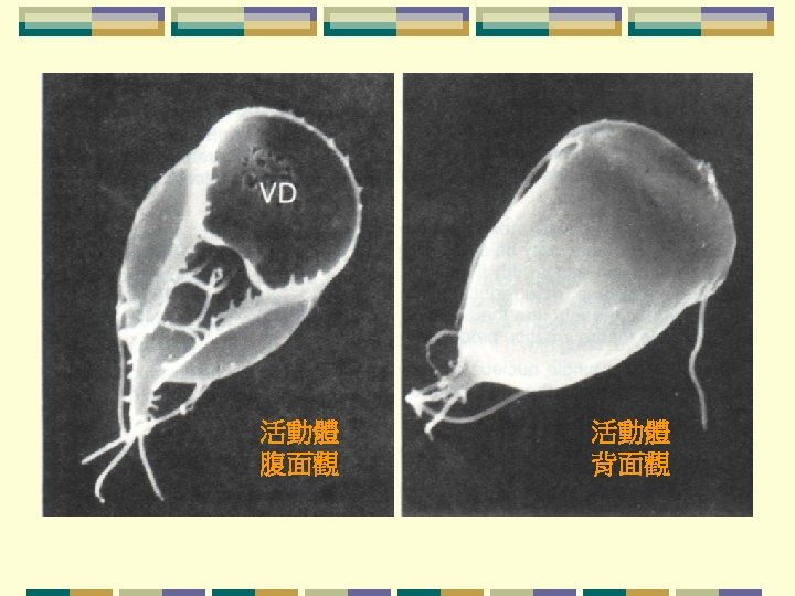
Ventral disc Ventro lateral flange
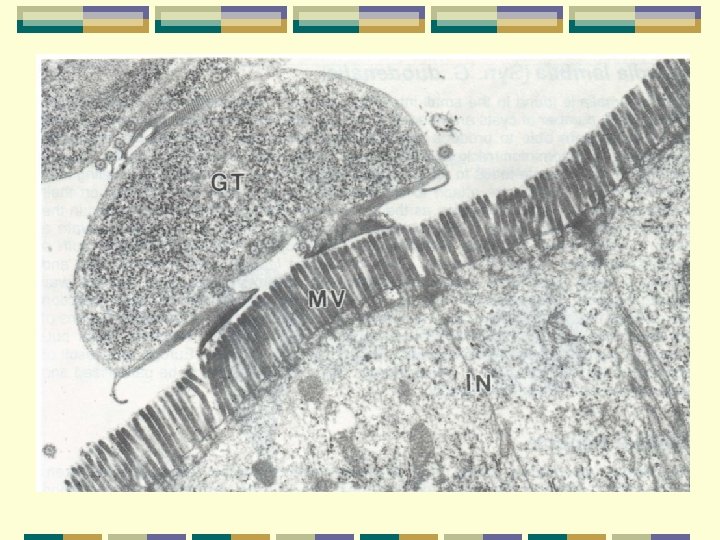
Giarida lamblis Life cycle Trophozoite: symmetrical in shape (badminton racket) 2 oval nuclei; 4 pairs of flagella; moving as “falling leaf” binding to tissue by striated disc (ventral surface) mechanical binding by host proteases activate the lectin of Giardia to promote attachment to enterocytes (腸細胞) multiple by binary fission
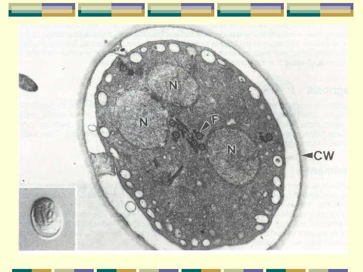
Giarida lamblis Life cycle Cyst: oval to ellipsoid in shape (11 -14 μm) 4 small oval nuclei distinct wall; shrunk cytoplasm produce a space under cyst wall
Giarida lamblis Clinical aspects Wide range of gastrointestinal symptoms (especial in children) Vomiting 嘔吐 Flatulence 腸胃脹氣; Diarrhoea 痢疾, 下痢 Malabsorption syndrome 吸收障礙 Cholecystitis 膽囊炎
Giarida lamblis Clinical aspects Histological aspects : mechanical & biochemical Shortening of villi Cellular infiltration of lamnia propria黏膜固有層 of mucous membrane functional impairment of enterocytes Abdominal cramps腹部絞痛 痙攣 Induce motility disturbance
Giarida lamblis Diagnosis Stool examination for trophozoites or cysts diagnosis may concentrated by zinc sulphate flotation硫酸鋅浮漂法 Duodenal aspiration “Enterotest” capsule腸內測試膠囊 a gelatin capsule containing a coiled thread for duodenal contents diagnosis
Giarida lamblis Treatment Mepacrine (quinacrine) hydrochloride(鹽酸奎那克林) 5 mg/kg/day in divided doses thrice daily for 1 week Nitroimidazoles Tinidazole 2 g single dose Albendazole
Giarida lamblis Prevention and control Food hygiene Fly control Sewage disposal Proper water supply Identification and treatment of carriers Chlorination of water can not kill the cysts Iodination of water can kill the cysts (13 ml saturated solution of iodine per liter of water
Trichomonas spp 滴蟲 Life cycle Trophozoite: ovoid or pyriform gliding motion 1 or 2 oval nuclei 4 flagella on anterior end 1 flagellum turns back and attach on undulating membrane axostyle軸柱 projects out of body for attach to host tissues and cause irritation divide by binary fission No cystic stage
Trichomonas hominis人毛滴蟲 Parasite in large intestine No medical significance
Trichomonas tenax口腔毛滴蟲 Parasite in the mouth No medical significance
Trichomonas vaginalis 陰道滴蟲 Urogenital infection No cystic stage Infected male acting as carrier
Trichomonas vaginalis Life cycle Trophozoite: ovoid or pyriform gliding motion single oval nuclei 4 flagella on anterior end 1 flagellum turns back and attach on undulating membrane axostyle projects out of body for attach to host tissues and cause irritation ingests food particles by cytostome Divide by binary fission
Trichomonas vaginalis Clinical aspects Vaginitis (陰道炎) resulting in a frothy and creamy white discharge Inflame of vulva and cervix Asymptomatic in males but some urethritis(尿道炎) or (prostatitis)前列腺炎
Trichomonas vaginalis Diagnosis Vaginal secretion (woman) trophozoites Semen or prostatic fluid examination (man) Artificial culture
Trichomonas vaginalis Treatment infected female and her male consort Drug treatment Nitroimidazoles Metronidazole; 200 mg thrice daily orally for 1 week 2 g for single doses Tinidazole ; 2 g thrice daily orally Ornidazole;
Trichomonas vaginalis Prevention and control Normal sexual behavior Fixed sexual partner Safety sexual behavior
Haemoflagellates 血液性鞭毛蟲 Leishmania spp 利什曼原蟲 L. donovani complex; L. tropica; L. mexicana complex L. braziliensis complex; L. major L. aethiopica L. peruviana Trypanosoma spp 錐蟲 T. rhodesiense; T. cruzi; T. gambiense T. rangeli
Leishmania spp infecting man Geographical distribution Visceral leishmaniasis (VL)內臟利什曼原蟲症 India, Bangladesh, scattered areas in the Middle East Mediterranean region, parts of East Africa, South and Central America Cutaneous leishmaniasis (CL)皮下利什曼原蟲症 N. India, Pakistan, Middle East, Southern Europe Northern Africa all Mediterranean Coast, parts of East and West Africa, South and Central America Muco-cutaneous leishmaniasis (MCL)黏膜利什曼原蟲症 South and Central America
Leishmania spp infecting man Taxonomic position is often confusing monoclonal antibody typing DNA probe hybidization RFLP karyotyping L. donovani complex; L. major L. tropica; 熱帶利什曼原蟲 L. aethiopica L. mexicana complex L. braziliensis complex; L. peruviana
General Classification of Important Leishmaniasis in Relation to Clinical Manifestation Species Involved Clinical Manifestation Old world New world VL L. donovani complex MCL Rarely occurs L. braziliensis complex CL L. tropica L. braziliensis complex L. major L. mexicana complex
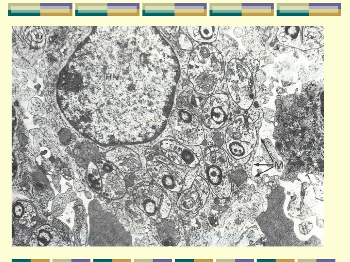
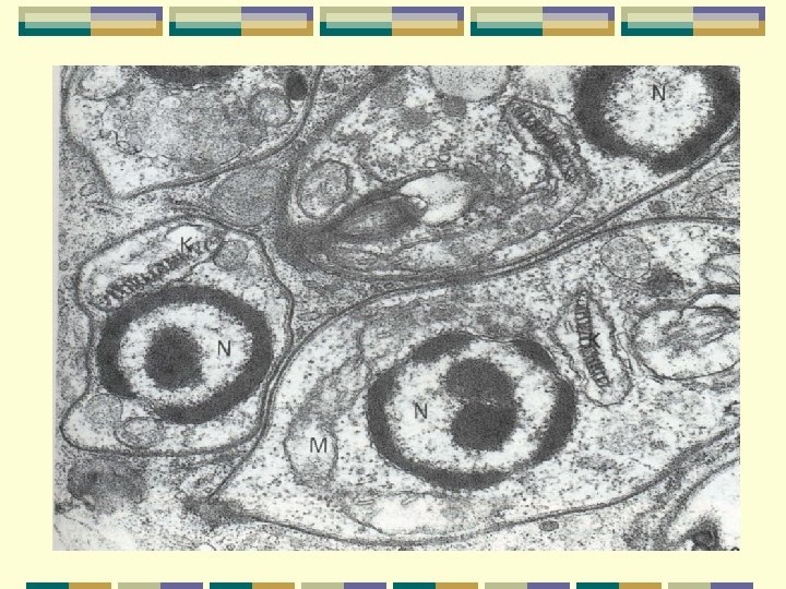
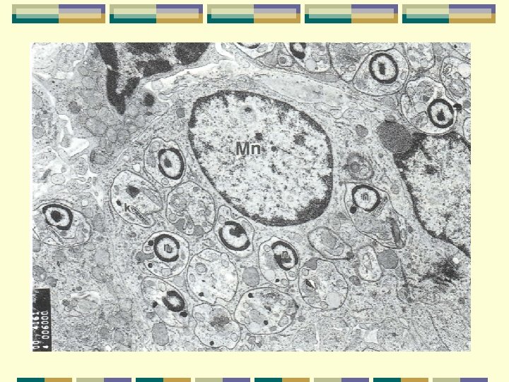
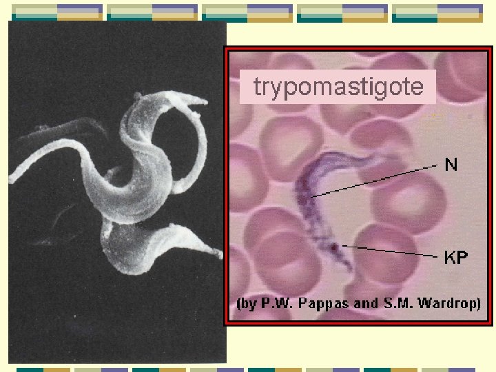
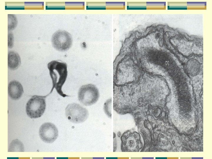
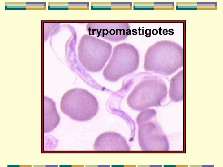
Leishmania spp Morphology 4 types fig. 30 Trypomastigote 錐鞭毛體 kinetoplast is at posterior end, locomotory flagellum, long undulating membrane(supported by microtubules), Epimastigote側鞭毛體 kinetoplast is anterior to the nucleus, short U membrane Promastigote前鞭毛體 kinetoplast is at anterior end, no U membrane Amastigote無鞭毛體(“Leishman-Donovan” body) ovoid, 2 -3 μm, no flagellum binary fission, starts by division of the kinetoplast, following nucleus division, ending the outer membrane splits
Leishmania spp Kinetoplast(動基體) Single mitochondrian Extends along the whole length of the parasite Reveals many cristae in the midgut of vector high degree of activity Relatively less cristae in blood stream activity is suppressed
Distribution of morphological types in haemoflagellates Trypomastigote Lesimania spp. Animal host Vector Trypanosoma Animal spp. (Afican) host Vector Trypanosoma Animal cruzi host Vector + + Epimastigote + + + Promastigote + - Amastigote + + -
Life cycle Vectors: Sandflies白蛉 Phlebotomus Lutzomyia Psychodopygus Western hemisphere : Lutzomyia ; Psychodopygus World wild: Phlebotomus Female sandfly pool feeder ; biting by proboscis or mouth part
Host cell infection by promastigotes flagellar attachment or aflagellar poles phagocytosis by host cells Macrophages Phagocytic cells Become amastigotes inside a parasitophorou vacuole Binary fission for multiplication 1. eliciting a small phagocytic oxidative response 2. possessing membrane-associated acid phosphotase 3. Possessing more catalase & glutathione peroxidase than promastigotes Toxoplasma gondii : preventing phagosome-lysosome fusion
Leishmania spp infecting man Clinical aspects Difference in virulence of various parasite species Difference in susceptible of various host Muco-cutaneous leishmaniasis (MCL)黏膜利什曼原蟲症 Starts as a pustular swelling in mouth or on nostrils Lesion become ulcerative after many months Then extend into the naso-pharyngeal mucous membrane 2 nd infection is very common with destruction of the nasal cartilage and the facial bone
Leishmaniasis利什曼原蟲症
Leishmania spp Visceral leishmaniasis (VL)內臟利什曼原蟲症: Kala-azar黑熱病 Incubation period is 4 to 10 months Early symptoms: low grade fever with malaise and sweating Later stages: fever become intermittent hyperplasia of lymphoid-macrophage system liver and spleen become grossly enlarged no inflammatory changes lymphadenitis is common in Chinese form but not Indian form post Kala-azar dermal lieshmaniasis (PKDL) after 1 – 2 years hypopigmented areas; nodular as lepromatous leprosy skin biopsy shows many parasites PKDL is of epidemiological significance vector transmission easily
Leishmania spp Cutaneous leishmaniasis (CL)皮下利什曼原蟲症 Starts as a painless papule(丘疹) on exposed parts of body generally on the face Circular or oval ulcer produce Lesion ulcerates after few months dry type lesion: ulcer remains dry and heals readily wet type lesion: ulcer may spread with inflammatory zone around it, and heal slowly Diffuse cutaneous leishmaniasis (DCL) occur in parts of Africa Nodules and thickening of the skin without any ulceration Leishmaniasis recidivans (LR): chronic infection with very few parasites, not heal spontaneously
Leishmania spp Diagnosis Low WBC count Anaemia Chronic symptom gama-globulin coagulation: formaldehyde Immuno-assay Biopsy tissues: bone marrow, spleen; lymph gland Culture
Leishmania spp Treatment Visceral & Muco-cutaneous leishmaniasis Pentavalent antimony compounds sodium stibogluconate Diamidines pentamidine isethionate; hydroxystilbamidine Cutaneous leishmaniasis sodium stibogluconate or mepacrine
Leishmania spp Prevention and control Vector control Residual insecticides spread for control sandflies High susceptible to insecticide of sandflies Reservoir animal control Vaccination with promastigotes in cutaneous leishmaniasis U. S. S. R and Israel Personal protection long sleeves and trousers; insect repellents; fine mesh nets impregnated with insecticides
Trypanosoma spp infecting man Sleeping sickness非洲睡眠病 : Africa Trypanosoma rhodesiense羅德西亞錐蟲 Trypanosoma gambiense甘比亞錐蟲 Chagas’ disease : America Trypanosoma cruzi枯西式錐蟲 Non pathogenic : Central and South Africa Trypanosoma rangeli
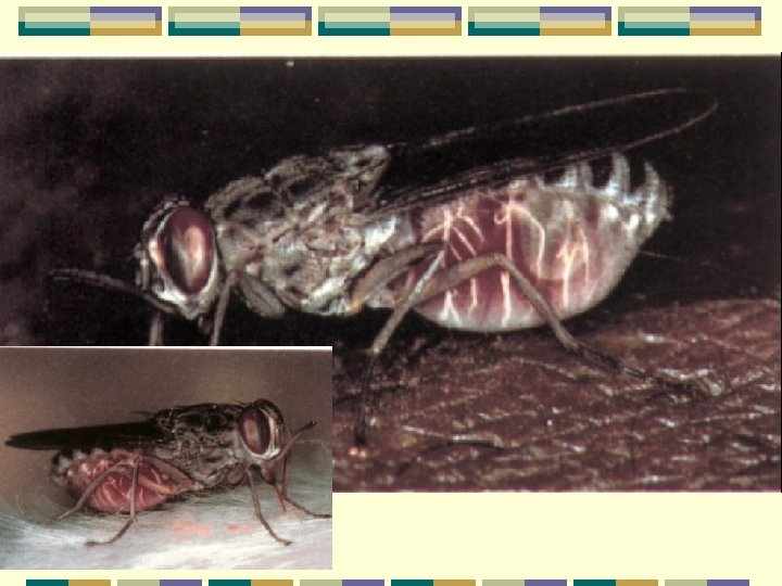
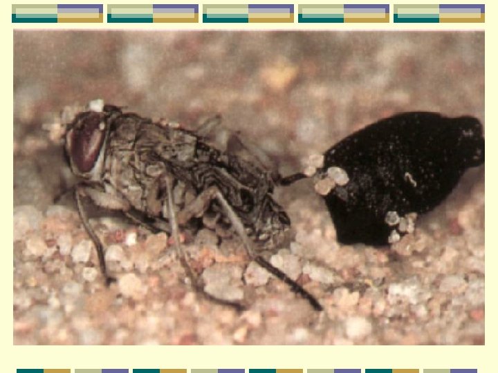
African trypanosomes (sleeping sickness) Pathogens:Trypanosoma rhodesiense 羅德西亞錐蟲 Trypanosoma gambiense 甘比亞錐蟲 (morphologically indistinguishable) Vector:biting flies; tsetse fly 采采蠅; Glossina parasite undergoes a complex development in fly Host:host in human blood with binary fission movement in the direction of flagellum pleomorphic; slender; intermediate; stumpy nucleus, kinetoplast, undulating membrane, flagellum can be recognised with stain
Geographical distribution Trypanosoma rhodesiense 羅德西亞錐蟲 East Africa Rhodesian form sleeping sickness; zoonosis Trypanosoma gambiense 甘比亞錐蟲 Western and Central Africa Gambian form sleeping sickness: human
Life cycle Page 46 fig. 36 Host:divided by binary fission slender; intermediate; stumpy Vector:stumpy form initiated in midgut surrounded with PM (peritrophic membrane ) Change to elongated form (longer) Migrate to the space between PM and midgut, become a shorter elongated form Penetrate PM and forward movement to proboscis Turn back migrate to salivary gland, become a epimastigotes further develop to metacyclic form Infect host via biting injection with saliva may be penetrate into haemocoel via midgut
Antigenic variation抗原變異作用 Fascination mechanism for escape host immunity Avoid host immune response Protecting the parasites from antibodies Change and replacing outer variant surface glycoprotein (VSG) Weekly or 10 -day cycle Antigenically different Serotype; heterotype Limitation : unlimitation ?
Clinical Aspects Primary reaction 初始感染反應 Small sub-cutaneous nodule 皮下瘤結 occurs at the site of inoculation of Tyrpanosoma become a larger nodule, 25 to 100 mm in diameter persist about 2 -3 weeks
Clinical Aspects Systemic manifestation 系統性病變 Fever with headache Winterbottom’s sign: T. gambiense lymph gland become enlarged (cervical & suboccipital) Fever becomes intermittent in later stages CNS involvement daytime sleeping; psychological changes; tremors震顫; convulsions全身痙欒; coma昏迷 Death generally occurs from intercurrent infection併發感染
Clinical Aspects T. rhodesiense infection fast with CNS involvement occurring within a few months Pathology of CNS leptomeningitis軟腦膜炎 cellular infiltration組織浸潤 : mononuclear cells perivascular cuffing圍管現象 : around the blood vessels plasma cells and lymphocytes
Diagnosis • Lymph gland puncture: T. gambiense • Blood diagnosis: T. rhodesiense • DEAE column separation: parasites concentration form blood • Immuno-assay: CFT; complement fixation test • FAT; fluorescence antibody test • Ig. M level rise: blood & CNS (trypanosomiasis’ pathognomonic) • Rats inoculation with T. rhodesiense : heavy parasitaemia • Card agglutination trypanosomasis test (CATT) •
Differences Between T. gambiense and T. rhodesiense T. gambiense T. rhodesiense Virulence Less virulent to humans and laboratory animals More virulent to humans and laboratory animals Reservoir Mainly humans Mainly animals Vector Mainly Glossina palpalis Mainly Gglossina morsitans Geographical distribution Mainly West Africa Mainly East Africa
Treatment Depending on CNS condition Early stage: suramin (germanin) 0. 2 g intravenously injection for side reaction test 0. 8 g apply on day 2 1 g every 4 th day until a total 10 g is given • Blood-brain barrier effect; BBB • Enphrotoxic (kidney failure patient avoid use)
Treatment Late stage: melarsoprol with dimercaprol (reduce toxicity) cells and protein levels higher than 40 mg/100 ml 20 mg/kg; intravenously injection; 3 course treatment each course 3 days; separated by 7 days rest Side effect: stop to rescue Encephalopathy : arsenical encephalopathy 砷腦病變 reactive encephlopathy 反應性腦病變 Eflornithine : early Gamabian trypanosomiasis
Prevention and control Gambian form sleeping sickness: human Riverine tsetse : for collecting water or for washing people Rhodesian form sleeping sickness; zoonosis Reservoir host reducing Wild game destruction Vector control : tsetse fly elimination color attract: blue & black residues application of insecticide
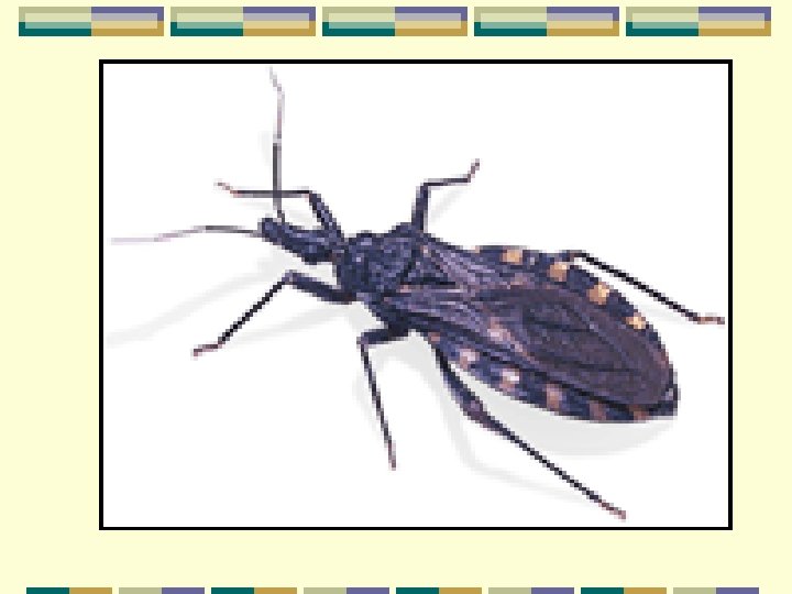
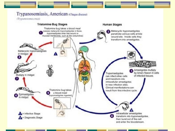
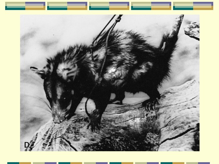
American trypanosomes (Chagas’ disease) Geographical distribution South America Central America Vectors Reduviid bugs: Triatoma, Panstrongylus. Rhodnius Reservoir animal Armadillos犰狳 , opossums小袋鼠, cats, dogs, pigs
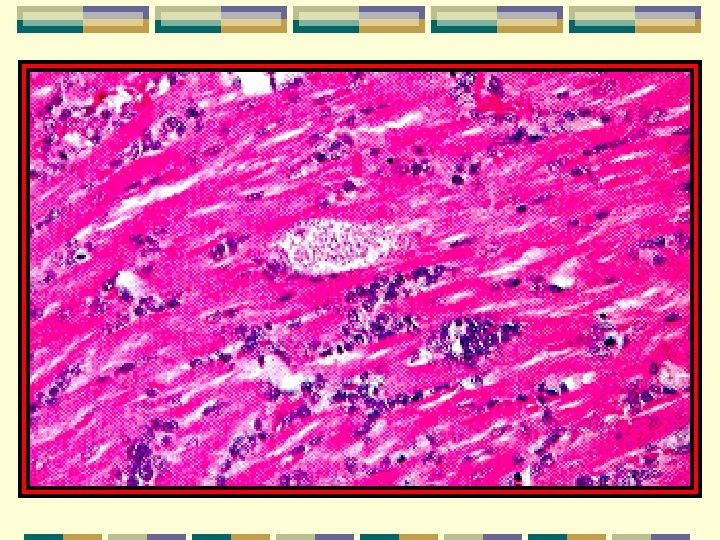
Life cycle of Trypanosoma cruzi Metacyclic form is infectious form Pass through the faeces of infected vector Parasites via skin damaged by the bite of assassin bug via the contamination mucous membranes Penetrate into tissue cells from blood stream mesenchymal origin間質細胞 cardiac muscle mainly 心肌細胞 Change into amastigote , epimastigote, trypomastigote Replication only occur in amastigote Trypomastigote emerge form muscle cells and enter blood stream Re-invasion : cardiac muscle cells circulate without division
Life cycle of Trypanosoma cruzi Vector Trypomastigote picked up by insect Change to epimastigotes Becoming metacyclic form after 8 -10 days in the hindgut of vector T. cruzi also transmissable by the congenital route or blood transfusion Large kinetoplast and curved appearance distinguish with T. gambiense & T. rhodesiense Congenitial route or blood transfusion infection
Life cycle of Trypanosoma cruzi T. Cruzi survive in the cytoplasm of macrophages not in the parasitophorous vascuole of macrophages (Leishmania & Toxoplasma)
Clinical aspects Chagoma as primary lesion: on the face near eyelids swelling of the eye; conjunctivitis; Romans’s sign Chronic disease is cardiomyopathy mild case : extra systole心收縮 slight tachycardia心悸, 心搏過速 Severe case: heart block partially or completely cardiac failure Some case: megaoesophagus巨食管; dysphagia 吞嚥困難 megacolon巨結腸
Diagnosis Blood examination: isolation of T. cruzi from blood direct examination; centrifugation help parasites culture; NNN medium rats inoculation; 10 days: blood diagnosis 60 days: cardiac tissues examined (amastigotes) Xenodiagnosis: uninfected laboratory-breed reduviid bugs 動物接種診斷法feeding on patient; 2 weeks inoculation examined for epimastigotes Serological diagnosis: indirect haemagglutination test FAT
Amastigotes in the heart muscle Pseudocyst
Epimastigote
Trypanosoma cruzi Treatment Effective chemotherapy is not available nitrofurans pyrimethamine primaquine nitroimidazoles allopurinol riboside Symptomatic treatment for cardiac failure by standard drugs Pace-maker implantation Surgery in megaoesophagus and megacolon cases
Trypanosoma cruzi Prevention and control Vector control successful in Brazil Urbanization: vector habitat elimination Gentian violet 龍膽紫 (1: 4000) preventing blood (donor) transfusion infection
Flagellates 鞭毛蟲 Terms Used in Relation to Flagellates(Intestinal, Genital and Blood) Flagellum鞭毛 –roma An elongated, hair-like organelle used for locomotion. At the ultrastructural level, 1 pair of central tubules and 9 pairs of peripheral tubules are visible. Undulating membrane 波動膜 – A membranous structure which connects the flagellum to the body of the parasite. It is thrown into folds as the parasite moves, giving itan undulating appearance. Costa 肋– A cytoplasmic thickening seen at the base of the undulating membrane in some flagellates. Axostyle 軸柱 – A central supporting rod seen in some flagellates.
Amastigote 無鞭毛體– Also known as the leishmanial stage. It is round or oval in shape without any free flagella. Promastigote 前鞭毛體 – Also known as the leptomonad stage. It is elongated with kinetoplast anterior and distal to the nucleus. The flagellum emerges from the anterior end. There is no undulating membrane. Epimastigote 側鞭毛體 – Also known as the crithidial stage. It is elongated with the kinetoplast anterior and close to the nucleus. There is a short undulating membrane. Trypomastigote 錐鞭毛體 – Also known as the trypanosome stage. It is elongated with the kinetoplast posterior and distal to the nucleus. There is a long undulating membrane.
Axoneme 軸絲 – A delicate filament extending from the region of the kinetoplast to the cell membrane. It represents the cytoplasmic part of the flagellum. Kinetoplast 動基體– An oval or rod-shaped body seen in haemoflagellates. It stains with nuclear dyes and contains DNA. It is regarded as a modified part of the mitochondrium. Pleomorphic 多形性 – When a number of morphological types occur in one life cycle. Monomorphic 單形性 – When a single morphological type occurs in one life cycle. Metacyclic trypanosome – Infective forms of trypanosomes which develop in the vector.
Xenodiagnosis 病媒接種診斷 – A method of diagnosis in which a vector is fed on a suspected case and is later examined for the presence of the parasite. Peritrophic membrane 圍食膜 – A membrne which is secreted from the anterior end of the midgut in some bloodfeeding arthropods. This membrane encloses the blood meal. Stercorian trypanosomes 糞內錐蟲 – Infective forms which develop in the faeces of the insect vector and enter the vertebrate host by contamination of the bite area. This is also known as the anterior station development. Salivarian trypanosomes 唾腺錐蟲 – Infective forms which develop in the mouth parts or salivary glands and enter the vertebrate host by inoculation during biting. This is also known as the anterior station development.
Volutin granules – Small inclusions seen in the cytoplasm of trypanosomes which stain red or purple with Romanovsky stains. They are probably lysosomes. Kissing bugs 錐鼻蟲 – Name given to biting bugs of the family Redurviidae which transmit Trypanosoma cruzi, become they frequently biet the face of sleeping people. Tsetse fly 采采蠅 – Name given to biting flies of the genus Glossina which transmit Trypanosoma gambiense and T. rhodesiense. Morula (mulberry) cells of Mott – These are altered plasma cells with cytoplasm filled with proteinaceous droplets. These are seen in brain tissues and characteristic of African trypanosomiasis.
Kerandel’s sign – Seen in African trypanosomiasis and in elicited by putting pressure on the palm of the hand. Severe pain occurs shortly after the pressure has been removed. Chiclero’s ulcer – A non-metastizing and long lasting skin lesion which is usually located on the ear. Caused by Leishmania mexicana. Espundia 鼻咽性 – A skin lesion which subsides spontaneously but later metastasizes to other areas. Caused by Leishmania braziliences. Uta – A skin lesion caused by Leishmania peruviana. It is usually mild and self healing. Oriental sore 東方瘡 – Cutaneous leishmaniasis of the old world cased Leishmania tropica and Leishmania major. Kala-azar 黑熱病 – Means black fever in Hindi (Indian). Refers to infection caused by Leishmania donovani.