Flagellates Luminal intestinal urogenital oral flagellates Haemblood tissue
 flagellates Haem(blood & tissue) flagellates")
Flagellates Luminal intestinal, urogenital& oral) flagellates Haem(blood & tissue) flagellates
 flagellates • Intestinal Flagellates: – Giardia lamblia – Dientamoeba fragilis")
Luminal (intestinal, urogenital& oral) flagellates • Intestinal Flagellates: – Giardia lamblia – Dientamoeba fragilis – Chilomastix mesnili – Trichomonas hominis – Enteromonas hominis – Retortamonas intestinalis • Urogenital flagellates – Trichomonas vaginalis • Oral flagellates – Trichomonas tenax

Learning Objectives: • Upon completion of this unit of instruction and lecture, the student will be able to: – Discuses epidemiology of Giardiasis – Explain transmission and Illustrate the life cycle of Giardia lamblia – Discuses the clinical feature of Giardiasis – Discuss prevention and control of Giardia lamblia infection •

Laboratory Learning Objectives: • Upon completion of this unit of instruction and laboratory experience, the student will be able to: – Distinguish trophozoite and cyst forms of Giardia lamblia from other non pathogenic intestinal flagellate and specimen artifacts – Differentiate cyst forms from trophozoite forms of Giardia lamblia – Identify cellular components of Giardia lamblia

Outline of Lecture • Characteristics of luminal protozoa • Important terminology • Giardia lamblia – Geographic distribution – Morphology, – Transmission and Life cycle of Giardia lamblia – Clinical features of Giardia lamblia – Prevention and control

Outline of Lecture • Laboratory Diagnosis: – Macroscopic – Microscopy – Immunological methods – Molecular methods
 flagellates • General characteristis – Usually posses 2 -6 flagella(~")
Luminal (intestinal, urogenital& oral) flagellates • General characteristis – Usually posses 2 -6 flagella(~ 8 flagella. ) – Inhabit the intestinal, urogenital and oral cavity – Direct life cycle/no biological vector – All have trophozoites and cyst stage except the Trichomonas species – All are comensal/ non-pathogenic/except G. lamlia and T. vaginalis

Giardia lamblia Synonym: • Cercomonas intestinalis • Megastoma enterica • Lamblia intestinalis • Giardia duodenalis
 –")
Epidimology • worldwide distribution – higher prevalence in tropical or developing countries (20%) – 1 -6% in temperate countries • most common and easly recognized protozoa in stools – ~200 million cases/yr • giardiasis – often asymptomatic – acute or chronic diarrhea


• Was fully described by Lambl in 1859 • He named it Cercomonas intestinalis Named Giardia lamblia by Stiles in 1915, in honor of Professor A. Giard of Paris and Dr. F. Lambl of Prague.

Morphology CYST Infective stage passed in feces TROPHOZOITE Replicative stage inhabiting sm. intestine

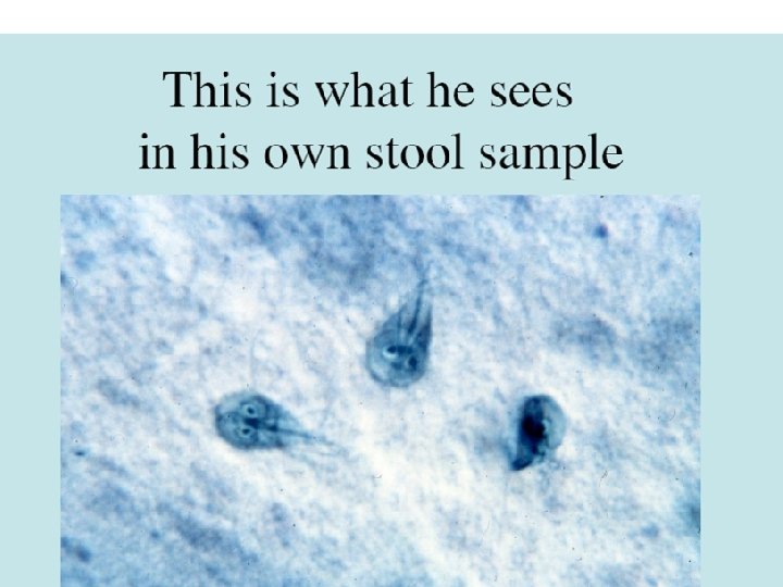
Giardia lamblia Trophozoite stage Typical Characteristics: Size range: 8 -20 um Shape: Pear/teardrop Motility : “Falling leaf” Appearance: Bilaterally symmetrical Nuclei: Two ovoidal-shaped, each with a large Karyosome No peripheral chromatin Flagella: Four pairs arising from the ventral side : One pair anterior end One pair posterior end Two pairs central laterally
 occupies 1/3 to ½ of")
Other Features of Trophozoites Adhesive disk (not always evident) occupies 1/3 to ½ of the area (used for attachment) Axoneme (axostyle) arising from the anterior end and protrudes beyond the posterior end dividing the body into 2 halves = 2 pairs of blepharoplast 1 pair near anterior end 1 pair at caudal end 2 parabasal body (median bodies) posterior to the sucking disc

Giardia lamblia Cyst • Typical Characteristics: • Size range: 11 -14 um • Shape: Ovoid ellipsoidal or may appear round • Nuclei: -2 -4 located at one end young cyst 2 nuclei, mature cyst 4 nuclei - Central Karyosome - No peripheral chromatin

Key Features of Cysts • Cyst wall- smooth, thick and doubly • contour set apart from cytoplasm • Other structures: • Two or four median bodies, axonem and other remnant organelles of the trophozoite are also found inside

• Transmission – Giardia is transmitted via the cyst stage 1 - ingestion of faecally contaminated food or water with cyst 2 - Person to person 3 - sexually among homosexual (feco-oral contact)
 •")
Fecal-Oral Transmission Factors • Poor personal hygiene • Children (eg, day care centers) • Food handlers • Developing countries • Poor sanitation • Endemic • Water-borne epidemics • Male homosexuality • Oral-anal contact

• Is giardiasis a zoonosis? • no definitive documentation • transmission between humans and dogs rare • person-to-person trans-mission is most prevalent
")
Habitat Small intestine ( duodunum & jejunum)

GIARDIASIS

Life Cycle. 1. Life cycle is direct - no intermediate host. 2. When cyst is ingested, the organisms escape from the cyst in the duodenum. 3. Attach via adhesive disk to microvillar surface of epithelium of upper two-thirds of the small intestine. 4. They divide by longitudinal fission. 5. Encystment occurs in lower intestinal tract. 6. Freshly passed cysts are infective 7. Trophozoites are found in diarrheic stools; cysts are found in formed stools.

• Cyst survive – cool, moist conditions • for long periods – in cold water: • 2 months at 8°C • 1 month at 21°C • some cysts can survive – 13°C for 2 weeks • relatively resistant to chlorination, particularly if the water is cold • the amount of chlorine in drinking water is not sufficient to kill G. lamblia

• Cysts are susceptible to: – desiccation and direct sunlight • Cysts and trophozoites are susceptible to: – 1% sodium hypochlorite – 2% glutaraldehyde or quaternary – ammonium disinfectants. • They can also be killed by boiling for at least 1 minute.

Pathogenesis • Epithelial damage • Villus blunting • Crypt cell hypertrophy • Cellular infiltration • Malabsorption develops • Enzyme deficiencies • Lactase (lactose intolerance) Possible Mechanisms • mechanical irritation • obstruction of absorption

Pathogenesis • Girdia Elicits localized hypersensitivity

Clinical Features and Symptoms • one of the more common diarrheal disease seen among travelers to foreign countries • Outcomes may be – Asymptomatic/latent – Acute short-lasting diarrhea – Chronic/nutritional disorders

Acute Symptoms êDevelops after an incubation period of 1 to 14 days (average of 7 days) and usually lasts 1 to 3 weeks êSudden explosive, watery diarrhea, bulky, frothy, greasy, foul-smelling stools, no blood or mucus êUpper gastro-intestinal uneasiness, bloating, flatulence, belching, cramps, nausea, vomiting, anorexia êUsually clears spontaneously (undiagnosed), but can persist or become chronic êIrregular excretion of cysts: ê 10 days lasting negative phases possible (>1 stool analysis

• Subacute/Chronic • recurrent diarrheal episodes • cramps uncommon • sulfuric belching, anorexia, nausea frequent • can lead to weight loss and failure to thrive • The more chronic stage is associated with vitamin B 12 malabsorption, disaccharidase deficiency and lactose intolerance • stools become steatorrheic (fatty stool) containing large amount of fats and mucus but no blood
,")
Laboratory Diagnosis: • Macroscopic – Stool: • is usually offensive, bulky, pale, mucoid (fatty), diarrheic (watery) but • there is no blood in the stool. • Microscopy • Immunological methods • Molecular methods
 should be tested before a")
Basic guidelines A. Multiple stool samples (at least 3) should be tested before a negative result is reported. B. Use concentration technique prior to microscopic examination for cysts – Exception: Specimens to be used for EIA should NOT be concentrated because antigens are lost during the procedure! C. Choice of diagnostic techniques depends on • available equipment and reagents, experience, • and considerations of time and cost.

• not all of the characteristics of flagellates can be seen in a single type of preparation • both unstained and stained preparations may be necessary. • Nuclei are not visible in unstained saline mounts • In fresh feces, trophozoites can be readily distinguished by their characteristic motion in saline mounts.

• Microscopy 1. Finding the trophozoite stages in fresh diarrhoeic stool • wet mounts or stained • Identity or confirm in stained smeer 2. Finding the trophozoite stages in duodenal aspirate 3. Finding cyst stage in formed stool. • wet mounts or stained • Concentration of cyst » Formol-ether/zink-sulphate – trophozoites and cysts are excreted irregularly – Collect stool at 3 non-consecutive days

Examination of stool for trophpzoites • • Make a thin preparation of faeces Examine with 1 st -10 X, then with 40 X Detect motile flagellates Confirm the identity in stained preparation using fields modified thin film staining technique • if Girdiasis is suspected and no flagellates are found in wet mount, always do stained preparation

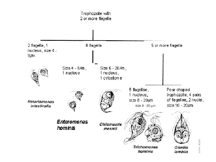
Characteristics used to distinguish flagellate species Trophozoites – Motility – Shape – Number of nuclei. • The appearance of the nucleus is not commonly used to distinguish species. – Other features such as undulating membrane, sucking disk, and prominent cytostome. – Number and location of flagella. • Although vary with the species, they are often difficult to see and count. • Therefore, the number and location of flagella are not practical diagnostic features for routine identification.

Examination of stool for trophpzoites • Trophozoite: -Size: 10 -21 by 5 -15 m » Shape: pear-shaped » Motility: Progressive, rapid, tumbling and spinning linked to a“falling leaf” » typical shape with 8 flagella » concavity at the anterior end » Stained Two nuclie Axonems Parabasal body

Trichrome stain • Trophozoites – appear as pear-shaped organisms, – Measure 12 to 15 μm (range: 10 to 20 μm). – contain two anteriorly placed nuclei and – 8 flagella (rarely seen because they stain poorly). • Cysts – appear ovoid to ellipsoid in shape. – Nuclei and intracytoplasmic fibrils are visible.

trophozoites stained with trichrome trophozoites stained with iron hematoxylin

Gardia lamblia trophozoite
 are")
Giardia intestinalis in culture. In these preparations, the flagellae (four pairs per cell) are clearly visible.

Examination of stool for cyst • Note: – Several specimen need to be examined –. Use concentration technique whenever possible – Make saline/eiosine preparation in one end and Dobell’s iodine on the other end of slide – Identify the cysts with 40 x objective – Examine the overall • • shape, size and contents of cyst- Fibrils Number of nuclei

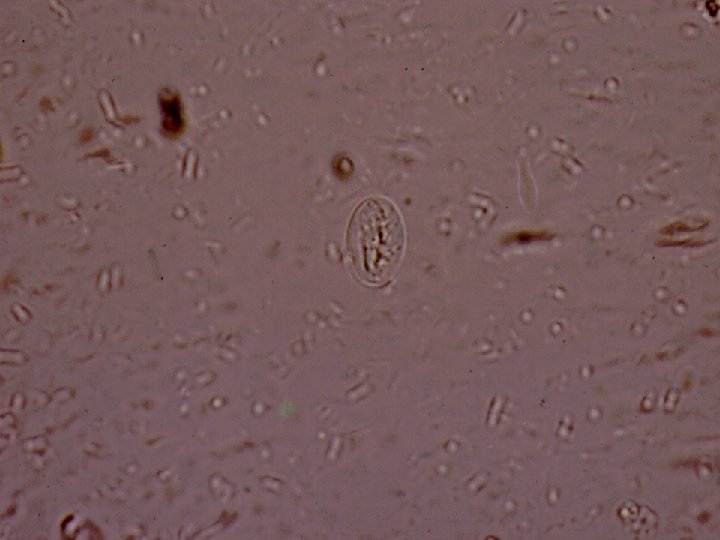
Examination of stool for cyst. diagnostic feature; oval shape, small size & contents • Cyst – oval shape with thick cyst wall – Size: 8 -12 m, . – Contains : thread-like remains of flagella; axonemes and parabasal bodies folded as S-shaped placed length wise in the center of the cyst (stained with iodine) -Nuclei: 2 -4 nuclei at one pole( difficult to see). – Cytoplasm: » clear when unstained; yellowish green or bluish in iodine solution. » Finely granular cytoplasm clearly separated from cyst wall. .

Unstained cyst Cyst stained with iodine

Cysts of Giardia intestinalis, stained with iron hematoxylin

 and oocysts of C. parvum (upper left) labeled")
Cysts of Giardia intestinalis (lower right) and oocysts of C. parvum (upper left) labeled with commercially available immunofluorescent antibodies

G. lambilia trophozoites in duodenal aspirate • Enterotest. • A gelatin capsule attached to a long string. • The end of the string remains outside the mouth and is taped to your cheek. • The gelatin capsule is then swallowed • The capsule dissolves in the stomach and the string passes into the upper part of the small intestine (duodenum). • The string is left in place for 4 to 6 hours or overnight. Then it is withdrawn and the end is examined under the microscope for parasites that have become attached.

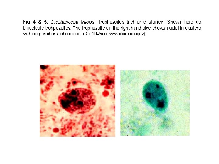
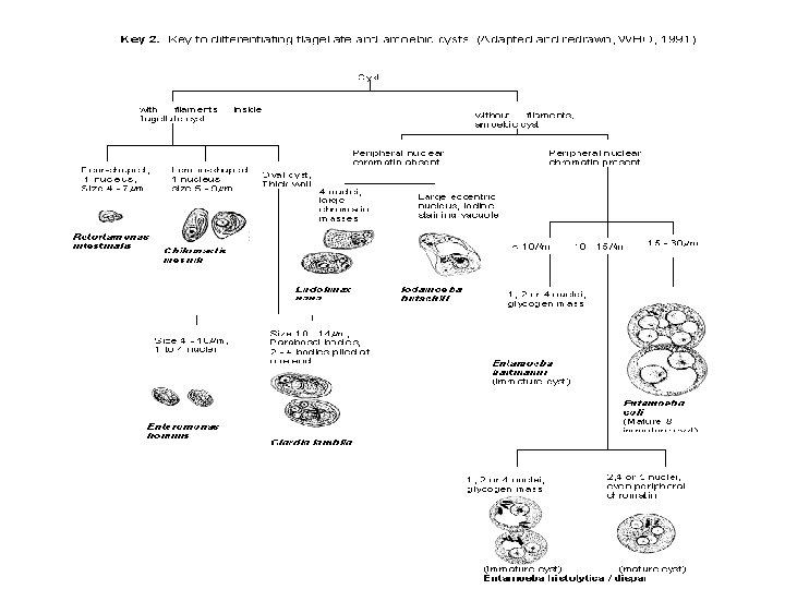
Differentiation of G. lamblia from non-pathogenic intestinal flagellates that can be fond in stool • Trophozoites: • can be easily differentiated from the nonpathogenic by their shape and movement (in fresh sample) • because they have two nucleus and more no of flagella. (stained) • D. fragilis has two nuclei is but this organism has no flagella or median bodies and looks like a small amoeba.

• Cyst: • Cyst of non pathogenic intestinal flagellates can be easily differentiated from those of G. lamblia(8 -12 m) » they are smaller and do not have the same characteristic appearance of G. lamblia (do not contain remains of flagella). » C. mesnili(5 -7 m) cysts have remains of flagella and cytosome but they are lemon shape. » D. fragilis does not have cyst stage.

Immunodiagnosis • Antigen test • Detection of Girdia specific antigen in faecal specimen using monoclonal antibody • EIA tchnique, ELISA

Molecular Diagnosis • 2% agarose gel analysis of a PCR diagnostic test for detection of Giardia DNA. • PCR was performed using primers JW 1/JW 2. • 1 Lane S: Molecular base pair standard (100 -bp ladder). – Black arrows show the size of standard bands. • Lane 1: Giardia intestinalis positive fecal specimen. – The red arrow shows the diagnostic band for G. intestinalis
 • 750 mg/tid/5 d • >90% cure")
Treatment Drug of Choice • metronidazole (Flagyl) • 750 mg/tid/5 d • >90% cure rate Alternatives • tinidazole (single dose) • paromomycin (pregnancy) • quinicrine • furazolidone Prognosis is good with no sequelae Control • avoid fecal-oral transmission • improve personal hygiene • especially institutions • treat asymptomatic carriers • eg, family members • health education • hand-washing • sanitation • food handling • protect water supply How to get rid of Giardia in water? : * boiling (at least 1 minute) ☺ * filtration (1μm pores) �� * chemical treatment (Iodine, …) ��

Summary • Giardia lamblia: – First discovered by Leeuwenhoek 1697 – 2 nd most frequent intestinal Protozoon – exist as both trophozoite, cyst – Reproduce by binary fission – Have fecal-oral transmission – Colonize duodenum, small intestine but no invasion – Less harmful, but chronic damage to intestinal wall can lead to malabsorption syndrome – Similar lifecycle to E. histolytica, but reservoir hosts may play role in transmission. – Diagnosis commonly by finding trophozoites and cyst in stool

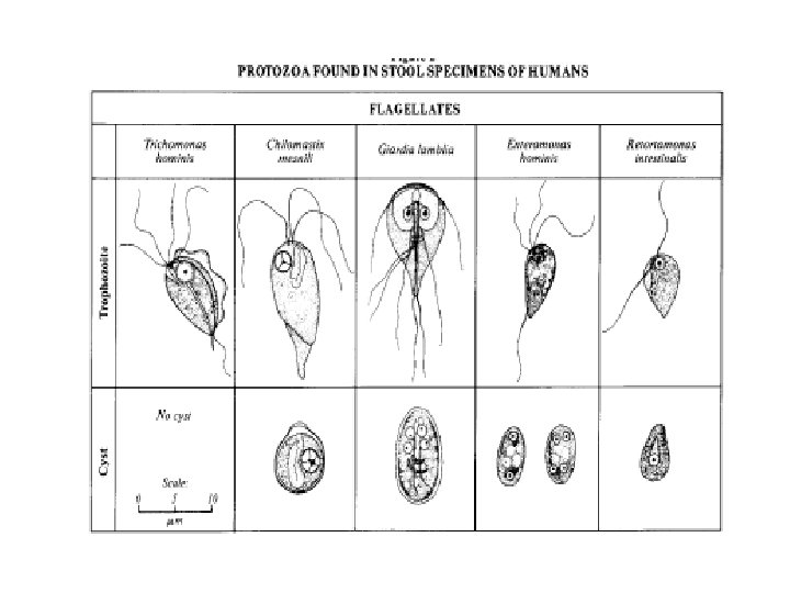
Other Flagellates Found in Human Feces • Chilomastix mesnili • Pentatrichomonas hominis • formerly called Trichomonas hominis • Enteromonas hominis • Retortamonas intestinalis • Dientamoeba fragilis • no flagella (discuss with amebas)
 • Morphology")
Chilomastix mesnili • Distribution: cosmopolitan • Habitat: large intestine (colon & caecum) • Morphology – the largest flagellate found in man – Has both trophozoites and cyst stage

• Trophozoite : – Size: 6 -20 by 3 -10 m – Shape: Triangular and tapered at one end – Motility: spiral in one definite direction. – Cytoplasm: – Spiral groove that makes asymmetrical flagellate – cytostome (mouth-like cleft) at the rounded end. – Nucleus: one nucleus, easily visible in unstained preparation – Flagella: Six flagella. Three anterior free flagella, Morphological distinctions: -symmetric, -4 flagellar, -Spiral groove - Single nucleus -cytostome.

• Unstained – – Pointed tail 3 anterior filagella Spiral groove Motility-rotary • Stained – 1 nucleus – Cytosome with fibril Chilomastix mesnili

• Cyst: - Size: 6 -8 by 46 m • Shape: pear or lemon shaped • Cystostome and remains of locomotory organelles can be seen. • Nucleus: single; .

Pentatrichomonas hominis • Distribution: – it is probably the most common intestinal flagellates next to Giardia lamblia. • Habitat: Large intestine. • Morphology: has trophozoite stage only
 •")
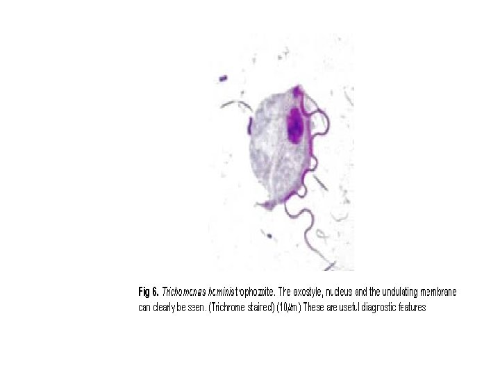
Trophozoite unstained • Size: 10 -15 m, pyriform (oval with two pointed poles) • 4 anterior flagella(3 -5) • Motility: whirls and turns (jerky) in all directions, seeming to vibrate. • Undulating membrane and costa reach full length of the body. • Axostyle extend beyond the body Stained • Single nucleus • Costa • Conspicuous cytostome • semi- rigid axostyle

Enteromonas hominis • Cosmopolitan in distribution • Is a small flagellate and is rarely encountered in man.

Morphology of the trophozoite • Oval in shape and 4 -10 m m in length. • Have 4 flagella, 3 anterior flagella and one adheres to the body ending in a tail, • motility: jerky rotational movement. • Have one nucleus with a large karyosome that is evident in a stained preparation.

Morphology of the cyst • oval in shape • Size: 6 -8 m m • They have up to 4 nuclei with a bipolar tendency.

Retortamonas intestinalis • Retortamonas intestinalis like E. hominis is a small flagellate • and is rarely encountered. • It is found in both warm and temperate climates

Morphology of trophozoites • is small, 4 -9µm. • Its movement is jerky and rotational • has 2 anterior flagella and a prominent cytosome that can be seen in an unstained preparation. • It has a relatively large nucleus at the anterior end with a small compact karyosome

Morphology of the cyst • small and pear shaped. • Size: 4 -7µm • Has one large nucleus frequently near the centre. • The fibril arrangement from the nucleus is suggestive of a birds beak. • This is characteristic of R. intestinalis cysts.

Dientamoeba fragilis • Trophozoite: – Size: 3 -22 m – Motility: Either non-motile (most often), or very actively motile in very fresh fluid stools with fan-like multiple pseudopodia. -It becomes non-motile under the cover slip or disintegrates immediately. – Cytoplasm: clear ectoplasm. – Nucleus: Usually one or two nuclei but 3 or 4 nuclei may be found - rarely. Karyosomes split into 4 -6 granules. – Inclusion bodies: Bacteria
 Shape Motility Number of Nuclei Number of Flagella* Other features")
summary Species Size (length) Shape Motility Number of Nuclei Number of Flagella* Other features Trichomonas hominis 8 -20 m m Usual range, 11 -12 m m Pear Shaped Nervous Jerky 1 Not visible in unstained mounts 3 -5 anterior. 1 posterior. Undulating membrane extending length of body. Chilomastix mesnili 6 -24 m m. Usual range, 10 -15 m m Pear Shaped Stiff Rotary 1 Not visible in unstained mounts 3 anterior. 1 in cytostome Prominent cytostome extending 1/3 – 1/2 length of body. Spiral groove across ventral surface. Giardia lamblia 10 -20 m m. usual range, 12 -15 m m. Pear Shaped "Falling Leaf" 2 Not visible in unstained mounts 4 lateral. 2 ventral. 2 caudal. Sucking disc occupying 1/2 -1/3 of ventral surface. Enteromonas hominis 4 -10 m m. usual range 8 -9 m m. Oval Jerky 1 Not visible in unstained mounts 3 anterior. 1 posterior. One side of body flattened. Posterior flagellum extends free posteriorly. Retortamonas intestinalis 4 -9 m m. Usual range, 6 -7 m m. Pear Shaped Or Jerky 1 Not visible in unstained mounts 1 anterior. 1 posterior. Prominent cytostome extending
- Slides: 75