BIOL 204 Lab For Week 11 Digestive System

BIOL 204 Lab For Week 11 Digestive System Anatomy

Activity 1 Microscopic Anatomy of the GI Tract The GI tract consists of a tube that extends from the mouth to the anus. It has four tissue layers (tunics):

• � • There are structural modifications of the tunics in various areas of the GI tract

Another, detailed view of GI tract tunics

Gastroesophageal Junction Different microscopic views

Histology of The Stomach Wall
 Micrograph")
Histology of the Small Intestine (Duodenum) Micrograph

Intestinal Villus

Activity 2 Histology of Selected GI Tract Organs A. The Pancreas – Gross View

Microscopic View of the Pancreas: Islet Cells: endocrine; secrete insulin and glucagon Acinar Cells: exocrine; secrete digestive enzymes

B. The Liver C. Gross View of the Liver

Liver – Microscopic Anatomy

Activity 3 Gross Anatomy of the GI Tract

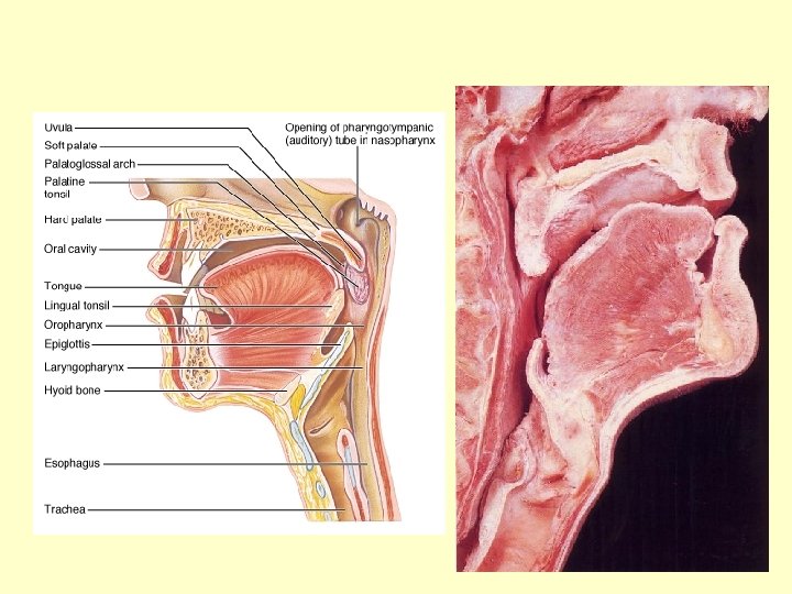
The GI Tract: The Mouth

The Esophagus

The Stomach

The Duodenum of the Small Intestine Receives the Ducts of the Liver, Gall Bladder and Pancreas

Portions of the Large Intestine

Extrinsic Salivary Glands

Types of Permanent Teeth
 6 molars 2 lateral incisors")
Each jaw has: 2 central incisors 2 canines (eyeteeth) 6 molars 2 lateral incisors 4 premolars

Internal Anatomy of a Tooth

1. Diaphragm 2. Liver – left lobe 3. Falciform Ligament 4. Liver right lobe 5. Stomach 6. Greater Omentum 7. Ascending Colon 8. Cecum 9. Small Intestine 10. Descending Colon 11. Sigmoid Colon

An Appendix, Unfixed

Gall Bladder on the Underside of the Liver

Liver – Gross View Serosal Surface

Human Duodenum, Unfixed

Abdomen of the Cat

Cat Abdomen – Greater Omentum Removed

Gall Bladder and Lobes of the Liver

Small and Large Intestine Removed

More Images

1. Parotid Gland 2. Sublingual Gland 3. Submandibular Gland 4. Esophagus 5. Tongue 6. Masseter Muscle 7. Larynx 8. Trachea

1. Liver 2. Stomach 3. Duodenum 4. Ascending Colon 5. Pancreas 6. Spleen
- Slides: 35