INTESTINAL OBSTRUCTION Classify Intestinal Obstruction Discuss the DD

INTESTINAL OBSTRUCTION

�Classify Intestinal Obstruction. Discuss the DD and management of AIO in a 70 yr old male. �Describe the cf , diagnosis and management of AIO. �Enumerate causes of dynamic IO. Discuss the diagnosis and management of acute small bowel obstruction.

The common Scenario A 50 year old gentleman presents with abdominal pain, distension and absolute constipation. With repeated episodes of vomiting. His vital sign were stable, abdomen distended with diffuse tenderness but minimal peritonism. Bowel Sounds are hyperactive. The plain abdominal xray was taken on admission.

What are your objectives? You should be able to address the following questions 1. Is this bowel obstruction or ileus? 2. Is this a small or large bowel obstruction? 3. Is this proximal or distal obstruction? 4. What is the cause of this obstruction? 5. Is this a complex or simple obstruction? 6. How should I start investigating my patient? 7. What is the role of other supportive investigations? 8. What is my immediate/ intermediate treatment plan? 9. What are the indications for surgery? 10. What are the medico-legal and ethical issues that I should address?

Definition � Any condition that interferes with normal propulsion and passage of intestinal contents. � Can involve the small bowel, colon or both.

TYPES �Dynamic: where peristalsis is working against a mechanical obstruction. �Adynamic: mechanical element is absent - Peristalsis may be absent(paralytic ileus) -May be present in non propulsive form. (mesenteric vascular occlusion or pseudo-obstruction)

Mechanical Obstruction � Acute vs Chronic �Partial vs Complete �Simple vs Closed loop �Gangrenous vs Nongangrenous

PATHOPHYSIOLOGY �Small Intestine Function � Transport -�Distension stimulates contractions � Mixing and peristalsis �Absorption � �Colon Function � Water Absorption � �Transport � Storage

PATHOPHYSIOLOGY �Dependent Upon: � �Degree of Obstruction � � Duration of Obstruction � �Presence and Severity of Ischemia �Results in: � �Accumulation of fluid and air � �Bacterial overgrowth �Maximal by 24 hrs after obstruction �Gut translocation to nodes and portal system � �Once Distended : � Impaired fluid and nutrient absorption �Secretion of isotonic fluid (intravascular to intraluminal)
")
PATHOPHYSIOLOGY �Systemic symptoms are secondary to: � �Hypovolemia � �Bacterial translocation (typically E. coli) � � Ischemia worsens general inflammatory state, bacterial translocation, and fluid requirements �Large Bowel Obstruction � �Ileocecal valve plays prominent role in pathophysiology of LBO � �If competent valve = closed loop obstruction � �Caecal ischemia around 10 -13 cm

Causes- SBO Luminal Mural Extraluminal F. Body Bezoars Gall stone Food Particles Neoplasims lipoma polyps leiyoma hematoma lymphoma carcimoid carinoma secondary Tumors Crohns TB Stricture Intussusception Congenital Postoperative adhesions A. lumbricoides Congenital adhesions Hernia Volvulus

Intussusception Gall stone Ileus IBD

Large Bowel Obstruction • Distinguishing ileus from mechanical obstruction is challenging • According to Laplace’s law: maximum pressure is at the it’s maximum diameter. Cecum is at the greatest risk of perforation • Perforation results in the release of formed feaces with heavy bacterial contamination Aetiology: 1. Carcinoma: The commonest cause, 18% of colonic ca. present with obstruction 2. Benign stricture: Due to Diverticular disease, Ischemia, Inflammatory bowel disease. 3. Volvulus: 1. Sigmoid Volvulus: Results from long redundant, faecaly loaded colon with a narrow pedicle 2. Caecal Volvulus 4. Hernia. 5. Congenital : Hirschusbrung, anal stenosis and agenesis

Sigmoid Volvulus Colonic Obstruction

�COMMON CAUSES: �Adhesions- 40% �Tumors -15% �Inflamatory- 15% �Obstructed hernia-12% �Intraluminal-10% �Miscellaneous -8%

SYMPTOMS The four cardinal features of intestinal obstruction: -abdominal pain -vomiting -distension -constipation Vary according to: location of obstruction Duration of obstruction underlying pathology intestinal ischemia
")
HISTORY �Did this ever happen before? � Change in bowel habits (acute vs chronic) � Progressive vs acute abd. distention � Weight loss � Flatus? � Prior Surgeries � Hx of abdominal CA? � Hx of Inflammatory Bowel Dz � Prior abdominal XRT? � Meds: Anticoagulants, Anticholinergics, Opioids, Antihistamines, Alpha-agonists, Catecholamines

SYMPTOMS Abdominal pain: - colicky in nature, around the umbilicus in SBO while in the lower abdomen in LBO - if it becomes continuous, think about perforation or strangulation. - does not usually occurs in paralytic ileus. Vomiting -starts early in SBO and late in LBO -As obstruction progresses vomitus alters from digested food to faeculent due to enteric bacterial overgrowth Distension -more with lower obstruction

SYMPTOMS Constipation -more with lower or complete obstruction - constipation is either absolute (no feces or flatus) or relative (flatus passed). -diarrhea may be present with partial obstruction

SYMPTOMS Dehydration �More common in small bowel obstruction, due to repeated vomiting. �Secondary polycythemia due to raised B. urea & hematocrit. Pyrexia �Onset of ischemia. �Intestinal perforation. �Inflammation associated with int. obst.

SYMPTOMS In strangulation: � � � severe constant abdominal pain fever tachycardia tenderness with rigidity/rebound tenderness. shock

CLINICAL EXAMINATION General examination. Vital signs Signs of dehydration –tachycardia, hypotension dry mucus membrane, decreased skin turgor, decreased urine output Inspection distension, scars, peristalsis, masses, hernial orifices Palpation tenderness, masses, rigidity Percussion tympanitic abdomen Auscultation high pitched bowel sound or silent abdomen *Examine rectum for mass, blood, feces or it may be empty in case of complete obstruction
 �Hyper kalemia, hyperamylasemia & raised LDH may be")
INVESTIGATIONS � Hemogram - WBC (neutrophilia-strangulation) �Hyper kalemia, hyperamylasemia & raised LDH may be associated with strangulation. �Plain AXR �Sigmoidoscopy (carcinoma, volvulus) � Contrast x-ray �CT abdomen.

�The most helpful diagnostic radiographic procedure in suspected SBO is: A. CT of Abdomen and Pelvis B. UGI gastrograffin contrast study C. Supine and Erect plain films of abdomen D. U/S of the abdomen

Imaging – Plain Films �Dilated loops, SB dilation? AFLs? �Are AFL’s and bowel loops in same place on supine and upright films? �Is there gas throughout the entire colon? �Ileus or p. SBO �Paucity of distal colonic gas or abrupt cutoff of colonic gas w/proximal distention and/or AFLs? �Suggesting complete or near-complete LBO �Massive dilation of colon, especially of the cecum or sigmoid? �Suggestive of volvulus or pseudoobstruction �Evidence of strangulation? �Thickened SB loops, mucosal thumb printing, pneumatosis cystoides intestinalis, or free air �Are there biliary calculi? Pneumobilia? �GS ileus?

AXR When distended by gas: �Jejunum is characterized by valvulae conniventes. �Ileum is featureless. �Caecum is shown by rounded gas shadow in RIF. �Colon shows haustral folds. �Fluid level appears later than gas shadow �Two fluid level in small bowel considered normal. �No. of fluid level is proportional to degree of obstruction and distal site in small bowel.

The Difference between small and large bowel obstruction Large bowel • Peripheral ( diameter 8 cm max) • Presence of haustration Small Bowel • Central ( diameter 5 cm max) • Vulvulae coniventae • Ileum: may appear tubeless

Adjunctive Tests �Sigmoidoscopy � �When large amounts of air extend down to the rectum � Flex or Rigid will exclude rectal or distal sigmoid obstruction • �CT Scan or U/S � � When normal plain films but history and exam are consistent with obstruction � �Plain films in SBO will be nondiagnostic approximately 30% of the time

Role of CT � � Used with iv contrast, oral and rectal contrast (triple contrast). Able to demonstrate abnormality in the bowel wall, mesentery, mesenteric vessels and peritoneum. � It can define � the level of obstruction � The degree of obstruction � The cause: volvulus, hernia, luminal and mural causes � The degree of ischaemia � Free fluid and gas � Ensure: patient vitally stable with no renal failure and no previous allergy to iodine

Role of barium gastrografin studies Barium should not be used in a patient with peritonitis � � � As: follow through, enema Limited use in the acute setting Gastrografin is used in acute abdomen but is diluted Useful in recurrent and chronic obstruction May able to define the level and mural causes. Can be used to distinguish adynamic and mechanical obstruction

TREATMENT �Three main measures- GI drainage - Fluid &Electrolyte replacement - Relief of obstruction, usually surgical

Treatment Conservative: -Nasogastric aspiration by Ryles tube -IV fluids- volume varies depending on dehydration -NPO -urinary catheter -check temp. and pulse 2 hourly -abdominal examination 8 hourly -Broad spectrum antibiotics initiated early- reduce bacterial overgrowth.

• Some cases will settle by using this conservative regimen, other need surgical intervention. • Surgery should be delayed till resuscitation is complete unless signs of strangulation and evidence of closed-loop obstruction. • Cases that show reasons for delay should be monitored continuously for 72 hours in hope of spontaneous resolution e. g. adhesions with radiological findings but no pain or tenderness • “The sun should not both rise and set” in cases of unrelieved obstruction.

Indication for surgery: - failure of conservative management - tender, irreducible hernia - strangulation �If the site of obstruction is unknown; laparotomy assessment is directed to-The site of obstruction. -The nature of obstruction. -The viability of gut.

Surgical treatment Operative decompression required-if dilatation of bowel loops prevent exposure, bowel wall viability is compromised, or if subsequent closure will be compromised. Savage’s decompressor used within seromuscular purse-string suture. Or large-bore NG tube maybe used for milking intestinal contents into stomach.

SURGERY The type of surgical procedure depend upon the cause of obstruction viz division of bands, adhesiolysis, excision , or bypass *Once obstruction relieved, the bowel is inspected for viability, and if non-viable, resection is required. Indicators of non-viability 1. absent peristalsis 2. loss of normal shine 3. loss of pulsation in mesentry 4. green or black color of bowel

SURGERY �If in doubt of viability, bowel is wrapped in hot packs for 10 minutes with increased oxygen and reassessed for viability. �Resection of non viable gut should be done followed by stoma. �Sometimes a second look laprotomy is required in 2448 hours e. g. multiple ischemic areas.

Adhesions �Most common cause of intestinal obstruction. �Peritoneal irritation results in local fibrin production, which produce adhesions. BANDS �Congenital : obliterated vitellointestinal duct. �A string band following previous bacterial peritonitis. �A portion of greater omentum adherent to parietes.

Causes of adhesions : �Abdominal operation : anastomosis, raw peritoneal surfaces �Foreign material: talc, starch, gauze, silk �Infection: peritonitis, T. B. �Inflammatory conditions: crohn’s disease. �Radiation entritis. Prevention �Good surgical technique. �Washing the peritoneal cavity with saline to remove the clots. �Minimizing contact with gauze. �Covering the anastomosis & raw peritoneal surfaces.
 �It should")
TREATMENT �Usually conservative treatment is curative. (i. v. rehydration & nasogastric decompression) �It should not be prolonged beyond 72 hrs. Surgery �Division of band. �Minimal adhesiolysis.

Tt. Of recurrent obstruction due to adhesions �Repeat adhesiolysis alone. �Noble’s plication : adjacent intestinal coils (15 -20 cms) are sutured with serosal sutures. �Charles-Phillips trans-mesenetric plication. �Intestinal intubation : initraluminal tube insertion via a WITZEL jejunostomy or gastrostomy.

INTERNAL HERNIA �When a portion of small intestine is entrapped in one of retropritoneal fossae or in a congenital mesentric defect. Sites of internal herniation: �Foramen of winslow. �A hole in mesentry / transverse mesocolon. �Defects in broad ligaments. �Congenital/ acquired diaphragmatic hernia. �Duodenal retroperitoneal fossae- Lt. paraduodenal & rt. Duodenoojejunal. � intersigmoid fossae.

�It is uncommon in the absence of adhesions. �Treatment : to release the constricting agent by division.

Gall stone ileus �It tends to occur in elderly. �Erosion of large gallstone into duodenum. �Present with recurrent obstruction. �X-ray: small bowel obstruction with air in billiary tree. -may show a radio opaque gall stone. �Treatment : laparotomy & removal /crushing of stone.

FOOD �After partial /total gastrectomy. �Unchewed food can cause obstruction. �Treatment similar to gall stone. �Trichobezoars �Phytobezoars BEZOARS WORMS �Ascaris lumbricoides �Frequently follows initiation of antihelminthic therapy. �Eosinophilia/worm with in gas filled bowel loops. �Laparotomy.

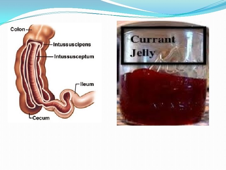
INTUSSUSCEPTION �One portion of gut becomes invaginated with in adjacent segment. �Most common in children(3 -9 months. ) �Idiopathic-70% �Associated gastroenteritis/UTI- 30% �Hyperlpasia of Peyer’s patches in terminal ileum can be initiating factor.

�In older children intussusception is usually associated with a lead point – meckel’s diverticulum, polyp, & appendix. �Adults: always with a lead point. - polyp, submucosal lipoma/ tumor. �It is composed of three parts: -Entering/ inner tube(Intussusceptum) - Returning/ middle tube -Sheath/ outer tube(intussuscipiens) �It is an example of strangulating obstruction with impaired blood supply of inner layer. �It may be ileoileal(5%); ileocolic(77%); ileo-colic(12%); colocolic (2%) & multiple.

CLINICAL FEATURES �Severe colic pain. � vomitting as time progress � blood & mucus (the ‘redcurrent’ jelly stool). �Abdominal lump(sausage shaped) � Emptiness in RIF(the sign of Dance). �Death may occur from bowel obstruction or peritonitis secondary to gangrene.

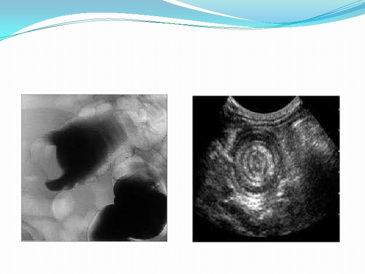
RADIOGRAPHY �Plain X-ray Abd. : Bowel obstruction with absent caecal shadow gas in ileo-ileal & ileo-colic cases. �Ba-enema: the claw sign in ileocolic & colocolic cases. �CT scan in equivocal cases of ileo-ileal intussusception. (small bowel mass may be revealed) Differential Diagnosis �Acute enterocolitis: faecal matter/ bile is always present. �Henoch-schoenlein purpura. �Rectal prolapse: projecting mucosa can be felt in continuity with perianal skin


TREATMENT �Theraputic Ba-enema : -in infants. - unlikely to succeed in lead points. - contrindications: peritonism, prolonged history (> 48 hrs. ). Operative �After resuscitation ; Laparotmy with reduction. �Cope’s method. �Irreducible/ gangrenous intussusception: excision of mass & anastomosis.

Volvulus of the Colon � Sigmoid most commonly involved � �� 180° or > counterclockwise twist � Cecal volvulus next most common �Transverse colon volvulus is rare � 4% of colonic volvulus �Splenic flexure volvulus VERY rare

Sigmoid Volvulus �Predisposing Factors �Long and highly-mobile sigmoid �Lengthy mesosigmoid, Narrow at its base � Adhesions between proximal sigmoid and rectosigmoid �with long loop of colon in between �Chronic constipation and high-fiber diet

Sigmoid Volvulus - Pathogenesis � Closed-loop type obstruction � Proximal colon dilates- �Extent depends on ileocecal valve competence �Simple or Strangulated � �Venous then arterial obstruction � �With simple form takes a few days for vascular compromise to develop ��In acute fulminating variant much more rapid course seen

Sigmoid Volvulus - Varieties �Acute Fulminating Type- �Mortality 37 -80% �, Younger patient, sudden onset, rapid course �Early vomiting, severe pain, peritonitis, and gangrene � Minimal distension often, hard to diagnose � Subacute Progressive Type- Generally older pt. , more gradual onset, �Hx prior attacks, chronic constipation, �Abdominal distension often extreme � Late vomiting, pain is minimal, no peritonitis

Sigmoid Volvulus - Diagnosis �Plain films of Abdomen-- �Massively dilated and distended bowel loop ��Both ends in pelvis, � Bow of loop cephalad � �“Bent inner tube” sign � �Air/fluid level within loop � � Proximal colon & SB may be dilated �Barium enema--�“Birds beak”

Sigmoid Volvulus - Treatment � Rigid sigmoidoscopic detorsion--decompression, & placement of rectal tube � � RT inserted past obstruction point � �Successful 77 - 90% of time � �Mortality rate 1. 2 - 5. 5% � � Preferred initial treatment �Colonoscopic decompression � Surgery if: � �Decompression not successful � �Ischemic or necrotic bowel encountered
 �Some")
Sigmoid Volvulus – Surgical Treatment � Recurrence rate is high (33 - 60%) �Some advise elective op after 1 st episode �Elective: �Resection of redundant colon �Small transverse LLQ incision �Sigmoid fixation another option � Emergent: Resection +/- stoma

Ogilvie’s Syndrome �Acute Colonic Pseudo obstruction � Massive dilation of cecum, right, and transverse colon (non-mechanical), to splenic flexure �Dilated SB loops in > 50% �Symptoms: �Distension, Nausea and vomiting, � Abdominal pain in 80% , Bowel sounds may be normal or increased
 �Pancreatitis,")
Conditions associated with Ogilvie’s syndrome � Non-operative Trauma �Non-GI surgery (Gyn, Ortho, Cardiac) �Pancreatitis, Cholecystitis �Diabetes, Malignancy �Narcotics, antidepressants, anticholinergics �Neurologic or Respiratory disease �Electrolyte (hypo K+, Ca+2), Acid / Base disorder � Radiation in past

Ogilvie’s Syndrome: DDx and Dx �Plain abdominal films to diagnose and follow cecal diameter (Q 12 -24 hrs) � � Normal is < 9 cm � �If ≥ 14 cm - perforation in 23% • �Differential Includes: �Fecal impaction, Cecal or Sigmoid Volvulus, Ischemic. Bowel, Mechanical Obstruction

Ogilvie’s Treatment : �Correct electrolyte problems and underlying condition �D/C narcotics and anticholinergics �NPO with NGT, +/- Rectal Tube �Colonoscopic decompression difficult but often successful, May need 2 nd decompression � Long colonic drains can be placed via colonoscope (old) �Neostygmine is the treatment of choice now � If signs of perforation then OR

STRANGULATION �The viability of the bowel is threatened. �Causes of strangulation �External �■ Hernial orifices �■ Adhesions/bands �Interrupted blood flow �■ Volvulus �■ Intussusception �Increased intraluminal pressure �■ Closed-loop obstruction �Primary �■ Mesenteric infarction

�The venous return is compromised before the arterial supply. �The resultant increase in capillary pressure leads to local mural distension with loss of intravascular fluid and red blood cells intramurally and extraluminally. �Once the arterial supply is impaired, haemorrhagic infarction occurs. �As the viability of the bowel is compromised there is marked translocation and systemic exposure to anaerobic organisms with their associated toxins. �The morbidity of intraperitoneal strangulation is far greater than with an external hernia?

Closed-loop obstruction �This occurs when the bowel is obstructed at both the proximal and distal points. �There is no early distension of the proximal intestine. �A classic form of closed-loop obstruction is seen in the presence of a malignant stricture of the right colon with a competent ileocaecal valve �The inability of the distended colon to decompress itself into the small bowel results in an increase in luminal pressure, which is greatest at the caecum, with subsequent impairment of blood supply. �Unrelieved, this results in necrosis and perforation.
- Slides: 67