Chapter 4 Viruses The concept of viruses Viruses


: 1)are tiny particles that are ultramicroscopic size, can go")
 Control and eliminate the aversive viruses, which")


 Capsid Virus particle Capsomer")


")
:identical to the m. RNA base sequence. antisense(-):complementary")

peplomere protein: are composed of peplos glycoprotein and matrix protein, located in peplos surface.")
 Virion enzyme: exist in virocapsomer, categorized as follow by function. One kind is")
: it means proteins encoded by virus genome have function")


 2 Nature")
 is the \"international court\" of experts")


")


 Nucleic acid Type Nucleic Acid Structure Virus Examples Single-Stranded")
The Reproduction of Double-Stranded DNA (T 4 Bacteriophages) Transcription Translation +m. RNA")
The Reproduction of ФX 174, a +Strand DNA Replication Transcription Bacterial Translation DNA ±DNA")
The Reproduction of Single-Stranded RNA Bacteriophages RNA replicase +RNA ±RNA (replicative form) +RNA genomes")
positive single-stranded RNA viruses(picornaviruses) +RNA Proteins(after processing by host")
 Double-stranded RNA viruses (reoviruses) RNA-dependent RNA polymerases* (Transcriptases) ±RNA +m. RNA Proteins RNA-dependent")
Negative single-stranded RNA viruses (paramyxovirusesmumps and measles; orthomyxoviruses-influenza RNA-dependent RNA polymerase+ (Transcriptase) -RNA +m.")
Retroviruses (Rous sarcoma virus, HIV) RNA-dependent DNA polymerase Ribonuclease H Reverse + Reverse")

")
 Eclipse period: it is the period from the virus penetrating host cell")


lysogenic conversion:The prophage will result in the phenotypic alternation")


Naked virus direct penetration. 2)Virus with envelope penetrates by endocytosis. 3)Virus with envelope penetrate")


The persistent infected animal cells can keep normal function for")

 (epstein-barr")

The integration of protovirus. (2)The expression of")

The environment are destroyed by human activity. 。 2)The")
,")
- Slides: 57
Chapter 4 Viruses
The concept of viruses Viruses are a unique group of tiny infectious particles that are obligate parasites of cells, are not cells but resemble complex molecules composed of DNA or RNA. Most of them are so small (0. 02 -0. 3µm) that an electron microscope is necessary to detect them.
Classification of the viruses: Viruses Non-cell organism Viroids Subviral agents Prions Satellites and Deltavirus
Characteristics of virions (virus particles): 1)are tiny particles that are ultramicroscopic size, can go through the bacteria filter. 2)are not cell but composed of protein and nucleic acid 3)Nucleic acid can be either DNA or RNA but not both. 4)use themselves nucleic acid to replicate and apply the assembling of viral protein and nucleic acid to multiply massively. 5)are obligate intracellular parasites and lack enzymes for most metabolic processes and synthesizing protein, multiply by taking control of host cell’s metabolism and synthetic machinery. 6)are abiotic macromolecular and contain the infectivity for a long time outside of the host cell. 7)are not sensitive to antibiotic but sensitive to interferon. 8)Some of them can be integrated into the host cell’s genome in provirus .
Significance of virus research : Significance 1) Control and eliminate the aversive viruses, which do harm to the aversive human health and the domestic animals. The national economy is severely affected by the virus diseases. In fermentation industry , contamination of phages will have bad influence in the fermentation production. 2) By study of the virus, we can upgrade the species, cultivate live virus vaccine strain, protect the ecological environment, apply virus as insecticida and use them as vectors for gene engineering.
Section 1. The General structure and Chemical Composition of Viruses 1. Size of the virus particle Most of them are so small and vary widely (0. 02 -0. 3µm)that an electron microscope is necessary to detect them, their size relegates them to the realm of the ultramicroscopic. The tiniest circoviridae, 17 nm in diameter,is a little bigger than ribosome, while the maximum Poxviruses, 200 nm in diameter,is as big as the minimum bacteria, even to be detected by light microscope.
2. Structure of viruses Nucleic acid Spike caps id Caps id capsomere The mode of naked nucleocapsid virus有囊膜病毒模式结构
Centrel core DNA/RNA Genome: Double/single strand sectile/unsectile Nucleocapsid (Basic Unit) Capsid Virus particle Capsomer Envelopewith Spike (nonbasic Unit) Component of virus particle 多肽 (结构亚单位)
3. Morphology and symmetry of viruses 3. 1 Morphology of Virus
3. 2 Architecture type of Virus habditiform:TMV etc helical symmetry Naked envelope Envelope silkiness:Bacterium coli M 13 phage etc coiling:Influenza virus etc bullet:Wut virus etc Architecture type of Virus icosahedral symmetry Naked envelope small:Poliovirus big:adenovirus Envelope : herper virus Naked envelope: Bacterium coli T phage (tadpole shape) complex symmetry Envelope :poxvirus(Brick shape)
1. Helical symmetry A typical virus helical symmetry is the tobacco mosaic virus( TMV). This is a RNA virus in which the 2130 protein subunits (each 158 amino acids in length) are arranged in helix. In TMV, the helix has 16 ½ subunits per turn and the overall dimensions of the virus particles are 18*300 nm. The lengths of helical viruses are determined by the lengths of the nucleic acid, but the width of the helical viruses particles is determined by the size and packing of the protein subunits.
2. Icosahedral symmetry Take adenovirus for example. The icosahedral capsid (70 to 100 nm) is made up of 252 capsomeres: 240 hexons forming the faces and 12 pentons at the vertices. Each penton bears a slender fiber and is composed of a pentamer(Ⅲ) and a trimer fibrin(Ⅳ) but each hexamere is not hexamer but trimer constituted by three 110 K polypeptide Ⅱ.
Sense and antisense single strand : sense(+):identical to the m. RNA base sequence. antisense(-):complementary to the m. RNA base sequence. Sense/antisence(±):complementary double strands. +RNA:as m. RNA template for translation to protein. -RNA: complementary to +RNA, can not as m. RNA template for translation to protein. +DNA: complementary to-DNA, not as template for transcription to m. RNA. -DNA: as template for transcription to m. RNA。 Segmentation and amerism: Some RNA viruses are not strictly ‘antisense' but ambisense, since they are part (-)sense & part (+)sense.
4. Proteins of viruses 1. Constitutive protein: constitute the morphological maturity and infectious virus particle, including capsid , peplos and enzymes accounting for 30 -70% per particle. (1)capsid protein: the protein covering of a virus’s nucleic acid core , exhibit symmetry due to the subunits called capsomers. Functions of capsid protein:constitutes the virus capsid to shield the nucleic acid; naked virus’s capsid participates the absorption, penetration, determination the host tropsim and surface antigen.
(2)peplomere protein: are composed of peplos glycoprotein and matrix protein, located in peplos surface. Peplos glycoprotein: are the chief surface antigen (SA) of virus; most of them are virus attachment protein by interaction with cell receptor to switch to the infection. Some of them may mediate the penetration of virus, agglutinate vertebrate animal akaryocyte, cell fusion and enzyme activity. Matrix protein: is the submembrane structure between lipid bilayer and nucleocapsid for supporting the peplos and virus structure. It can also mediate the recognition between nucleocapsid and peplos glycoprotein and is very important in virus budding off.
(3) Virion enzyme: exist in virocapsomer, categorized as follow by function. One kind is mediate penetration and release of virus, such as T 4 phage lysozyme, flu virus neuramidinase. Another is involved in virus macromolecule synthesis, such as retrovirus, hepadnavirus, the ds. RNA virus, antisense RNA virus. As for some complicate viruses , for instance the poxvirus duplicated in cytoplasm have many enzymes involved in DNA duplication and RNA transcript fabrication.
2. Non-structure protein (NS protein): it means proteins encoded by virus genome have function but do not bind the virus particles in duplicate process. The quantities and quality of them are dependent on the virus species, complex of virus genome and the periods of virus duplication. Mostly NS proteins have enzyme activities and involved in regulating virus replication and transcription. Recently data show that part of viruses have the ability to antiapoptosis and inhibition antigen presentation. For example, the 3 ABC protein in foot and mouth disease virus is used to classify the wild virus and attentuated vaccine immune.
3. Virus lipoid: virus lipoid is formed from the host cell during the release of virus, mainly resides in virus peplos. The lipoid accounts for 20 -35% in the structural composition, the poxvirus lipoid is 5% and the rabies virus is up to 50%. 4. Viral carbohydrate Some of viruses contain a few carbohydrate. It is present in virus carbohydrate and glucolipide by oligosacharide lateral chain or mucopolysaccharides. Except for the enveloped viruses ’ glucoprotein ecphyma, some of complex virus particles have internal glucoprotein or glycosylated capsid protein. Glucoprotein is significant immunigen, for instance, anti - influenza virus hemagglutinin serum have conspicuous virus neutralization. 5. Other composition There are some positive ionic compounds(such as tetramethylenediamine, apermidine, spermine) in some animal viruses , plant viruses and phage viruses. It is reported that there is metal positive ion in several plant viruses. These minimal organic positive ion or inorganic positive ion bind randomly with virus nucleic acid and affect the conformation of nucleic acid. The combining weight of them is relevant to the correlated ion concentration in environment and acquire the unconstant ingredient from environment when virus assembly.
Section 3 Reproduction and taxnomy of viruses
1. Principle groups and classification system Number character 1 Nature of host(animal、plant、bacteria、insect、mycetes) 2 Nature of nucleic acid(DNA or RNA;single or double strands; molecular mass;segmentation or not,how many segments(RNA virus);sense or antisense genome 3 Capsid symmetry——icosahedron、helical、complex symmetry 4 With or without envelope , sensitive to ether 5 Diameter of virus particle of nucleocapsid 6 Numbers of icosahedron 7 Immune character 8 Number of genome and genetic map 9 Location of virus in cell duplication 10 DNA replicative intermediate(ss. RNA virus);reverse 11 Mode of release 12 Specific clinical symptom、propagative pathway
International Committee on the Taxonomy of Viruses( ICTV) is the "international court" of experts that rules on names and relationships of all viruses , but only to the level of species, . The ICTV virus classification system apply order, family, subfamily, genus and species as categorization units, Below this level, the nomenclature of serovar, genotype, toxicity, variant, isolation are determined by received international experts. It is reported that all the recoganized viruses are categorized into 233 families, 1550 species and 204 families are belonged to 64 orders. Since the sixth report, the viruses are categorized into DNA viruses, DNA and RNA retroviruses and RNA viruses. On the basis of virus evolution genetics, ICTV set up 3 orders based on part families in the 7 th report, which are Caudovirales, Mononegavirales , Nidovirales. The classification of different genera in a subfamily is based on the virus’s immunology properties and host specificity. But the viral specie is a kind of uncertain taxonomical unit. In 1990, ICTV make a definition that the species is a group of viruses with generation relation and occupancy of some niche. It also means that , under the preconditions having same family and genus features, the virus having some similar secondory features but not same are classified into the same kind if viruses.
Virus Nomenclature: The principal style changes are that the virus family, subfamily, and genus names should be capitalized and printed in italics. When such a name is used formally, the term for the taxon should precede the term for the taxonomic unit, for example, “the family Bunyaviridae” and “the genus Tospovirus”. When used formally, a virus species name should also be printed in italics, with the first word of any subsequent proper noun capitalized, for example, Avian leukosis virus, Lucerne Australian latent virus, Queensland Kashmir bee virus. Generally, the designation of the taxonomic unit “species” need not precede the species name, for example, Rous sarcoma virus need not be written as “the species Rous sarcoma virus”. The first use of a virus species name in a paper should usually be treated formally and therefore should appear in italics with the first word capitalized. Subsequent reference to the same virus should be an accepted acronym, which is not italicized, for example, AMV for Alfalfa mosaic virus. Virus names presented in tables should be written formally. The name of a tentative species whose taxonomic status is uncertain should not be written in italics, but its first word (and any proper nouns) should be capitalized.
2. Bacteriophage Phages are a kind of virus that infect protocaryon microorganism (bacteria, ray fungus and cyanobacteria et al) and distributes widespread. They are divided into ds. RNA virus, ss. DNA virus, ds. RNA virus and ss. RNA virus without envelope. They are in the shape of tadpole, microballoons and silk(Fig 4 -19). The quantities and qualities of fermentation badly contaminated by phages will result in heavy loss. It is extensively used in molecular biology research, such as the colibacillary λ phage as gene engineering vector; colibacillary M 13 phage as phage display technique for expression of polypeptide and protein as well as protein-protein interaction.
Bateriaphage pantomorphia(source from Richard )
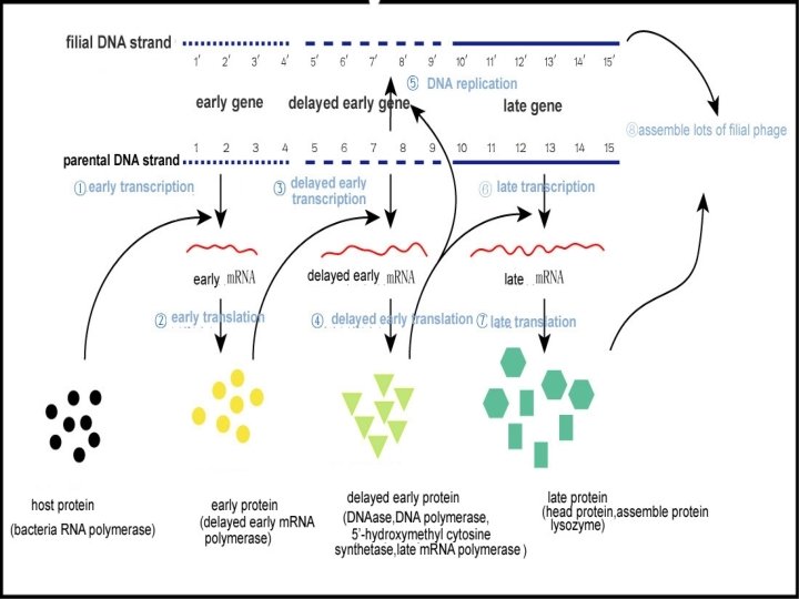
1. Virulent bacteriaphage and reproductive mode This kind of phages makes cell release virion by lysis cell when they penetrate host cell. Therefor they are called virulent phage, such as T 4 phage. The cycle of virus mutiplication: The cycle is separated into adsorption, penetration, duplication, assembly , maturation and release. This process iscalled lytic cycle.
1. Adsorption 2. Penetration
Types of Viral Nucleic Acids(RNA) Nucleic acid Type Nucleic Acid Structure Virus Examples Single-Stranded Linear, single stranded, positive strand Linear, single stranded, negative strand Linear, single stranded, segmented, positive strand Linear, single stranded, segmented, diploid (two identical single strands), positive strand Linear, single stranded, segmented, negative strand Linear, double stranded, segmented Picornaviruses(polio, rhinoviruses), togaviruses, RNAbacteriophages, TMV, and most plant viruses Rhabdoviruses(rabies), paramyxov -iruses(mumps, measles) Brome mosaic virus(individual segments in separate virions) Retroviruses(Rous sarcoma virus, human immunodeficiency virus) Paramyxoviruses, orthomyxovirus es(influenza) Retroviruses, wound-tumor virus of plants, cytoplasmic polyhdrosis virus of insects, phage Ф 6, many mycoviruses Double-stranded
3. Biosynthesis (a)The Reproduction of Double-Stranded DNA (T 4 Bacteriophages) Transcription Translation +m. RNA Proteins Replication ±DNA New virions
(b)The Reproduction of ФX 174, a +Strand DNA Replication Transcription Bacterial Translation DNA ±DNA polymerase +m. RNA +DNA (Replicative form) +DNA Proteins New virions
(c)The Reproduction of Single-Stranded RNA Bacteriophages RNA replicase +RNA ±RNA (replicative form) +RNA genomes m. RNA activity Replication Translation Virus Proteins New virions
RNA Animal virus Reproductive Strategies: (a)positive single-stranded RNA viruses(picornaviruses) +RNA Proteins(after processing by host and virus-coded proteinases New virions Viral replicase ±RNA Replicative form +RNA
(b) Double-stranded RNA viruses (reoviruses) RNA-dependent RNA polymerases* (Transcriptases) ±RNA +m. RNA Proteins RNA-dependent RNA polymerase (Replicase) ±RNA New virions
(c)Negative single-stranded RNA viruses (paramyxovirusesmumps and measles; orthomyxoviruses-influenza RNA-dependent RNA polymerase+ (Transcriptase) -RNA +m. RNA proteins RNN-dependent RNA polymerase (viral replicase) ±RNA -RNA Replicative form New virions
(d )Retroviruses (Rous sarcoma virus, HIV) RNA-dependent DNA polymerase Ribonuclease H Reverse + Reverse transcriptase + Component of transcriptase Proviral DNA -DNA +RNA/-DNA ±DNA RT Transcription +RNA(virion) +m. RNA Proteins New virions
4. Assembly and maturation
5. Release(lysis)
( 1) Eclipse period: it is the period from the virus penetrating host cell to the releasing of virion. Latent prophase or called as eclipse phase means the period during which virus nucleic acid penetrates host cell and assembly the first virion. At that time there are not complete, infectious mature virion. Latency anaphase: During this period, the titer of active virus particles inside cell rises dramatically, but are not release from host cell, which marks the end of latency. (2)Burst phase:During this period, the number of virion increases rapidly with lysis of host cell. (3)Platform phage:It is the period that all of the host cells are lysated and the valence of virus reaches the apogee.
Two characteristic data Eclipse period: :it is the minimum course from the penetration of virus to virion release. It is different from different viruses, such as phage’s eclipse period is counted by minute, animal and plant viruses are by hour or day. Burst size: it is the average number of released virion, which is equal to the number of all the released virion divided by infected cell numbers in eclipse period and is also equal to the ratio between viral valence in stable phage and viral valence in eclipse period. Determined by one step growth curve, the phage’s burst size is generously from a few to a few hundred and plant and animal viruses’ burst size are up to a few hundred even to ten thousand.
3. Template phage and lysogeny Some of phages integrate their genome into the host genome and duplicate along with host material at the time of cell division but do not induce host cell lysis after they penetrating the host cell. Therefore, they are called temperate phage. The concurrent state is called lysogeny, the infected cells called lysogen,the virus integrated into cell genome called as prophage.
裂解途径 诱导 溶原途径 细胞反复分裂 The mode of λphage lysis and lysogenic pathway
The characters of of lysogeny: (1)lysogenic conversion:The prophage will result in the phenotypic alternation of host cell, which is not directly related to the completion of life cycle and are known as lysogenic conversion. (2)Immunity:Although a lysogenic bacterium may be susceptible to other viruses, it can not be infected by virus particles of the type for which it is lysogenic. The λ repressor protein expressed by prophage gene can inhibits the duplication of virus in host cell.
3. Pathogenicity conversion:Some of prophages result in pathogenicity conversion. For instance, Corynebecterium diphtheriae and Clostridium botulinum are not toxic to human. Once infected by template phages, they are converted into toxogenic strains, resulting in damage to infected tissue and diphtherea and botulism toxic symtom. 4. Lysogeny is reversible : The prophages can be reactivated spontaneously of under outside stimulus, and enter lytic cycle with lysis of host cells. The probability of this process is very low(1/10000 -1/100000 or so).
3. Vertebrate animal virus There many virus parasitize in all kind s of vertebrate animals,but only those associated with human healthy and domestice livestock poultry are investigated much more. There are more than 300 kind of viruses associated with human health and more than 900 with other vertebrate animals. 70 -80% of human contagious diseases are caused by viruses. The vertebrate animals viruses are categorized into ds. RNA, ss. DNA, da. DNA, ds. RNA and ss. RNA. Some of them have envelope or not; Some of them have spike protein outside envelope or not.
1. Adsorption. The virus attaches to its host cell by specific binding of its spikes to cell receptors. 1. Vertebrate animal Viruses multiplication 2. Penetration. The virus is engulfed into a vesicle and its envelope is uncoated, thereby freeing the viral RNA into cell cytoplasm. Duplication. Under the control of viral genes, the cell synthesizes the basic components of new viruses: RNA molecule, capsomers, spikes. 5. Release. Enveloped viruses bud off of the membrane , carrying away an envelope with spikes. This complete virus of virion id ready to infect another cell. 4. Assembly. Viral spike proteins are inserted into the cell membrane for viral envelope; nucleocapsid id formed form RNA and capsomers.
The mode by which animal viruses adsorb to the host cell membrane.
1)Naked virus direct penetration. 2)Virus with envelope penetrates by endocytosis. 3)Virus with envelope penetrate by fusion of cell membrane The mode by which animal viruses penetrate
The mode by which animal viruses bud off and release:
2. Vertebrate animal virus persistence Some of virus do not kill host cell nut integrated its DNA into host genome or exist in host cell as plasmids after infecting the host. This kind of viral nucleic acid is called as protovirus.
The characters of persistence: 1)The persistent infected animal cells can keep normal function for many years. 2)The virus dose not duplicate and stay in persistence state befoe re activated. 3) The expression of integrated protovirus gene sometimes assign host cell new features. 4)Protoviruses are activated by environmental agents such as UV, then they are duplicated , matured and released , departing from the chrosome. 5)Some herper virus can “turn off/on” multiplication of virion. Under stimulus such as cold , heat, press or immune suppression , herper virus staying in organism cell all the life time will be reactivated to duplication and lysis of host cell, resulting in deasees, then come back to persistence state. Thereby, this kind of persistence os also celled as latency. 6) Some viruses lead to slow virus infections. Measles virus can hidden in brain cells for mang years, causing progressive damage and loss of function.
3. Virus and tumorgenesis Tumor : An abnormal mass of tissue that results from excessive cell division. Tumors perform no useful body function. They may be either begin or malignat. Benign tumors: do not invade neighboring tissues and do not seed metastases, but may locally grow to great size. They . usually do not return after surgical removal Malignant tumors are called cancer: . Cancer has the potential to invade and destroy neighboring tissues and create mtastases. There many complex factors, such as physical, chemical and biological factors , causing the human and vertebrate animal tumorgenesis. Some animal viruses enter their host cell and permanently alter its genetic material, leading to cancer. These viruses are termed oncovirus those are mainly ds. RNA viruses and retroviruses. Their effect on the cell is called transformation.
Some human cancers which may be caused by viruses Oncovirus cancer type Epstein-Barr(EB) (epstein-barr virus) Burkitt leucoma hepatitis b virus hepatitis c virus retrovirus htlv papovavirus hepatoma many animals leucemia huaman adenolymphoma each animal tumors; huamnuternin cervix cancer It is estimated that 15 % of human cancers are caused by virus infection.
Conspicuous character of oncovirus: can integrated its nucleic acid into host cell genome like lysogenic phage. The transformed cells’ growth rate increases uickly. The chromosome and cell surface molecules are changed. The cell can infinite division and lost the contact
The mechanism of cell transformation by oncovirus (1)The integration of protovirus. (2)The expression of specific gene related with transformation. These special transformation-related gene is termed Viral oncogenes,which is homologue of human and vertebrate animals proto-oncogene and is mutation of normal cell proto-oncogene. It is considered that the proto-oncogenes in normal host cell are very important regulating genes, which encoding proteins are very important in regulating cell proliferation and multiplication. The mutation proteins encoded by mutant proto-oncogenes will lead to cell aggressive growth. Some of human cancers are induced by several proto-oncogenes mutating at the same time, such as colon carcinima. Viruses may also induce cell abnormal growth. For example, the tumor viruses alter the proto-oncogenes activities or abnormal expression , therefor resulting in cell transformation and tumorgenesis.
4. Emerging viruses are the viruses those are regional low lever of infection and have interspecies barrier enlarge their host to other species , resulting in wide-ranging human infectious diseases. Such as ebola virus, lassa virus, AIDS virus. Recently emerging SARS virus and birds influenza virus are belonged to this kind of virus. We should pay much attention to these defection diseases caused by them.
The causes of emerging virus: 1)The environment are destroyed by human activity. 。 2)The modern transportation, development of tourism and large scale population migration. 3)The mutation of virus gene change the host range. 4)The intimate contact between human and animals.
References Prescott LM, Harley JP, and Klein DA. , Microbiology (5 th ed. ), Higher education press and Mc. Graw-Hill Companies, Inc. 2002 Michael TM, John MM, Jack P. Brock. , Biology of Micoorganisms,International edition, Pearson Education, Inc. 2003 Talaro K. P. Foundations in Microbiology (Fifth Edition), Higher education press and Mc. Graw-Hill Companies, Inc. 2005 Zhou De-qing,Microbiology textbook,second edition,high education publishing house,2002