Protein Structure Why protein folds Secondary structure Alphahelix
 Beta-sheet (beta-conformation) Beta-turn (b-turn) Ramanchandran")


 • Linked by peptide bonds •")

 f = -57°, y = -47° • Discovered by")
 • Some")
 – – • • •")


")
 Stick")
")


. “Mol. Script: A Program to Produce Both")
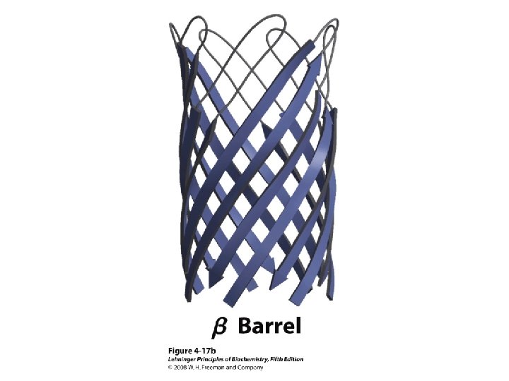
 β Barrel")
 α Helix")


 • Swiss-PDB viewer is an application that provides a user")


 are entire proteins or regions of proteins")


- Slides: 72
Protein Structure Why protein folds? Secondary structure Alpha-helix (a-helix) Beta-sheet (beta-conformation) Beta-turn (b-turn) Ramanchandran plot Tertiary & Quaternary Structure Motif & Domain Stable conformation Visualization of protein Denature of protein
Much More About Structure Function Structure Mechanism Structure Origins/Evolution Structure-Based Drug Design Solving the Protein Folding Problem
Protein Conformation Native protein Why Protein Folds? Peptide bond Disulfide bond Hydrogen bonding Ionic interaction Hydrophobic-lipophilic interaction Van der Waals interaction Rotational prohibition
Levels of Protein Structure • Primary – Linear AA sequence – Covalent bonds • Secondary – Local structure; certain “motifs” are common – Mostly H-bonds • Tertiary – Complete 3 D shape – H-bonds, hydrophobic interactions, ionic bonds, van der Waals interactions, disulfide bonds • Quaternary – >1 peptide chain – Mostly H-bonds
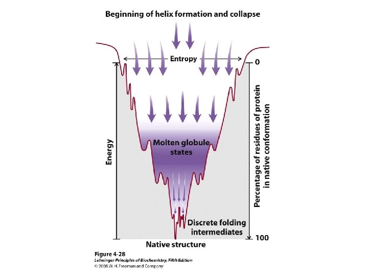
Protein Folding • Folded shape = conformation • Three-dimensional, functional structure = native – Energy of native conformation? • Molecular chaperones • There are thousands of possible conformations, but not an infinite amount… • Conformations are restrained by – planarity of peptide bond – “allowed” angles • No algorithm predicts the 3 D shape with high accuracy
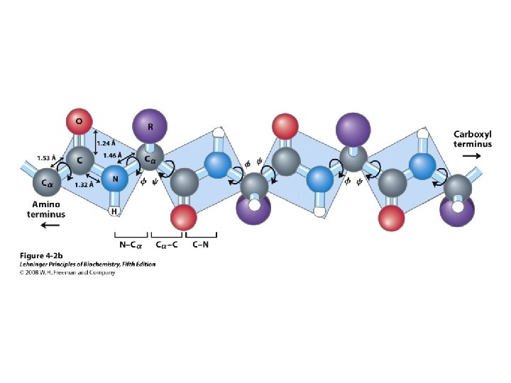
Primary Structure • AA sequence of polypeptide chain(s) • Linked by peptide bonds • Linear sequence • Predication of primary structure? • Experimental determination: protein sequencing
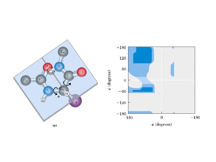
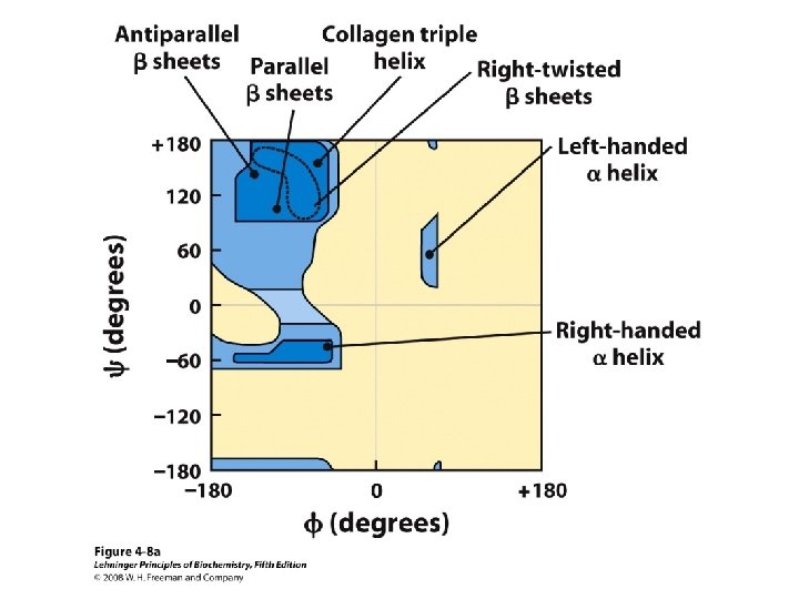
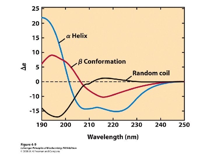
Secondary Structure • Regular repeating structure – Helices – Sheets • Torsion/dihedral angles – – Angles of rotation around Ca Clockwise (+) and counterclockwise (-) f = rotation around Ca-N y = rotation around Ca-C • How free is rotation? – Not very (sterics) – Avoid collision of C=O, N-H, R – Calculations of allowed values = Ramachandran diagram
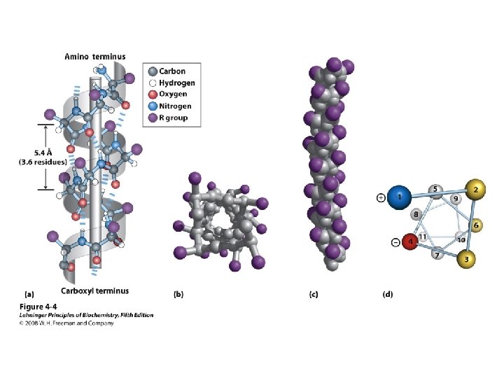
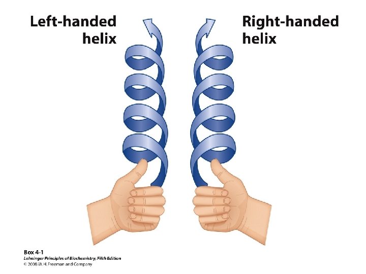
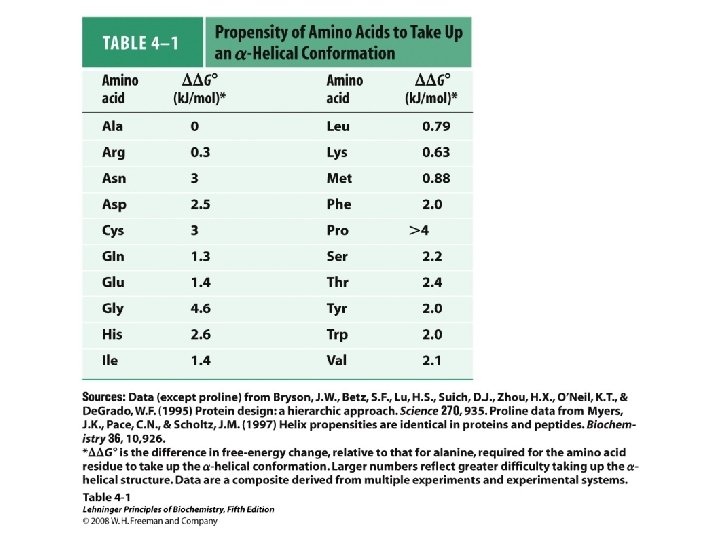
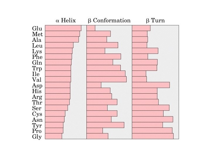
Alpha Helix • (30 -35%) f = -57°, y = -47° • Discovered by Pauling: 1951 • α-helix formers: A, C, L, M, E, Q, H, K • Tightly wound, repeating sequence • “Right-handed” • Each twist ∼ 5. 4 Å; 3. 6 residues • Average length = 18 residues • R-groups are on outside of helix • Stabilized by H-bonds between C=O (i) and N-H (i + 4)
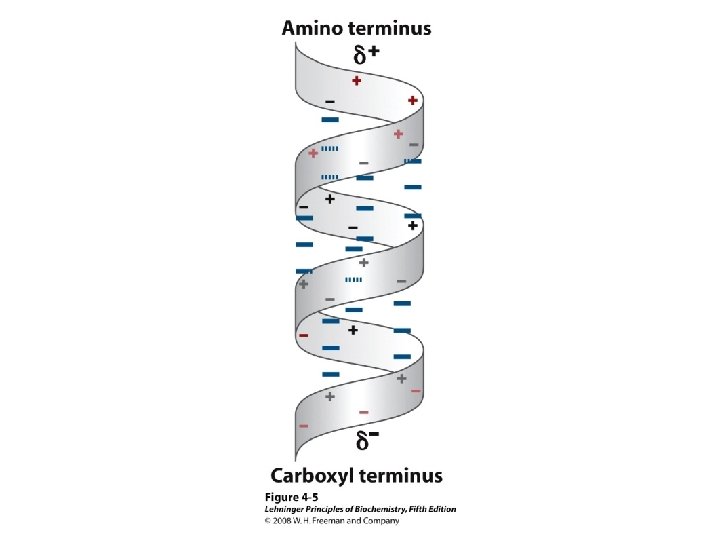
Alpha helix, cont. • Deviates from ideal conformation at ends (less H-bonding) • Some amino acids are “α-helix breakers” – Repeating like-charges – Repeating “bulky” groups – Pro and Gly • Effects on helical stability: – Electrostatic interactions between adjacent residues – Steric interference between adjacent residues – Interactions between residues 3 -4 amino acids away – Polarity of residues at both ends of helix (positive at amino end; negative at carboxyl)
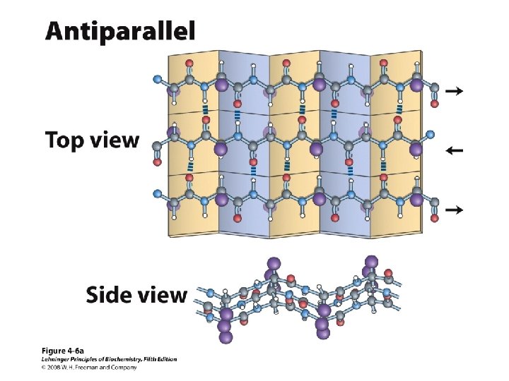
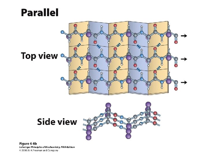
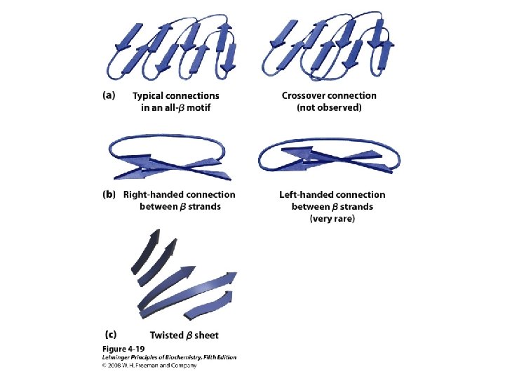
b-Sheet / b-Strand • Extended, zigzag conformation (20 -25%) – – • • • Hydrogen bond between groups across strands Forms parallel and antiparallel pleated sheets Residues alternate above and below β-sheet formers: V, I, P, T, W Intrastrand H-bonding Average 6 residues/strand; up to 15 2 -12 strands/sheet; average 6 R-groups alternate on opposite sides of sheet Distortions: – Beta-bulge = extra residue – Kink = Pro
Anti-parallel vs. Parallel • Anti-parallel b-sheet – Opposite orientation f = -140°, y = 135° – More stable – Can be twisted – 6. 5 Å per two amino acid residues – Can withstand distortions and exposure to solvent • Parallel b-sheet – Same amino-carboxyl direction – Less twisted – Tend to be buried f = -120°, y = 115° – 7. 0 Å per two amino acid • Can have mix of parallel and anti-parallel
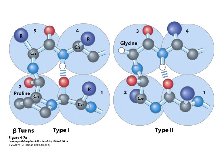
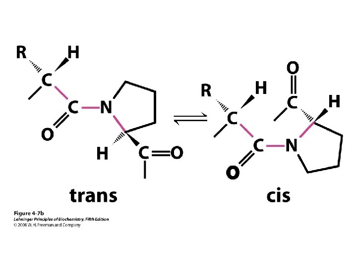
b-turns • Interacting strands can be many amino acids apart • Turns are 180°; “connect” strands in folded (globular) proteins • Interaction is between carbonyl oxygen of AA 1 and amino hydrogen of AA 4 – Short turn (4 residues) – Hydrogen bond between C=O & NH groups within strand (3 positions apart) – Usually polar, found near surface – β-turn formers: S, D, N, P, R • Pro and Gly are often present – Gly: small and flexible (Type II turns) – Pro: cis conformation makes inclusion in tight turn favorable
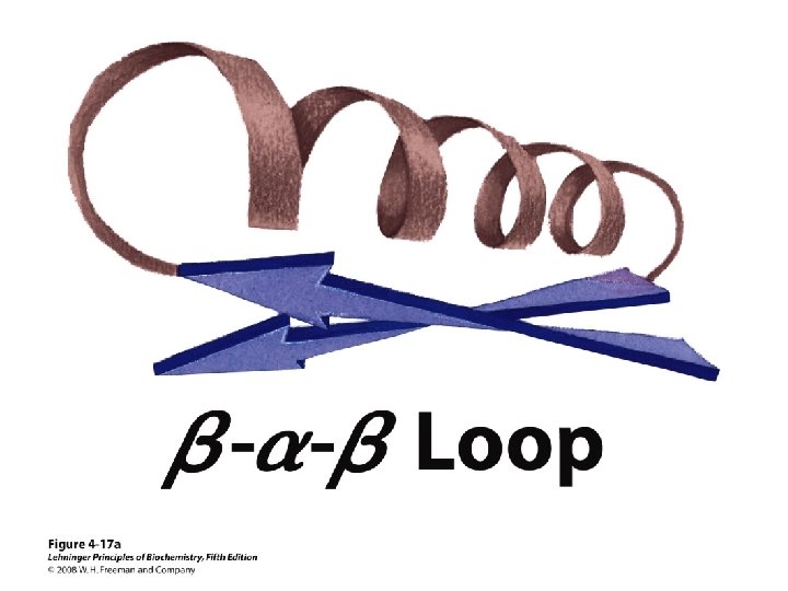
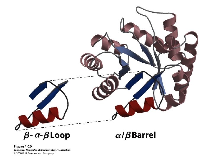
Others • Loop – Regions between α-helices and β-sheets – On the surface, vary in length and 3 D configurations – Do not have regular periodic structures – Loop formers: small polar residues • Coil (40 -50%) – Generally speaking, anything besides αhelix, β-sheet, β-turn
Proteins are Complex • • Average residue contains 8 “heavy” atoms Average protein contains 300 amino acids Average structure contains 2400 atoms First structure (sperm whale myoglobin) took ~5 years with a team of ~15 key punch operators working around the clock to solve • Most structures still take 1 year to solve
Solving Protein Structures • Only 2 kinds of techniques allow one to get atomic resolution pictures of macromolecules • X-ray Crystallography (first applied in 1961 - Kendrew & Perutz) • NMR Spectroscopy (first applied in 1983 - Ernst & Wuthrich)
X-ray Crystallography • • Crystallization Diffraction Apparatus Diffraction Principles Conversion of Diffraction Data to Electron Density • Resolution • Chain Tracing
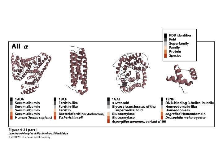
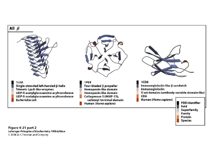
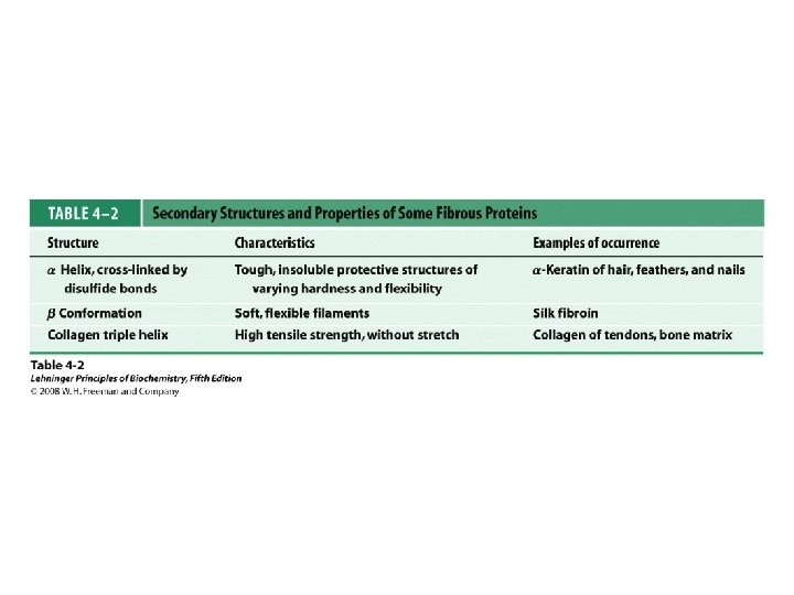
Classification of Proteins by Secondary Structure • Fibrous – High composition of single secondary structure – Strong and flexible – Collagen • Triple helix (lefthanded helices, righthanded super helix) – Silk fibroin • Anti-parallel a-sheet a-Keratin • Left-handed coil • Globular – Majority of all proteins – Contain several types of secondary structure (regular and non-regular) – Percentage of protein (on average): • • 31% a-helix 28% b-sheet 13% turns/bends 28% loops and random coil
Aspects Which Determine Tertiary Structure • Covalent disulfide bonds from between closely aligned cysteine residues form the unique Amino Acid cystine. • Nearly all of the polar, hydrophilic R groups are located in the surface, where they may interact with water • The nonpolar, hydropobic R groups are usually located inside the molecule
Protein Visualization Model CPK Lin e Trace Cartoon
Protein Visualization Model Cylinder Ribbon (N-C gradient)
Protein Visualization Model Ribbon (2 o structure) Stick
Protein Visualization Model Space Filling Wire Frame (Vector)
Protein Visualization Model Wireframe Ball and stick
Visualization Software • • • • • Biodesigner CACTVS Chemdraw net Plugin Chemical 2 vmd Chimera Cn 3 D CONSCRIPT Dino Flex. V Garlic Gdis g. Open. Mol GRASP Hyperactive Molecules Using Chemical MIME ICMLite Image. Magick INTERCHEM modelling software Jmol n n n n n n Kinemage Mac. Molecule 2 and PCMolecule 2 Maestro Marvin Applets and Java. Beans Mercury Mind. Tool Molden MOLEKEL MOLMOL Mol. Pov Mol. Script Mol. View and Mol. View Lite MSP NIH Image O ORTEX POV-Ray Pov 4 Grasp Pov. Chem Protein Explorer PSI 88 Py. MOL n n n n n Qmol QTree Ras. Mol Raster 3 D Ribbons RNA Movies Rna. Viz Spock Swiss-Pdb. Viewer Tachyon VEGA Viewmol VMD Web. Lab Viewer. Lite Web. Mol Win. MGM XMol
Mol. Script • Kraulis, P. J. (1991). “Mol. Script: A Program to Produce Both Detailed and Schematic Plots of Protein Structure. ” J. Appl. Cryst. 24: 946 -950 • Web Site: http: //www. avatar. se/molscript/ • Mol. Script is a program for displaying molecular 3 D structures, such as proteins, in both schematic and detailed representations. • Helps from scripting language, we can render the representation by ourselves.
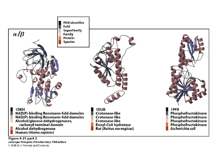
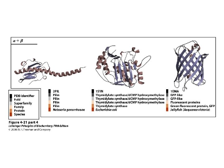
Molscript Representations of Typical Protein Architectures. β 7 Propellor (2 bbk. H) β Barrel (2 por) α 4 -Layer Sandwich (2 dnj. A) Sandwich (2 hla. B)
Molscript Representations of Typical Protein Architectures. α β Barrel (4 tim. A) α Helix Bundle (2 ccy) 2 Solenoid (1 tsp) α Horseshoe (1 bnh)
Ras. Mol/Open. Ras. Mol • Ras. Mol is a program for molecular graphics visualization originally developed by Roger Sayle. • Web Site: http: //www. openrasmol. org/ • “Grand-daddy” of all visual freeware • The most popular one viewer.
Rasmol
Jmol • Jmol is a free, open source molecule viewer for students, educators, and researchers in chemistry and biochemistry. • It’s cross-platform, running on Windows, Mac OSX, and Linux/UNIX systems. • Web Site: http: //jmol. sourceforge. net/ • It has more detailed materials on how to write a script language and an interactive web UI to demonstrate. • It’s a Java based application.
1 ATP vs. 1 BO 1 PDBID: 1 ATP PDBID: 1 BO 1
Py. MOL • Py. MOL is a user-sponsored molecular visualization system on a open-source foundation. • Web Site: http: //pymol. sourceforge. net/ • It’s a Python based application.
Deep. View (Swiss-PDB Viewer) • Swiss-PDB viewer is an application that provides a user friendly interface allowing to analyze several proteins at the same time. • Web Site: http: //www. expasy. org/spdbv/ • Swiss-PDB viewer is not just a viewer and more powerful than the other viewers. • It also supports SWISS-MODEL, a fully automated protein structure homology-modeling server. • SWISS-MODEL: An Automated Comparative protein Modeling Server. • It also provides a function of structure alignment.
Swiss PDB Viewer
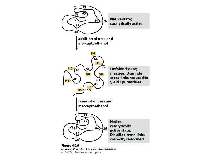
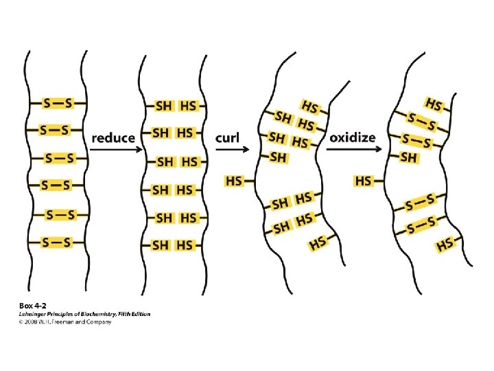
Protein Denaturation The loss of secondary, tertiary, or quaternary protein structure due to disruption of noncovalent interactions and/or disulfide bonds that leaves peptide bonds and primary structure intact.
Factors That Cause Protein to Denature Heat p. H change Hydrogen bonding reagents Detergents Non-polar solvents Extremely high or low salt concentration Oxidants/reductants Mechanical Agitation
What is protein disorder? Disordered regions (DRs) are entire proteins or regions of proteins which lack a fixed tertiary structure, essentially being partially or fully unfolded. Such disordered regions have been shown to be involved in a variety of functions, including DNA recognition, modulation of specificity/affinity of protein binding, molecular threading, activation by cleavage, and control of protein lifetimes. Although these DRs lack a defined 3 -D structure in their native states, they frequently undergo disorder-to-order transitions upon binding to their partners. Depiction of degrees of protein disorder: 0 – totally ordered 1 -5 – partial disorder 6, 7 – total disorder
• Heat: The weak side-chain attractions in globular proteins are easily disrupted by heating, in many cases only to temperatures above 50°C. • Mechanical agitation: The most familiar example of denaturation by agitation is the foam produced by beating egg whites. Denaturation of proteins at the surface of the air bubbles stiffens the protein and causes the bubbles to be held in place. • Detergents: Even very low concentrations of detergents can cause denaturation by disrupting the association of hydrophobic side chains.
• Organic compounds: Organic solvents can interfere with hydrogen bonding or hydrophobic interactions. The disinfectant action of ethanol results from its ability to denature bacterial protein. • p. H change: Excess H+ or OH- ions react with the basic or acidic side chains in amino acid residues and disrupt salt bridges. An example of denaturation by p. H change is the protein coagulation that occurs when milk turns sour because it has become acidic. • Inorganic salts: Sufficiently high concentrations of ions can disturb salt bridges. • Most denaturation is irreversible. Hard-boiled eggs do not soften when their temperature is lowered.