Figure 21 1 The major respiratory organs in


 0 mm Hg")
- Slides: 18
Figure 21. 1 The major respiratory organs in relation to surrounding structures. Nasal cavity Nostril Oral cavity Pharynx Larynx Trachea Carina of trachea Right main (primary) bronchus Right lung Left main (primary) bronchus Left lung Diaphragm © 2014 Pearson Education, Inc.
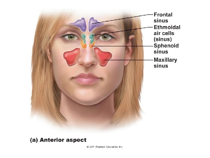
Figure 21. 3 b The upper respiratory tract. Cribriform plate of ethmoid bone Sphenoid sinus Frontal sinus Nasal cavity Nasal conchae (superior, middle and inferior) Nasal meatuses (superior, middle, and inferior) Nasal vestibule Posterior nasal aperture Nasopharynx Pharyngeal tonsil Opening of pharyngotympanic tube Uvula Nostril Oropharynx Palatine tonsil Isthmus of the fauces Hard palate Soft palate Tongue Lingual tonsil Laryngopharynx Esophagus Trachea © 2014 Illustration Pearson Education, Inc. Larynx Epiglottis Vestibular fold Thyroid cartilage Vocal fold Cricoid cartilage Thyroid gland Hyoid bone
Figure 21. 4 a The larynx. Epiglottis Thyrohyoid membrane Body of hyoid bone Thyroid cartilage Laryngeal prominence (Adam’s apple) Cricothyroid ligament Cricoid cartilage Cricotracheal ligament Tracheal cartilages © 2014 Pearson Education, Inc. Anterior superficial view
Figure 21. 4 b The larynx. Epiglottis Thyrohyoid membrane Body of hyoid bone Thyrohyoid membrane Cuneiform cartilage Fatty pad Corniculate cartilage Vestibular fold (false vocal cord) Arytenoid cartilage Thyroid cartilage Arytenoid muscles Vocal fold (true vocal cord) Cricoid cartilage Cricothyroid ligament Cricotracheal ligament Tracheal cartilages Sagittal view; anterior surface to the right © 2014 Pearson Education, Inc.
Figure 21. 4 c The larynx. Epiglottis Hyoid bone Thyroid cartilage Lateral thyrohyoid membrane Corniculate cartilage Arytenoid cartilage Glottis Cricoid cartilage Tracheal cartilages © 2014 Pearson Education, Inc. Photograph of cartilaginous framework of the larynx, posterior view
Figure 21. 5 a Movements of the vocal folds. Base of tongue Epiglottis Vestibular fold (false vocal cord) Vocal fold (true vocal cord) Glottis Cuneiform cartilage Corniculate cartilage Vocal folds in closed position; closed glottis © 2014 Pearson Education, Inc.
Figure 21. 6 a Tissue composition of the tracheal wall. Posterior Mucosa Esophagus Trachealis muscle Submucosa Lumen of trachea Seromucous gland in submucosa Hyaline cartilage Adventitia Anterior Cross section of the trachea and esophagus © 2014 Pearson Education, Inc.
Figure 21. 6 b Tissue composition of the tracheal wall. Goblet cell Mucosa • Pseudostratified ciliated columnar epithelium • Lamina propria (connective tissue) Submucosa Seromucous gland In submucosa Hyaline cartilage © 2014 Pearson Education, Inc. Photomicrograph of the tracheal wall (320 x)
Figure 21. 6 c Tissue composition of the tracheal wall. Scanning electron micrograph of cilia in the trachea (2500 x) © 2014 Pearson Education, Inc.
Figure 21. 7 Conducting zone passages. Trachea Superior lobe of left lung Left main (primary) bronchus Superior lobe of right lung Lobar (secondary) bronchus Segmental (tertiary) bronchus Middle lobe of right lung Inferior lobe of right lung © 2014 Pearson Education, Inc. Inferior lobe of left lung
Figure 21. 11 A cast of the bronchial tree. Right lung Right superior lobe (3 segments) Left lung Left superior lobe (4 segments) Right middle lobe (2 segments) Right inferior lobe (5 segments) © 2014 Pearson Education, Inc. Left inferior lobe (5 segments)
Figure 21. 8 Respiratory zone structures. Alveoli Alveolar duct Respiratory bronchioles Alveolar duct Terminal bronchiole Alveolar sac Respiratory bronchiole Alveolar duct Alveoli Alveolar sac © 2014 Pearson Education, Inc. Alveolar pores
Figure 21. 9 a Alveoli and the respiratory membrane. Terminal bronchiole Respiratory bronchiole Smooth muscle Elastic fibers Alveolus Capillaries Diagrammatic view of capillary-alveoli relationships © 2014 Pearson Education, Inc.
Figure 21. 9 c Alveoli and the respiratory membrane. Red blood cell Nucleus of type I alveolar cell Alveolar pores Capillary Macrophage Endothelial cell nucleus Alveolus Respiratory membrane Alveoli (gas-filled air spaces) Red blood cell in capillary Type II alveolar cell Type I alveolar cell Detailed anatomy of the respiratory membrane © 2014 Pearson Education, Inc. Alveolus Alveolar epithelium Fused basement membranes of alveolar epithelium and capillary endothelium Capillary endothelium
Figure 21. 10 a Anatomical relationships of organs in the thoracic cavity. Intercostal muscle Rib Lung Parietal pleura Pleural cavity Visceral pleura Trachea Thymus Apex of lung Right superior lobe Horizontal fissure Right middle lobe Oblique fissure Left superior lobe Oblique fissure Left inferior lobe Right inferior lobe Heart (in mediastinum) Diaphragm Cardiac notch Base of lung Anterior view. The lungs flank mediastinal structures laterally. © 2014 Pearson Education, Inc.
Figure 21. 12 Intrapulmonary and intrapleural pressure relationships. Atmospheric pressure (Patm) 0 mm Hg (760 mm Hg) Parietal pleura Thoracic wall Visceral pleura Pleural cavity Transpulmonary pressure 4 mm Hg (the difference between 0 mm Hg and − 4 mm Hg) – 4 0 Lung Diaphragm © 2014 Pearson Education, Inc. Intrapulmonary pressure (Ppul) 0 mm Hg (760 mm Hg) Intrapleural pressure (Pip) − 4 mm Hg (756 mm Hg)