Practical Clinical Hematology 11 Leukemia THE LEUKEMIAS l


 acute myeloblastic")


 High N: C ratio (3:")
 o High N: C")
 • N: C ratio (2: 1)")
 o N: C")


 • • lymphoblast predominant cell type most frequent in children")


 (ALL) showing lymphoblasts and one")
. The blast cells are")


")

and (B), Hypergranular acute promyelocytic leukemia, promyelocytes with prominent azurophilic")

 leukemia (CLL) o mature-appearing")


- Slides: 72
Practical Clinical Hematology 11 Leukemia
THE LEUKEMIAS l l malignancies involving lymphoid or hematopoietic cell lines increase in circulating leukocytes (not invariable) bone marrow always involved replacement/hypoproliferation of normal hematopoietic cells – – anemia, infection, hemorrhage leukemic infiltration in liver, spleen, lymph nodes, and other tissues
THE LEUKEMIAS l acute leukemias l chronic leukemias
ALL naïv e B-lymphocytes Lymphoid progenitor Plasma cells T-lymphocytes AML Hematopoietic stem cell Myeloid progenitor Neutrophils Eosinophils Basophils Monocytes Platelets Red cells
THE LEUKEMIAS l acute leukemias – – l acute lymphoblastic leukemia (ALL) acute myeloblastic leukemia (AML) chronic leukemias – – chronic lymphoid leukemia (CLL) chronic myelogenous leukemia (CML)
Morphological Features of Blasts in Acute Myeloid and Acute Lymphoblastic Leukemias Feature AML ALL Blast size Larger, usually uniform Variable, small to medium size Nuclear chromatin Usually finely dispersed Coarse to fine Nucleoli 1 -4, often prominent Absent or 1 or 2, often indistinct Moderately abundant, fine granules often present Usually scant, coarse granules sometimes present (~7%) Auer rods Present in 60%-- 70% of cases Not present Other cell types Often dyspastic changes in maturing myeloid cells Cytoplasm Usually not dysplastic
AML
Leukemias Acute leukemias Lymphoid L 1 L 2 L 3 Chronic leukemias Myeloid M 0 M 1 M 2 M 3 M 4 M 5 M 6 M 7 The original FAB-Classification system of Acute Leukemias, heavily based on morphologic findings
Maturation and Morphology of Immature Granulocytes myeloblast promyelocyte metamyelocyte MATURATION band neutrophil
Maturation and Morphology of Immature Granulocytes • Myeloblast: Ø The first and earliest granulocyte (15 μm) Ø High nucleus to cytoplasm (N: C) ratio (5: 1) Ø Round or oval nucleus with loose light staining euchromatin Ø 1 -2 nucleoli Ø Has minimal light blue cytoplasm Ø Contains no cytoplasmic granules Ø Begins to produce myeloperoxidase granules (MPO) Ø Comprises 1% of the nucleated cells in the bone marrow
• Promyelocyte: larger than a myeloblast (20 μm) High N: C ratio (3: 1) Loose chromatin with nucleoli Dark blue cytoplasm Contains large nonspecific cytoplasmic granules o Containing myeloperoxidase (MPO) o Comprises 3 -4% of nucleated bone marrow cells o o o
• Neutrophilic Myelocyte: o medium cell size (12 μm) o High N: C ratio (3: 1) o Round, oval, or slightly indented nucleus with darker o blue heterochromatin o Last stage of cell division o Has active RNA, therefore, the cytoplasm is blue o Contains MPO and secondary granules containing leukocyte alkaline phosphatase o Comprises 12% of bone marrow nucleated cells
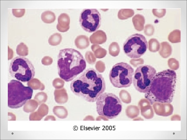
• Neutrophilic Metamyelocyte: • size (11 μm) • N: C ratio (2: 1) • last mononuclear stage, no mitosis • Nucleus is kidney or horseshoe shaped, and has condensed heterochromatin • Has a prominent Golgi apparatus – clear area located at the indentation site of the nucleus • Cytoplasm is similar to the mature cell • Comprises 18% of bone marrow cells
Band o Same size as a mature neutrophil (10 -12 μm) o N: C ratio has reversed (1: 2) o Nucleus is band- or sausageshaped without segmentation o Cytoplasm is filled with small neutrophilic granules o Last immature stage o Comprises 11% of bone marrow cells and 0 -3% of peripheral WBCs Stored in the bone marrow and released when there is an increased demand for neutrophils • Shift to the left is an increase in immature cells indicating increased demand for WBCs in peripheral blood
Neutrophils • • Also known as segmented neutrophils, segs, polymorphonuclear cells, polys, and PMNs N: C ratio is 1: 3, and the size is 10 -12 μm Average nucleus contains 3 -5 segments connected by narrow filaments Hyposegmented is less than 3 segments, and may indicate a shift to the left or an anomaly Hypersegmented is more than 5 segments and may indicate infection or megaloblastic anemia Cytoplasm contains very small neutral granules Makes up 55 -75% of all peripheral WBCs • Granules can become larger upon bacterial infection producing toxic granulation, which are numerous, large, basophilic granules
• Eosinophils o Average size is 13 μm o Nucleus is generally bilobed o Cytoplasm is bright red or orange which is due to large specific, secretory granules containing peroxidase, acid phosphatase, aryl sulfatase, betaglucuronidase, etc. that stain red with the eosin component of Wright’s stain
• Basophils o Is the smallest granulocyte at 10 μm o The nucleus is difficult to see due to heavy granulation o Cytoplasm contains large specific, secondary granules that contain heparin and histamines, which stain purple with Wright’s stain. These granules are water soluble and sometimes appear as holes in the cell if the cells are not fixed well during staining. o Makes up to 0. 5% of peripheral WBCs • Note: Tissue mast cells are similar to basophils but are larger and have no developmental relationship with basophils. Mast cells have a mesenchymal (connective tissue) origin and have granules containing serotonin (basophilic granules contain no serotonin).
Acute Non-Lymphoblastic Leukaemia Class M 0 M 1 Alternative AML with minimal differentiation AML without maturation M 2 AML with maturation M 3 APL M 4 AMMo. L M 5 AMo. L M 6 AEL M 7 AMeg. L Bone Marrow Appearance Identified by ultrastructural myeloperoxidase activity or immunophenotyping. Monomorphic with one or more distinct nucleoli, occasional auer rod and at least 3% myeloperoxidase positivity. 50% OR > myeloblasts & promyelocytes and common single auer rod. Dysplastic myeloid differentiation may also be present. Dominant cell type is promyelocyte with heavy azurophilic granulation. Bundles of Auer rods confirm diagnosis. Microgranular variant exist (M 3 v) As M 2 but > 20% promonocytes & monocytes. > 80% monoblasts is poorly differentiated (M 5 a) > 80% monoblasts, promonocytes or monocytes is well differentiated (m 5 b) >50% bizzar, dysplastic nucleated red cells with multinucleate forms and cytoplasmic bridging. Myeloblasts usually > 30%. Fibrosis, heterogeneous blasts population with cytoplasmic blebs. Platelet peroxidase positive.
AML
AML M 0 • the blasts are undifferentiated by morphology and cytochemistry • The cytoplasm is usually scant, and grey to light blue in color without granules, and Auer rods are not seen. • N: C ratio is high • The nucleus is round to oval or irregular and usually eccentrically placed • The nuclear chromatin ranges from being finely granular and evenly dispersed to being slightly clumped • One or more nucleoli may be visible
AML M 0 Bone marrow smear, May-Giemsa stain, x 1000 the enzyme MPO demonstrated by immunocytochemistry analysis
AML M 1 • The blasts vary in size, but are approximately the size of mature segmented neutrophils • The nuclei are round to slightly oval or irregular, and the nuclear chromatin ranges from finely granular and evenly distributed to slightly clumped. • One to two nucleoli are usually visible • The cytoplasm is scant and greyblue to light blue in color, and agranular. • N: C ratio is high • Auer rods are seen in a minority of blasts
M 1
M 1
M 1 Immunostain antibody: CD 33 Bone marrow smear, Peroxidase stain
FAB M 1
FAB M 1 AUER ROD
M 2
M 2
M 2 Peroxidase stain with eosinophilia
FAB M 2
FAB M 2
FAB M 3
FAB M 3
FAB M 3
FAB M 4
FAB M 4 Double esterase stain
FAB M 5
FAB M 5
FAB M 5
FAB M 6
FAB M 6
FAB M 6
FAB M 6 PAS Stain
ALL
ACUTE LYMPHOBLASTIC LEUKEMIA (ALL) • • lymphoblast predominant cell type most frequent in children better prognosis than AML FAB classification L 1 -L 3 o not used as a diagnosis, just as a descriptor o entirely based on morphology
Morphological Features of Acute Lymphoblastic Leukemia L 1 L 2 L 3 small to intermediate large, heterogeneous large, homogeneous Nuclear shape uniform pleiomorphic indentations common uniform Nucleoli small or absent large, prominent often single Cytoplasm scanty large, prominent moderate (basophilic) Cytoplasmic vacuoles none to few prominent Cell size
FAB L 1 type of ALL
FAB L 2 type of ALL
FAB L 2 type of ALL
FAB L 3 type of ALL
FAB L 3 type of ALL
FAB L 3 type of ALL
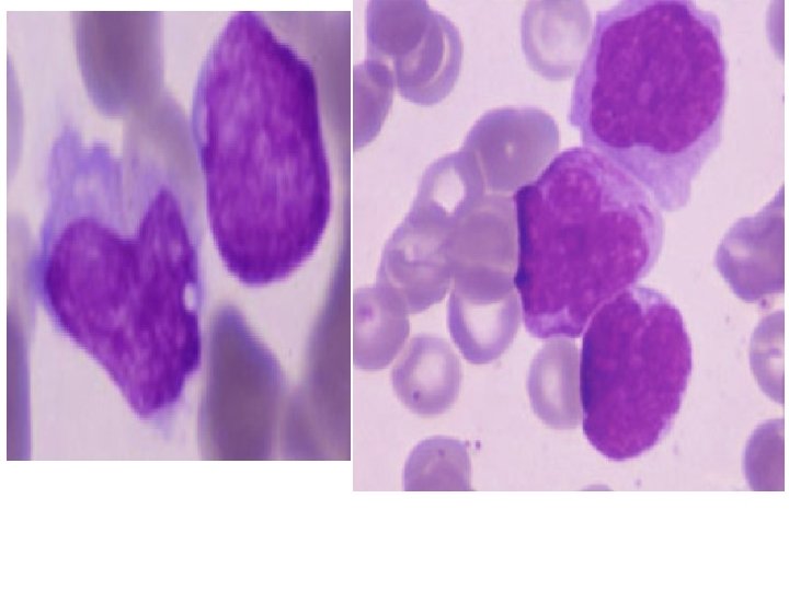
Peripheral blood film in L 1 (acute lymphoblastic leukaemia) (ALL) showing lymphoblasts and one nucleated red blood cell (NRBC). The lymphoblasts vary in size but are relatively uniform in morphology. The smaller blast cells show some chromatin condensation, which can be a feature of lymphoblasts but not of myeloblasts. This case was shown on immunophenotyping to be of B lineage.
Peripheral blood film in L 2 acute lymphoblastic leukaemia (ALL). The blast cells are larger and more pleomorphic than in LI ALL and in this case have a more diffuse chromatin pattern; one of the blasts has a hand-mirror conformation. This case was shown on immunophenotyping to be of T lineage
Other Lymphoid Leukemia
Hairy cell leukemia. Lymphocytes with filamentous cytoplasmic projections
Hairy cell leukemia
Hairy cell leukemia
Hairy cell leukemia (positive TRAP stain)
Prolymphocytic leukemia.
• • • (A)and (B), Hypergranular acute promyelocytic leukemia, promyelocytes with prominent azurophilic granules. (C) Hypergranular APL with multiple Auer rods. (D) Microgranular APLv. These abnormal promyelocytes have lobulated nuclei and absent or fine azurophilic granules.
Chronic Leukemia
CHRONIC LEUKEMIAS • longer clinical course • chronic lymphocytic (lymphatic) leukemia (CLL) o mature-appearing lymphocytes • chronic myelocytic (myeloid, myelogenous, granulocytic) leukemia (CML, CGL) o segs, bands, metamyelocytes, and myelocytes
CLL
CML
CML
CML
CML