
Advanced Hematology Medi 6304 Ahmad Silmi Second semester
- Slides: 64
Advanced Hematology (Medi 6304) Ahmad Silmi Second semester MAR 6 th 2017
Hemolytic anemia: Definition v Anemia results from an excessive hemolysis of RBC’s v BM compensatory capacity: 6 -8 folds of normal Several folds destruction rate Life span with erythrocytes less than 30 days Anemia developed (Signs & symptoms) v Two type of hemolysis: 1. Intra-vascular hemolysis 2. Extra-vascular hemolysis
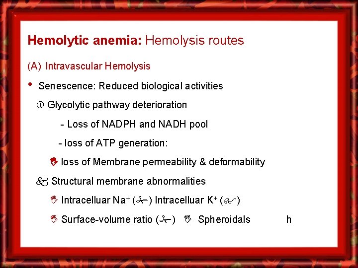
Hemolytic anemia: Hemolysis routes (A) Intravascular Hemolysis • Senescence: Reduced biological activities Glycolytic pathway deterioration - Loss of NADPH and NADH pool - loss of ATP generation: loss of Membrane permeability & deformability Structural membrane abnormalities Intracelluar Na+ ( ) Intracelluar K+ ( ) Surface-volume ratio ( ) Spheroidals h
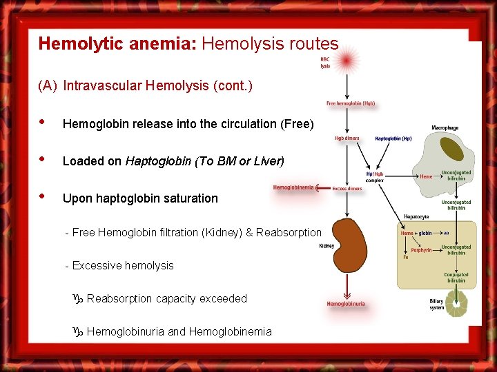
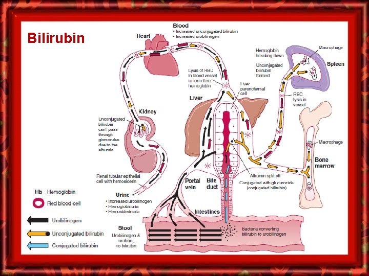
Hemolytic anemia: Hemolysis routes (A) Intravascular Hemolysis (cont. ) • Hemoglobin release into the circulation (Free) • Loaded on Haptoglobin (To BM or Liver) • Upon haptoglobin saturation - Free Hemoglobin filtration (Kidney) & Reabsorption - Excessive hemolysis Reabsorption capacity exceeded Hemoglobinuria and Hemoglobinemia
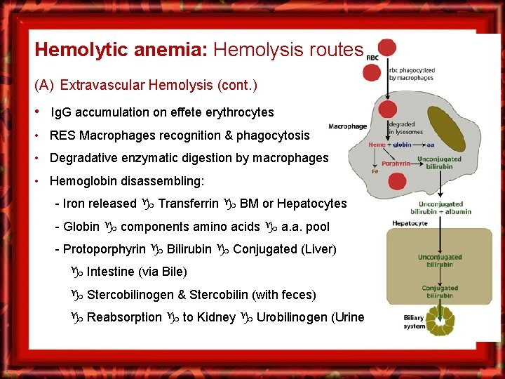
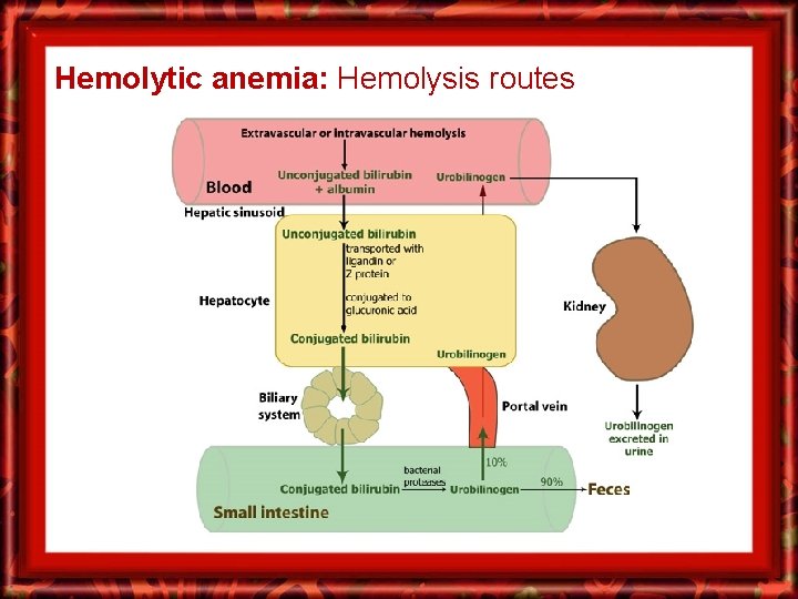
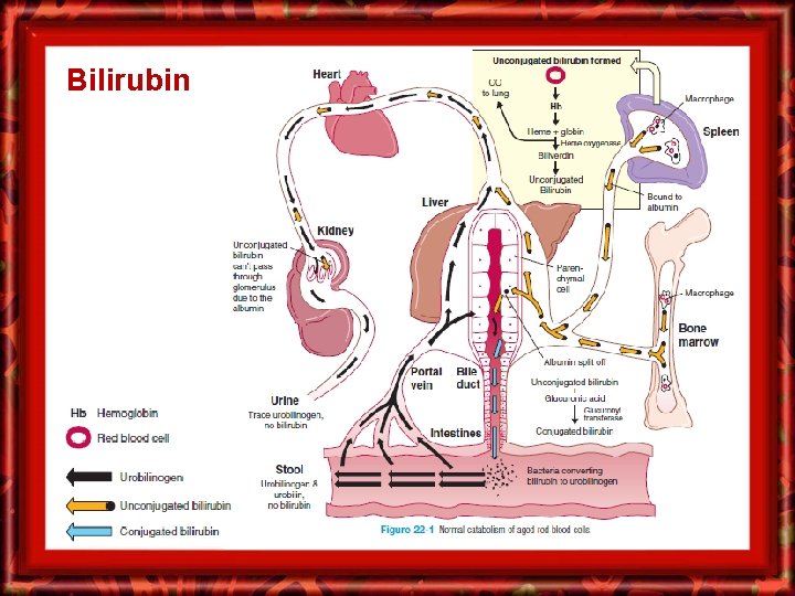
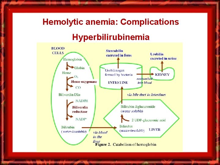
Hemolytic anemia: Hemolysis routes (A) Extravascular Hemolysis (cont. ) • Ig. G accumulation on effete erythrocytes • RES Macrophages recognition & phagocytosis • Degradative enzymatic digestion by macrophages • Hemoglobin disassembling: - Iron released Transferrin BM or Hepatocytes - Globin components amino acids a. a. pool - Protoporphyrin Bilirubin Conjugated (Liver) Intestine (via Bile) Stercobilinogen & Stercobilin (with feces) Reabsorption to Kidney Urobilinogen (Urine
Hemolytic anemia: Hemolysis routes
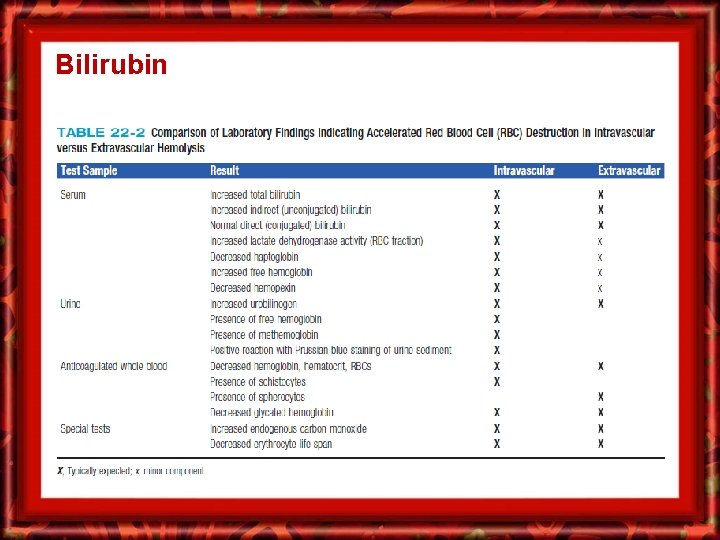
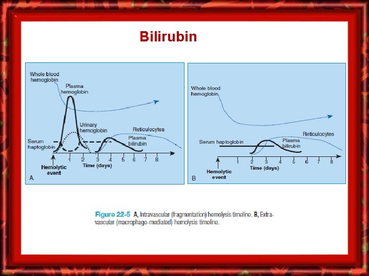
Bilirubin
Bilirubin
Bilirubin
Bilirubin
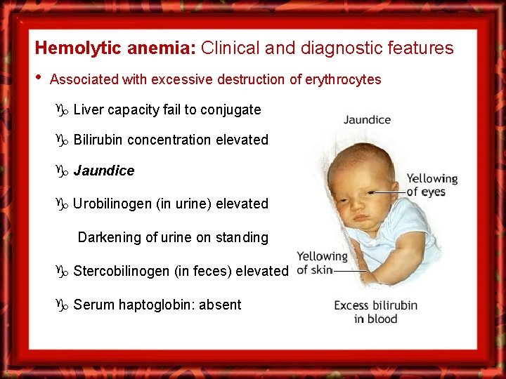
Hemolytic anemia: Clinical and diagnostic features • Associated with excessive destruction of erythrocytes Liver capacity fail to conjugate Bilirubin concentration elevated Jaundice Urobilinogen (in urine) elevated Darkening of urine on standing Stercobilinogen (in feces) elevated Serum haptoglobin: absent
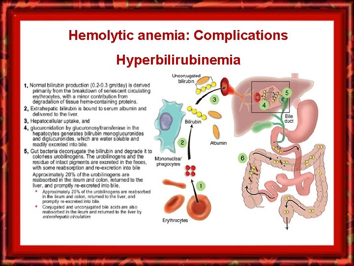
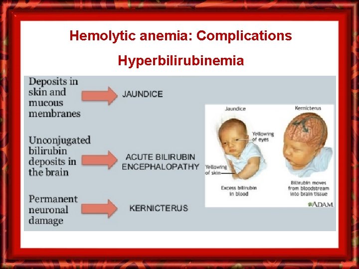
Hemolytic anemia: Complications Hyperbilirubinemia
Hemolytic anemia: Complications Hyperbilirubinemia
Hemolytic anemia: Complications Hyperbilirubinemia
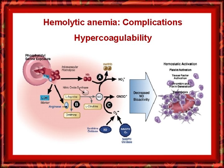
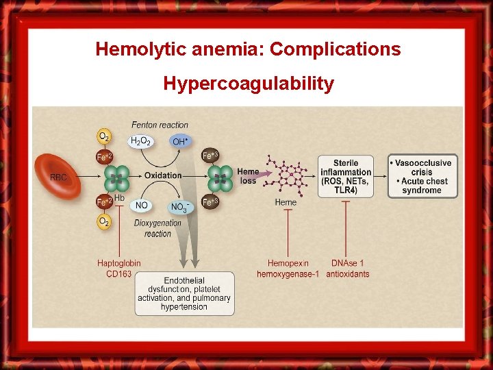
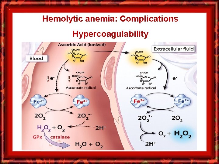
Hemolytic anemia: Complications Hypercoagulability
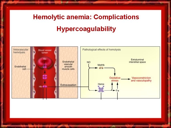
Hemolytic anemia: Complications Hypercoagulability
Hemolytic anemia: Complications Hypercoagulability
Hemolytic anemia: Complications Hypercoagulability
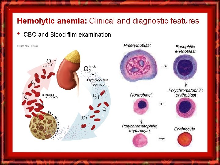
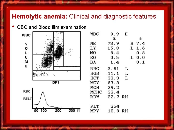
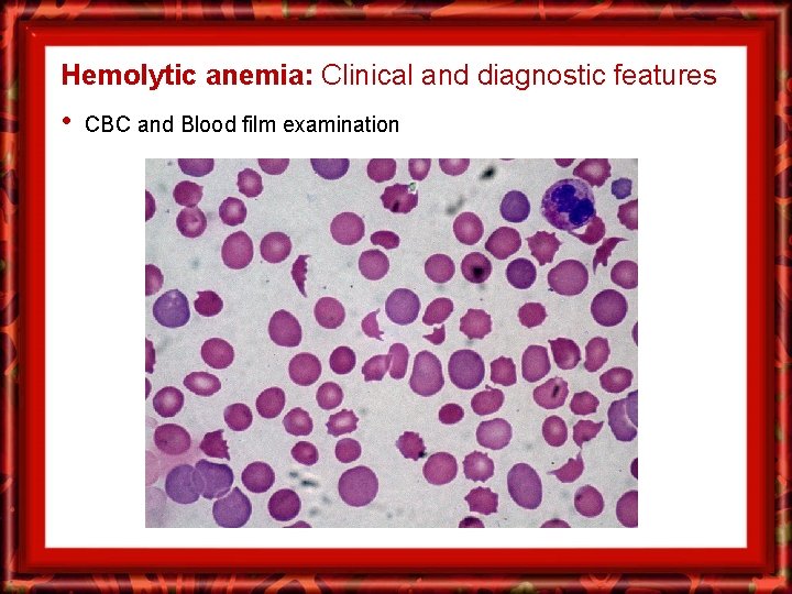
Hemolytic anemia: Clinical and diagnostic features • CBC and Blood film examination
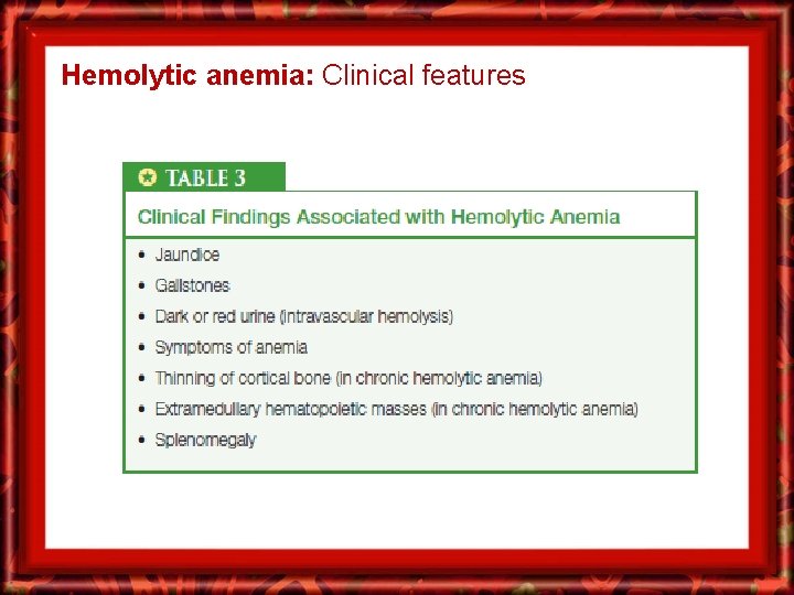
Hemolytic anemia: Clinical features
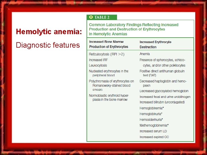
Hemolytic anemia: Diagnostic features
Hemolytic anemia: Clinical and diagnostic features • CBC and Blood film examination
Hemolytic anemia: Clinical and diagnostic features • CBC and Blood film examination
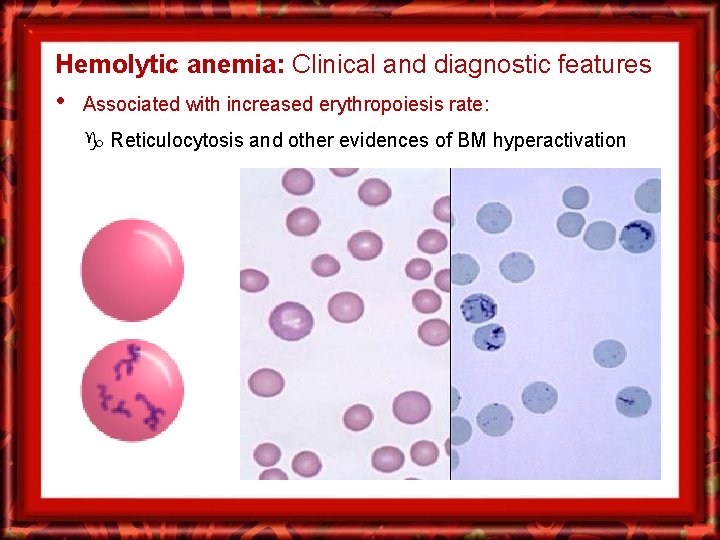
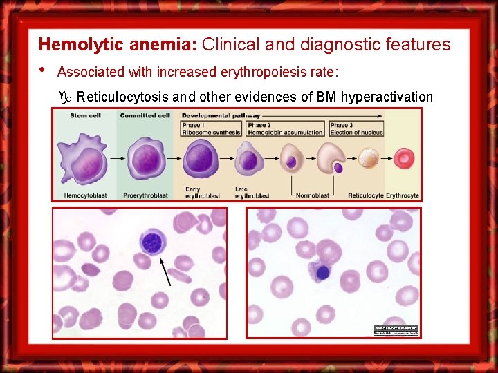
Hemolytic anemia: Clinical and diagnostic features • Associated with increased erythropoiesis rate: Reticulocytosis and other evidences of BM hyperactivation
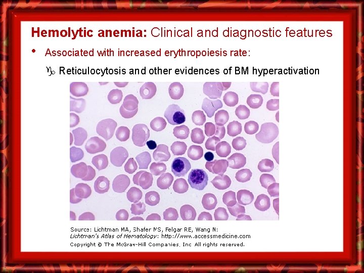
Hemolytic anemia: Clinical and diagnostic features • Associated with increased erythropoiesis rate: Reticulocytosis and other evidences of BM hyperactivation
Hemolytic anemia: Clinical and diagnostic features • Associated with increased erythropoiesis rate: Reticulocytosis and other evidences of BM hyperactivation
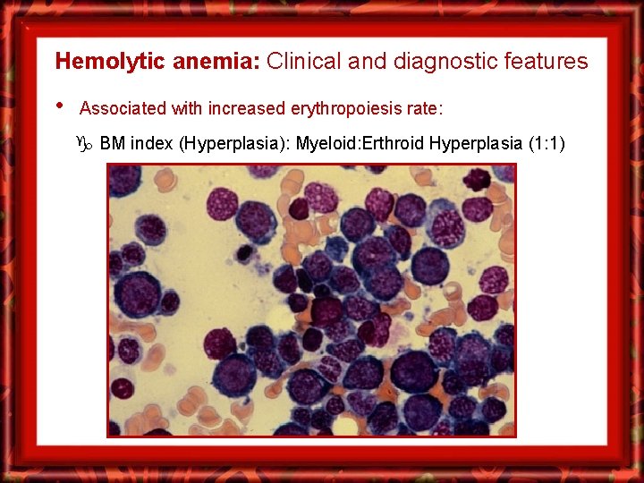
Hemolytic anemia: Clinical and diagnostic features • Associated with increased erythropoiesis rate: BM index (Hyperplasia): Myeloid: Erthroid Hyperplasia (1: 1)
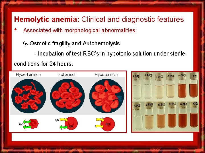
Hemolytic anemia: Clinical and diagnostic features • Associated with morphological abnormalities: Osmotic fragility and Autohemolysis - Incubation of test RBC’s in hypotonic solution under sterile conditions for 24 hours.
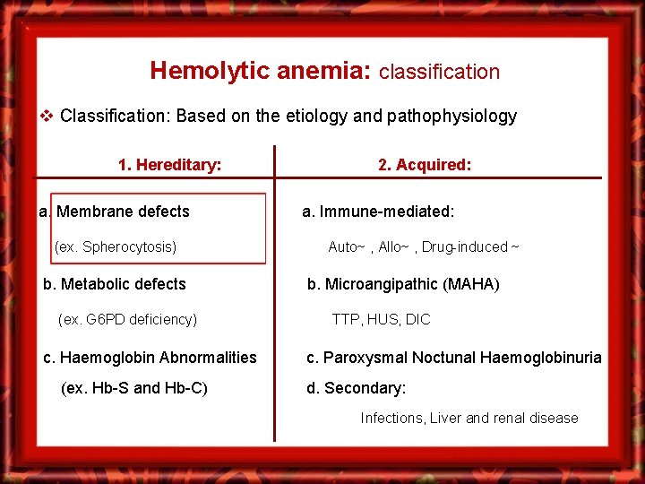
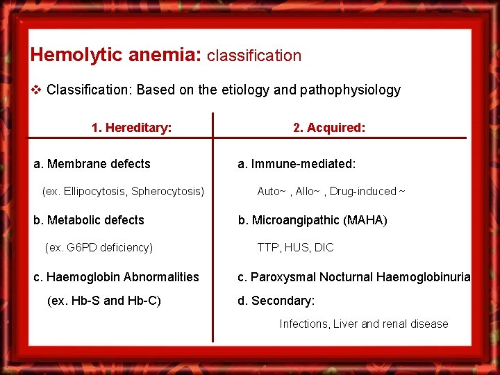
Hemolytic anemia: classification v Classification: Based on the etiology and pathophysiology 1. Hereditary: a. Membrane defects (ex. Spherocytosis) b. Metabolic defects (ex. G 6 PD deficiency) c. Haemoglobin Abnormalities (ex. Hb-S and Hb-C) 2. Acquired: a. Immune-mediated: Auto~ , Allo~ , Drug-induced ~ b. Microangipathic (MAHA) TTP, HUS, DIC c. Paroxysmal Noctunal Haemoglobinuria d. Secondary: Infections, Liver and renal disease
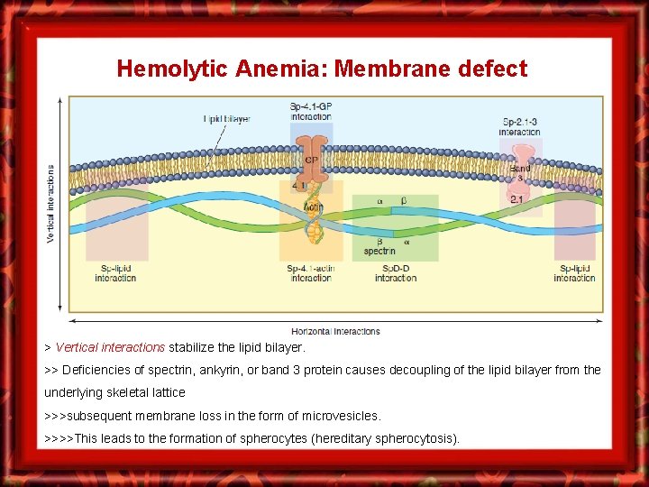
Hemolytic Anemia: Membrane defect > Vertical interactions stabilize the lipid bilayer. >> Deficiencies of spectrin, ankyrin, or band 3 protein causes decoupling of the lipid bilayer from the underlying skeletal lattice >>>subsequent membrane loss in the form of microvesicles. >>>>This leads to the formation of spherocytes (hereditary spherocytosis).
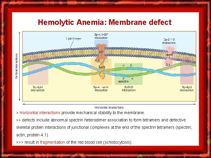
Hemolytic Anemia: Membrane defect > Horizontal interactions provide mechanical stability to the membrane. >> defects include abnormal spectrin heterodimer association to form tetramers and defective skeletal protein interactions of junctional complexes at the end of the spectrin tetramers (spectrin, actin, protein 4. 1) >>> result in fragmentation of the red blood cell (schistocytosis).
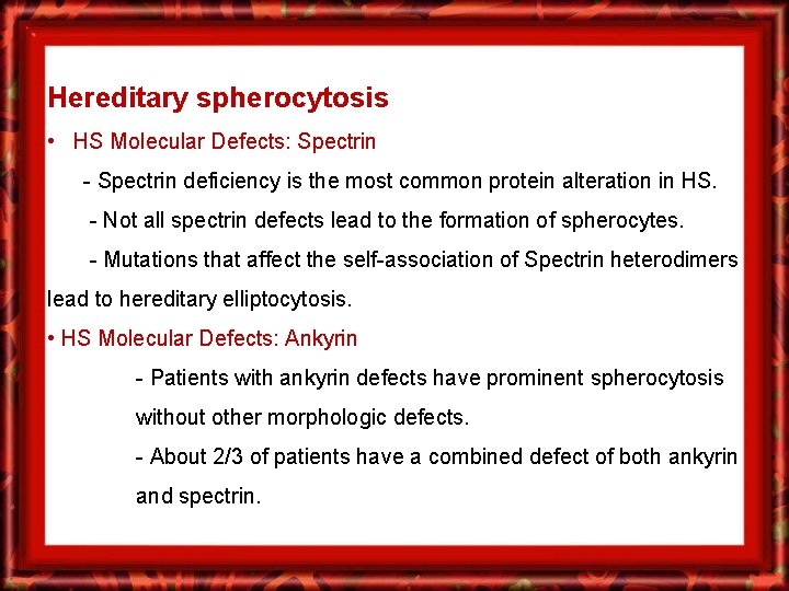
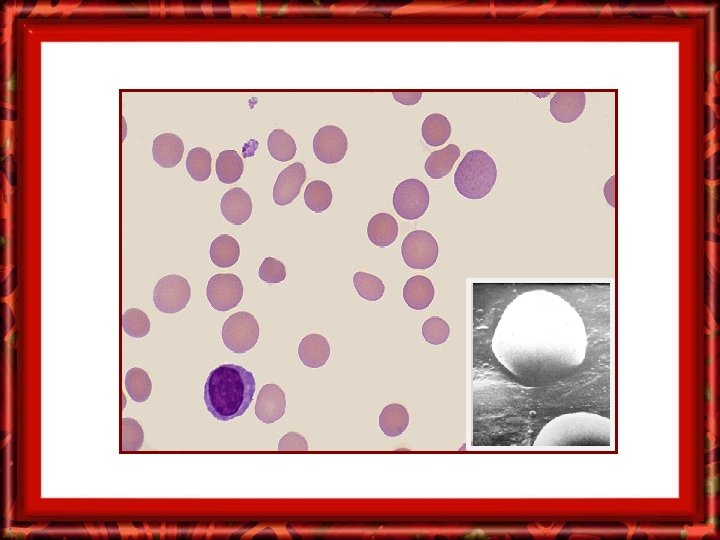
Hereditary spherocytosis • HS Molecular Defects: Spectrin - Spectrin deficiency is the most common protein alteration in HS. - Not all spectrin defects lead to the formation of spherocytes. - Mutations that affect the self-association of Spectrin heterodimers lead to hereditary elliptocytosis. • HS Molecular Defects: Ankyrin - Patients with ankyrin defects have prominent spherocytosis without other morphologic defects. - About 2/3 of patients have a combined defect of both ankyrin and spectrin.
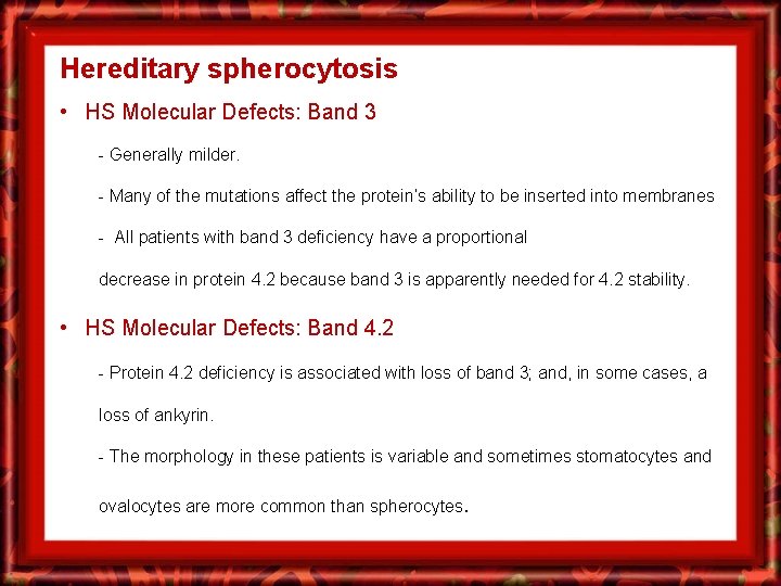
Hereditary spherocytosis • HS Molecular Defects: Band 3 - Generally milder. - Many of the mutations affect the protein’s ability to be inserted into membranes - All patients with band 3 deficiency have a proportional decrease in protein 4. 2 because band 3 is apparently needed for 4. 2 stability. • HS Molecular Defects: Band 4. 2 - Protein 4. 2 deficiency is associated with loss of band 3; and, in some cases, a loss of ankyrin. - The morphology in these patients is variable and sometimes stomatocytes and ovalocytes are more common than spherocytes.
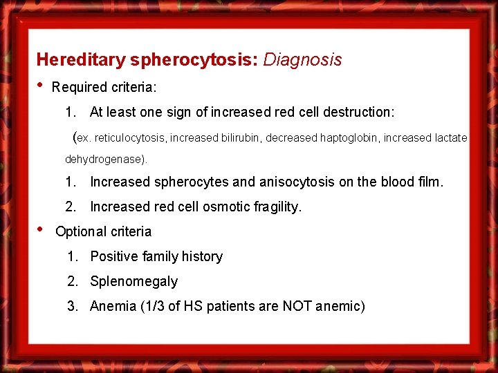
Hereditary spherocytosis: Diagnosis • Required criteria: 1. At least one sign of increased red cell destruction: (ex. reticulocytosis, increased bilirubin, decreased haptoglobin, increased lactate dehydrogenase). 1. Increased spherocytes and anisocytosis on the blood film. 2. Increased red cell osmotic fragility. • Optional criteria 1. Positive family history 2. Splenomegaly 3. Anemia (1/3 of HS patients are NOT anemic)
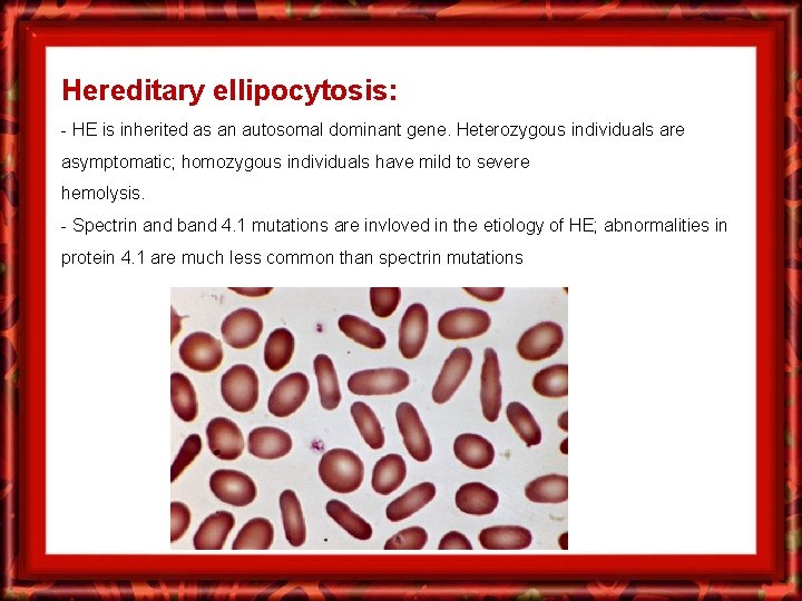
Hereditary ellipocytosis: - HE is inherited as an autosomal dominant gene. Heterozygous individuals are asymptomatic; homozygous individuals have mild to severe hemolysis. - Spectrin and band 4. 1 mutations are invloved in the etiology of HE; abnormalities in protein 4. 1 are much less common than spectrin mutations
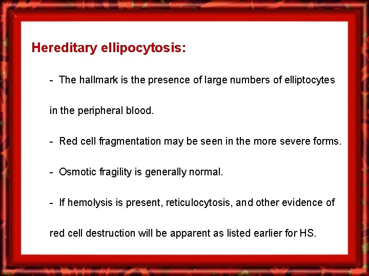
Hereditary ellipocytosis: - The hallmark is the presence of large numbers of elliptocytes in the peripheral blood. - Red cell fragmentation may be seen in the more severe forms. - Osmotic fragility is generally normal. - If hemolysis is present, reticulocytosis, and other evidence of red cell destruction will be apparent as listed earlier for HS.
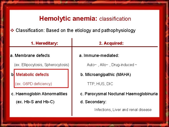
Hemolytic anemia: classification v Classification: Based on the etiology and pathophysiology 1. Hereditary: a. Membrane defects (ex. Ellipocytosis, Spherocytosis) b. Metabolic defects (ex. G 6 PD deficiency) c. Haemoglobin Abnormalities (ex. Hb-S and Hb-C) 2. Acquired: a. Immune-mediated: Auto~ , Allo~ , Drug-induced ~ b. Microangipathic (MAHA) TTP, HUS, DIC c. Paroxysmal Noctunal Haemoglobinuria d. Secondary: Infections, Liver and renal disease
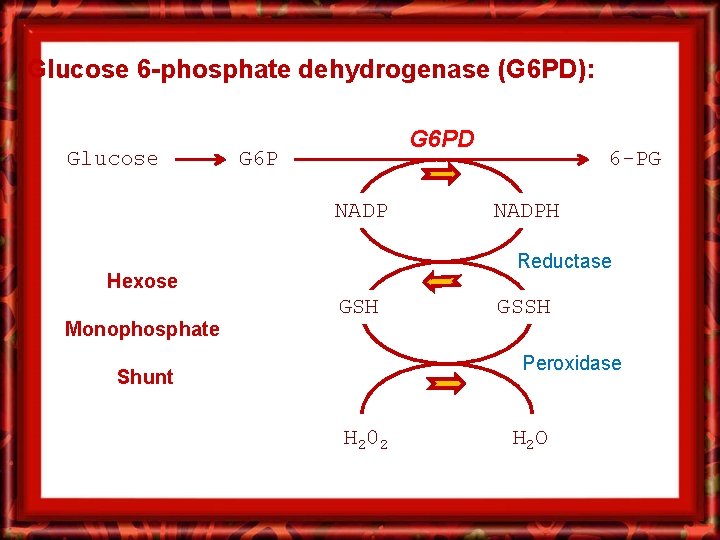
Glucose 6 -phosphate dehydrogenase (G 6 PD): Glucose G 6 PD G 6 P NADPH Reductase Hexose Monophosphate 6 -PG GSH GSSH Peroxidase Shunt H 20 2 H 2 O
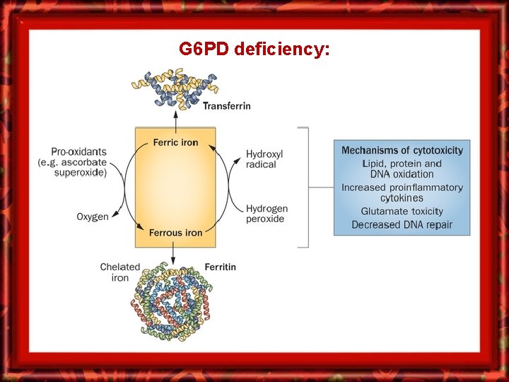
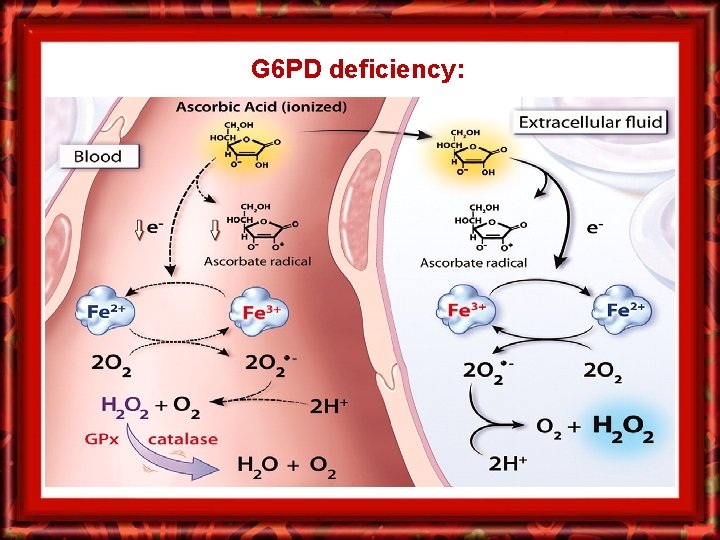
G 6 PD deficiency:
G 6 PD deficiency:
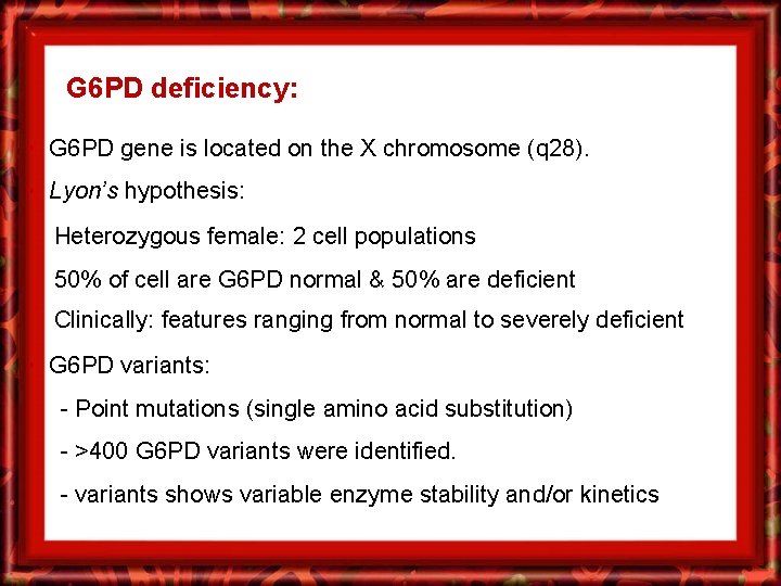
G 6 PD deficiency: • G 6 PD gene is located on the X chromosome (q 28). • Lyon’s hypothesis: Heterozygous female: 2 cell populations 50% of cell are G 6 PD normal & 50% are deficient Clinically: features ranging from normal to severely deficient • G 6 PD variants: - Point mutations (single amino acid substitution) - >400 G 6 PD variants were identified. - variants shows variable enzyme stability and/or kinetics
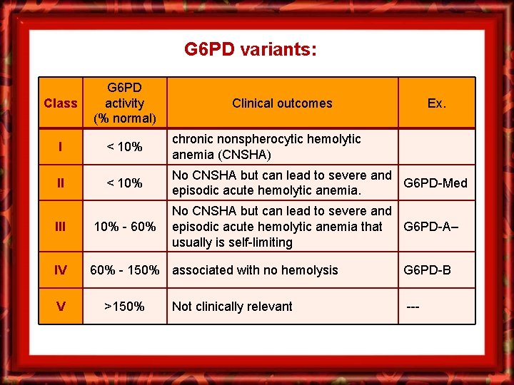
G 6 PD variants: Class G 6 PD activity (% normal) I < 10% chronic nonspherocytic hemolytic anemia (CNSHA) II < 10% No CNSHA but can lead to severe and G 6 PD-Med episodic acute hemolytic anemia. Clinical outcomes No CNSHA but can lead to severe and episodic acute hemolytic anemia that G 6 PD-A– usually is self-limiting III 10% - 60% IV 60% - 150% associated with no hemolysis V >150% Ex. Not clinically relevant G 6 PD-B ---
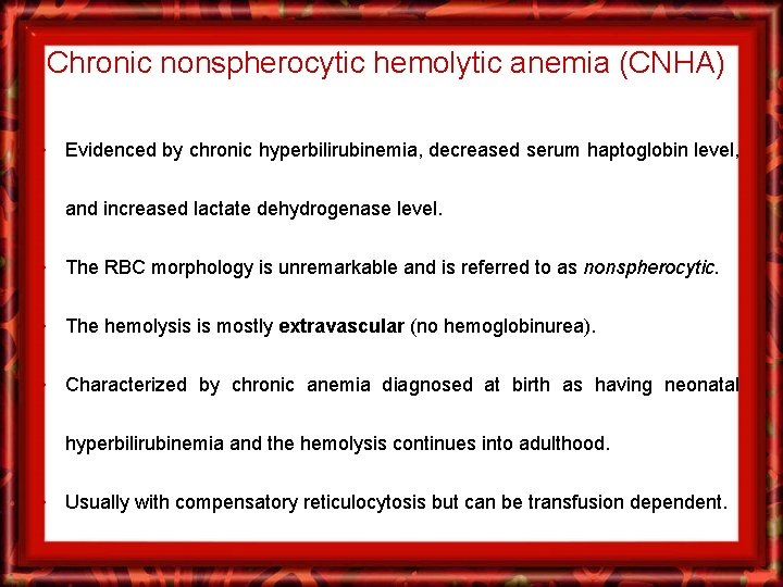
Chronic nonspherocytic hemolytic anemia (CNHA) • Evidenced by chronic hyperbilirubinemia, decreased serum haptoglobin level, and increased lactate dehydrogenase level. • The RBC morphology is unremarkable and is referred to as nonspherocytic. • The hemolysis is mostly extravascular (no hemoglobinurea). • Characterized by chronic anemia diagnosed at birth as having neonatal hyperbilirubinemia and the hemolysis continues into adulthood. • Usually with compensatory reticulocytosis but can be transfusion dependent.
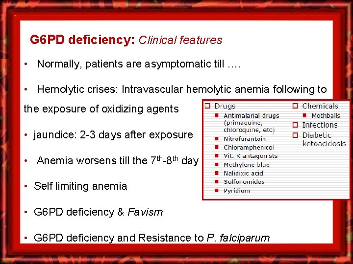
G 6 PD deficiency: Clinical features • Normally, patients are asymptomatic till …. • Hemolytic crises: Intravascular hemolytic anemia following to the exposure of oxidizing agents • jaundice: 2 -3 days after exposure • Anemia worsens till the 7 th-8 th day • Self limiting anemia • G 6 PD deficiency & Favism • G 6 PD deficiency and Resistance to P. falciparum
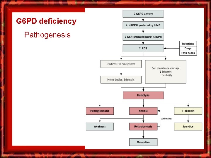
G 6 PD deficiency Pathogenesis
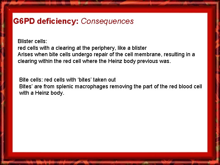
G 6 PD deficiency: Consequences Blister cells: red cells with a clearing at the periphery, like a blister Arises when bite cells undergo repair of the cell membrane, resulting in a clearing within the red cell where the Heinz body previous was. Bite cells: red cells with ‘bites’ taken out Bites’ are from splenic macrophages removing the part of the red blood cell with a Heinz body.
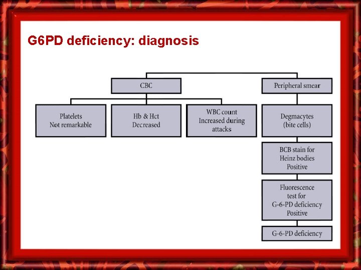
G 6 PD deficiency: diagnosis
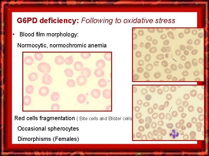
G 6 PD deficiency: Following to oxidative stress • Blood film morphology: Normocytic, normochromic anemia Red cells fragmentation ( Bite cells and Blister cells) Occasional spherocytes Dimorphisms (Females)
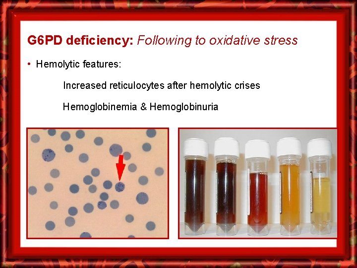
G 6 PD deficiency: Following to oxidative stress • Hemolytic features: Increased reticulocytes after hemolytic crises Hemoglobinemia & Hemoglobinuria
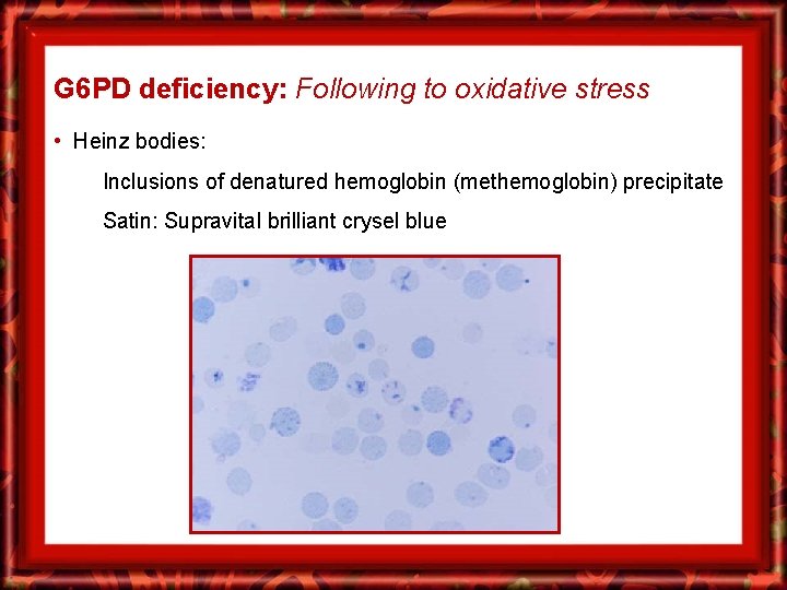
G 6 PD deficiency: Following to oxidative stress • Heinz bodies: Inclusions of denatured hemoglobin (methemoglobin) precipitate Satin: Supravital brilliant crysel blue
Acquired immune-mediated
Hemolytic anemia: classification v Classification: Based on the etiology and pathophysiology 1. Hereditary: a. Membrane defects (ex. Ellipocytosis, Spherocytosis) b. Metabolic defects (ex. G 6 PD deficiency) c. Haemoglobin Abnormalities (ex. Hb-S and Hb-C) 2. Acquired: a. Immune-mediated: Auto~ , Allo~ , Drug-induced ~ b. Microangipathic (MAHA) TTP, HUS, DIC c. Paroxysmal Nocturnal Haemoglobinuria d. Secondary: Infections, Liver and renal disease
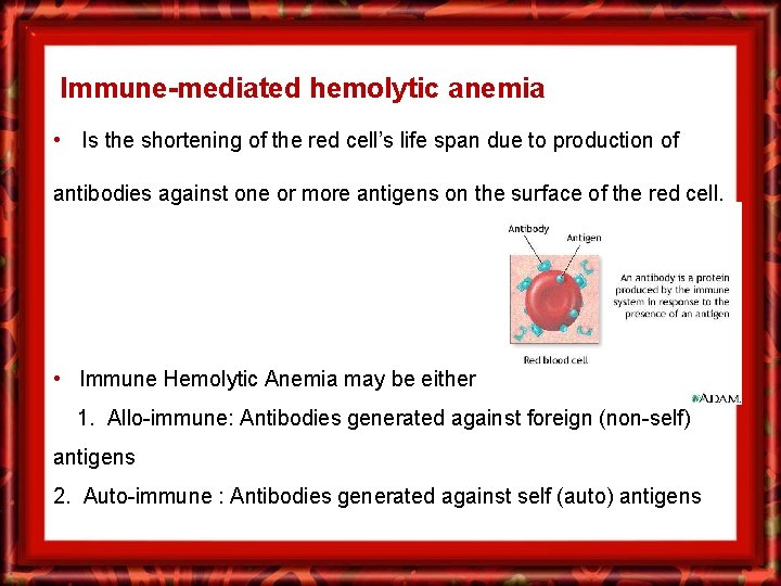
Immune-mediated hemolytic anemia • Is the shortening of the red cell’s life span due to production of antibodies against one or more antigens on the surface of the red cell. • Immune Hemolytic Anemia may be either 1. Allo-immune: Antibodies generated against foreign (non-self) antigens 2. Auto-immune : Antibodies generated against self (auto) antigens
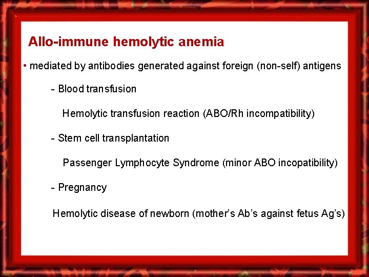
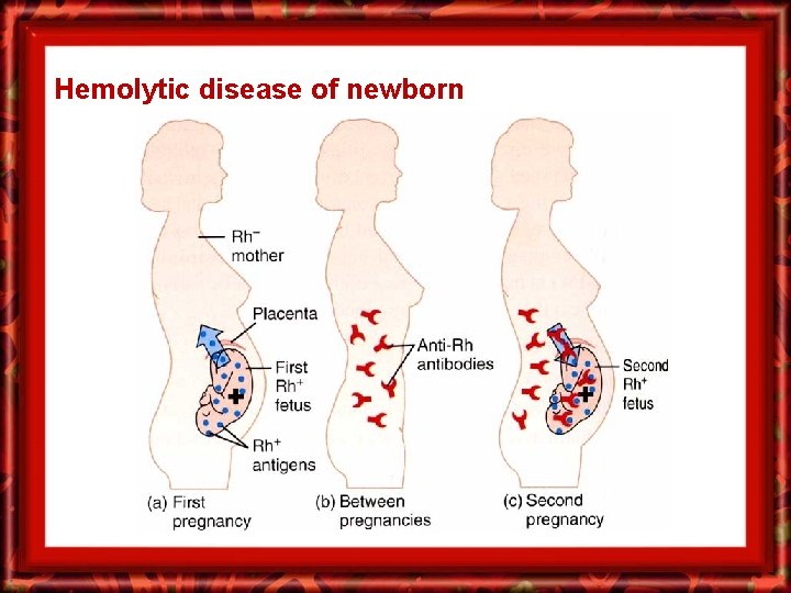
Allo-immune hemolytic anemia • mediated by antibodies generated against foreign (non-self) antigens - Blood transfusion Hemolytic transfusion reaction (ABO/Rh incompatibility) - Stem cell transplantation Passenger Lymphocyte Syndrome (minor ABO incopatibility) - Pregnancy Hemolytic disease of newborn (mother’s Ab’s against fetus Ag’s)
Hemolytic disease of newborn
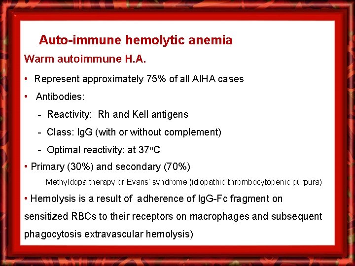
Auto-immune hemolytic anemia Warm autoimmune H. A. • Represent approximately 75% of all AIHA cases • Antibodies: - Reactivity: Rh and Kell antigens - Class: Ig. G (with or without complement) - Optimal reactivity: at 37 o. C • Primary (30%) and secondary (70%) Methyldopa therapy or Evans’ syndrome (idiopathic-thrombocytopenic purpura) • Hemolysis is a result of adherence of Ig. G-Fc fragment on sensitized RBCs to their receptors on macrophages and subsequent phagocytosis extravascular hemolysis)
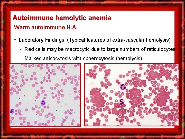
Autoimmune hemolytic anemia Warm autoimmune H. A. • Laboratory Findings: (Typical features of extra-vascular hemolysis) - Red cells may be macrocytic due to large numbers of reticulocytes - Marked anisocytosis with spherocytosis (hemolysis)
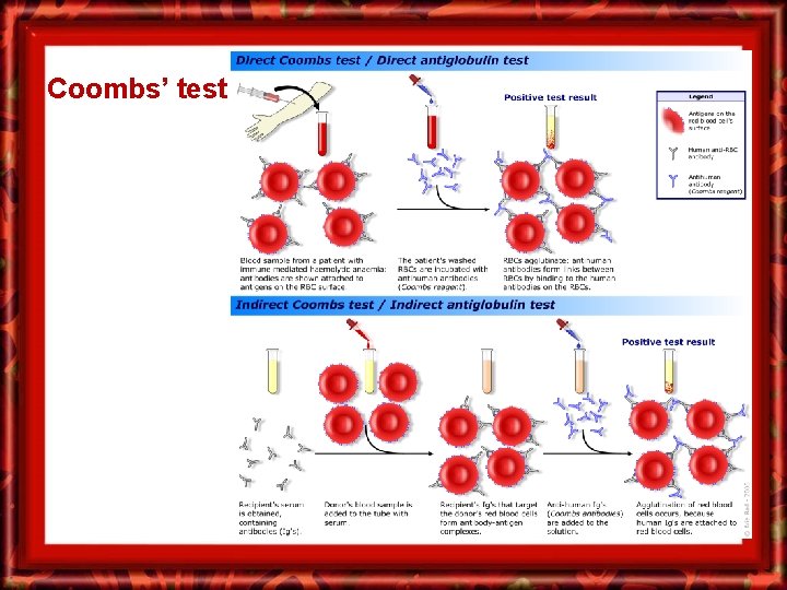
Autoimmune hemolytic anemia Warm autoimmune H. A. • Laboratory Findings: - Direct antibody test (DAT) is positive in the majority of cases. - The indirect antiglobulin test may demonstrate Autoantibodies
Coombs’ test
Auto-immune hemolytic anemia Cold-agglutinins autoimmune H. A. • Antibodies: - Reactivity: Ii blood grouping antigens - Class: Usually Ig. M (occasionaly Ig. G) - Optimal reactivity: at 0 - 4 Oc - Ab’s are capable of binding complement • Hemolysis is usually extra-vascular in the liver due to the C 3 b receptors on Kupffer cells. • When the patient is exposed to cold, agglutination of red cells in the small capillaries causes numbness, pain, and skin discoloration (acrocyanosis or Reynaud’s phenomenon). Gangrene may develop in severe cases.
Cold- agglutinins hemolytic anemia • Laboratory Findings: - Hemoglobin may fluctuate with seasons. - Degree of reticulocytosis and bilirubin concentration varies with severity of red cell hemolysis. - Clumping of red cells at room temperature may lead to erroneous laboratory data from automated instruments. - For patients experiencing hemolysis, the cold agglutinin titer at 4 o. C is over 1000 (it is generally under 64 in healthy individuals). - The DAT is usually positive when a polyspecific antiglobulin serum is used that detects both complement and antibodies on the red cell surface.
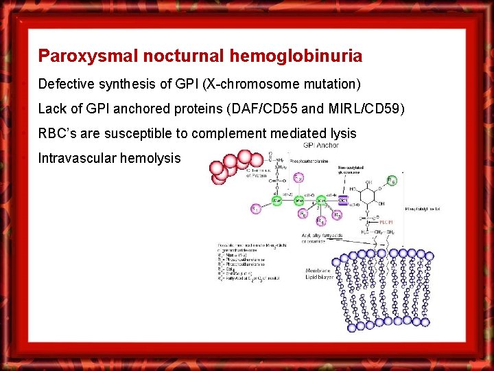
Paroxysmal nocturnal hemoglobinuria • Defective synthesis of GPI (X-chromosome mutation) • Lack of GPI anchored proteins (DAF/CD 55 and MIRL/CD 59) • RBC’s are susceptible to complement mediated lysis • Intravascular hemolysis
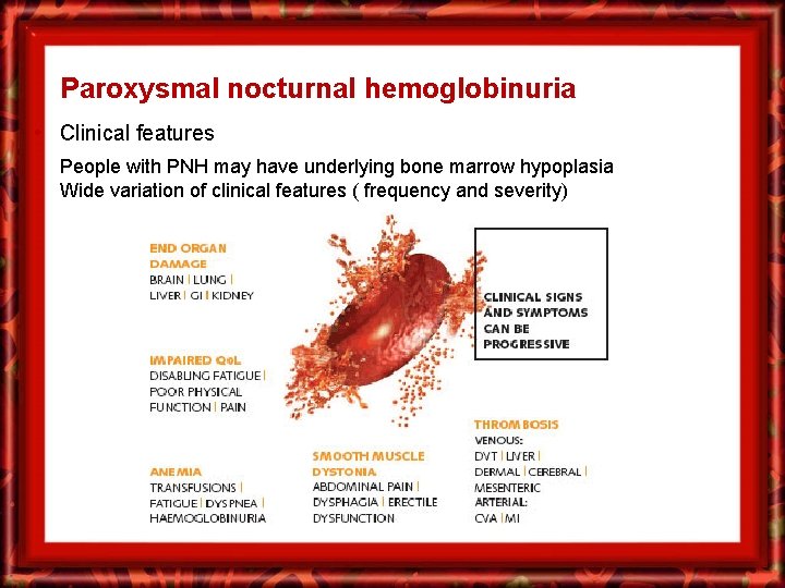
Paroxysmal nocturnal hemoglobinuria • Clinical features People with PNH may have underlying bone marrow hypoplasia Wide variation of clinical features ( frequency and severity)