
MLO 240 Hematology II Lymphoid Malignancies Leukemia vs
- Slides: 20
MLO 240: Hematology II Lymphoid Malignancies
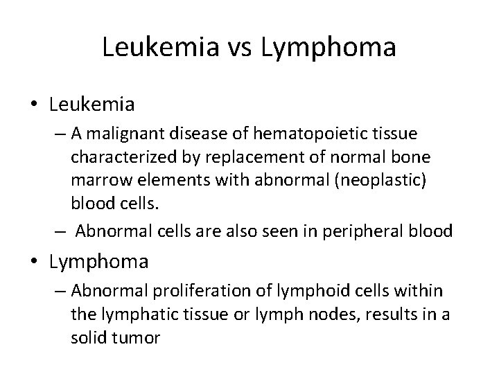
Leukemia vs Lymphoma • Leukemia – A malignant disease of hematopoietic tissue characterized by replacement of normal bone marrow elements with abnormal (neoplastic) blood cells. – Abnormal cells are also seen in peripheral blood • Lymphoma – Abnormal proliferation of lymphoid cells within the lymphatic tissue or lymph nodes, results in a solid tumor
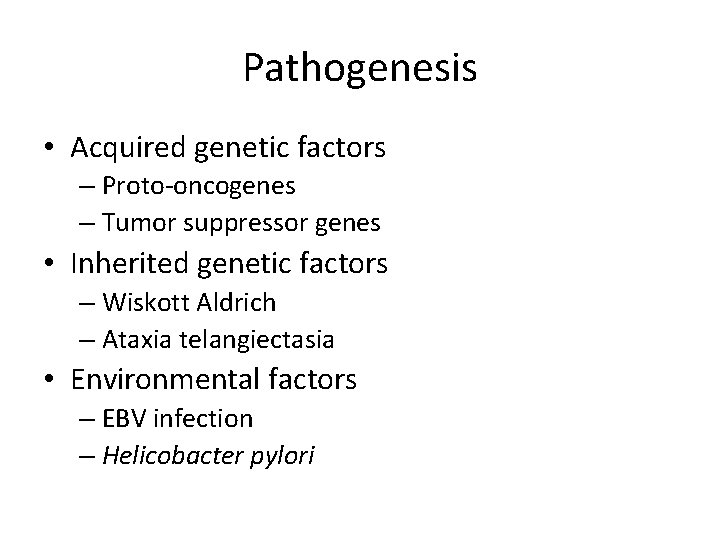
Pathogenesis • Acquired genetic factors – Proto-oncogenes – Tumor suppressor genes • Inherited genetic factors – Wiskott Aldrich – Ataxia telangiectasia • Environmental factors – EBV infection – Helicobacter pylori
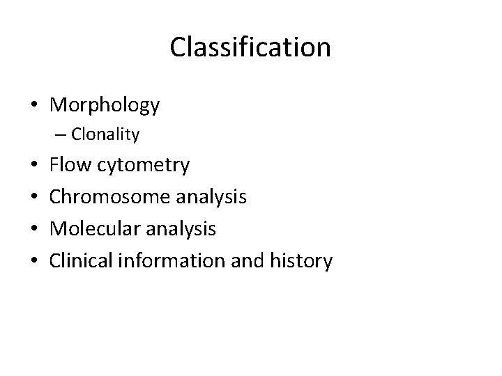
Classification • Morphology – Clonality • • Flow cytometry Chromosome analysis Molecular analysis Clinical information and history
CLL: Chronic Lymphocytic Leukemia • Clinical features – Occurs in persons >50 years old – Chronic fatigue, infection • Result of bone marrow replacement of normal cells with lymphocytes. – Skin and organ infiltration and enlargement – Median survival is 10 years
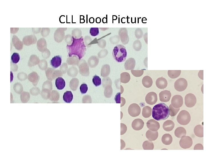
Chronic Lymphocytic Leukemia • General requirements for diagnosis – Peripheral blood (>5000 ALC) and bone marrow lymphocytosis (>30%) – Lymphocytes are small to slightly larger than normal with mature appearance • Nucleus is round, with block-type chromatin • Cytoplasm scarce – Smudge cells (bare nuclei) are common • Occur due to the cell’s fragility in making a smear – Prolymphocyte < 10%
CLL Blood Picture
Chronic Lymphocytic Leukemia • Treatment – Usually treatment is not required until lymphocytosis causes other cells to be crowded out resulting in infections. – Treatment depends on the stage at which the disease is diagnosed and is usually for the symptoms, not the disease. • Chemotherapy given according to stage of disease • IV gamma globulin for prevention of infection • Bone marrow transplant done on aggressive cases
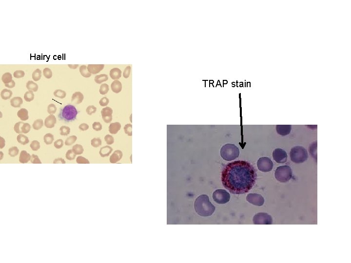
Hairy cell leukemia (HCL) – Presents in middle age – Affects males 7: 1 over females – Pancytopenia common • Increases opportunity for infections – Bone marrow aspirate can result in a “dry tap” due to marrow fibrosis. – Hairy cell leukemia can be treated with a one-time chemotherapy regimen with a good prognosis of long-term survival.
Hairy cell TRAP stain
Plasma Cell Disorders • Disorders that do not involve lymph nodes • Secrete monoclonal immunoglobulin into the serum and /or urine • Disorders – Plasma Cell Myeloma – Plasmacytoma – Monoclonal gammopathy of undetermined significance (MGUS)
Plasma Cell Myelomas • Overproduction of abnormal plasma cells which are the final stage in the development of B lymphocytes. • These cells secrete immunoglobulin, resulting in lytic bone lesions • Risk increases with age- rare under 40 – Median age 70 years old • Suspected cause is chronic stimulation of the immune system from environmental sources. – Ionizing radiation – Viruses
Features • Increased production of immunoglobulin heavy and light chains (monoclonal gammopathy) – Heavy chains: Ig. G, Ig. A, Ig. D, Ig. E, Ig. M – Light chains: kappa, lambda – Most common type of plasma cell myeloma is increased production of Ig. G. • Bence-Jones protein – Light chains spill into the urine and can be detected by lab test – Causes kidney damage • Hyperviscosity syndrome – Excess immunoglobulin causes viscous blood which sludges and causes fluid congestion. • Normal Immunoglobulin production decreased – Results in infections
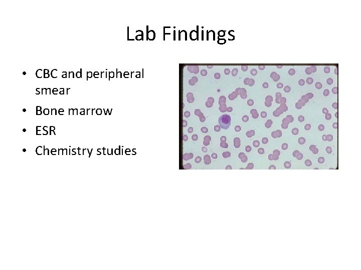
Lab Findings • CBC and peripheral smear • Bone marrow • ESR • Chemistry studies
Lymphomas • These are a heterogeneous group of diseases that arise from an overproliferation of abnormal cells of the lympoid tissue (lymphocytes, histiocytes, and reticulum cells). The spilling of these cells into the peripheral blood results in a leukemic phase of the disease.
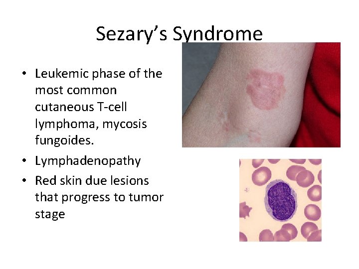
Sezary’s Syndrome • Leukemic phase of the most common cutaneous T-cell lymphoma, mycosis fungoides. • Lymphadenopathy • Red skin due lesions that progress to tumor stage
Hodgkin’s Lymphoma • Etiology and clinical features – Probable cause is Epstein-Barr virus. – Diagnosed between 15 and 35 years of age; also found in over 50 population – Nonpainful lymph node swelling
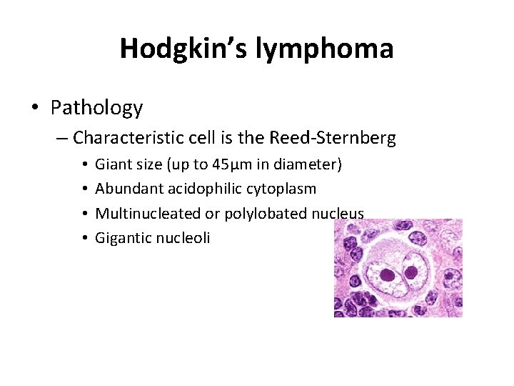
Hodgkin’s lymphoma • Pathology – Characteristic cell is the Reed-Sternberg • • Giant size (up to 45µm in diameter) Abundant acidophilic cytoplasm Multinucleated or polylobated nucleus Gigantic nucleoli
Hodgkin’s lymphoma • Treatment and prognosis – Radiation of localized involvement – Chemotherapy – Combination of above – With early diagnosis, long-term disease-free survival is seen in about 75% of cases.
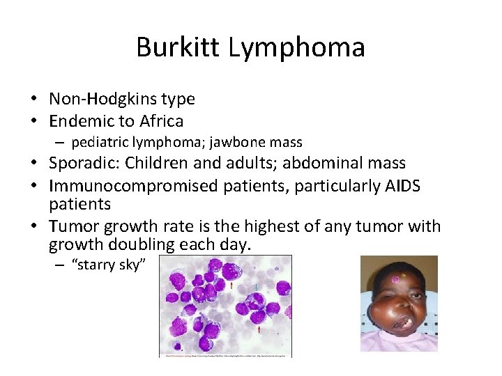
Burkitt Lymphoma • Non-Hodgkins type • Endemic to Africa – pediatric lymphoma; jawbone mass • Sporadic: Children and adults; abdominal mass • Immunocompromised patients, particularly AIDS patients • Tumor growth rate is the highest of any tumor with growth doubling each day. – “starry sky”