Nursing Care of Clients with Upper Gastrointestinal Disorders


 but")


 1. Definition l b. GERD common, affecting 15 – 20%")
 2. Pathophysiology l a. Gastroesophageal reflux results from transient relaxation")
 l c. Normally the peristalsis in esophagus and bicarbonate in")
 4. Complications l a. Esophageal strictures, which can progress to")
 6. Diagnostic Tests l a. Barium swallow (evaluation of esophagus,")
 7. Medications l a. Antacids for mild to moderate symptoms,")

 9. Surgery indicated for persons not improved by diet and")


 Definition and Risk factors l a. Break in mucous lining")
 2. Pathophysiology l A. Ulcers or breaks in mucosa of")

 3. Manifestations l a. Pain is classic symptom: gnawing, burning,")

 l. Blocks gastric acid")


 Complications l a. Hemorrhage: frequent in older adult: hematemesis, melena,")


 (spastic bowel, functional colitis) Definition l a. Functional GI tract")
 (spastic bowel, functional colitis) Pathophysiology l a. Appears there is")
 (spastic bowel, functional colitis) Manifestations l a. Abdominal pain relieved")
 (spastic bowel, functional colitis) 5. l l Diagnostic Tests: to")
 (spastic bowel, functional colitis) Medications l a. Purpose: to manage")
 (spastic bowel, functional colitis) Dietary Management l a. Often benefit")
 and")


 causing")
")


- Slides: 145
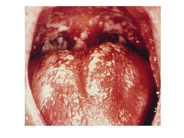
Nursing Care of Clients with Upper Gastrointestinal Disorders l I. Care of Clients with Disorder of the Mouth A. Disorder includes inflammation, infection, neoplastic lesions B. Pathophysiology 1. Causes include mechanical trauma, irritants such as tobacco, chemotherapeutic agents 2. Oral mucosa is relatively thin, has rich blood supply, exposed to environment C. Manifestations 1. Visible lesions or erosions on lips or oral mucosa 2. Pain
Nursing Care of Clients with Upper Gastrointestinal Disorders l D. Collaborative Care 1. Direct observation to investigate any problems; determine underlying cause and any coexisting diseases 2. Any undiagnosed oral lesion present for > 1 week and not responding to treatment should be evaluated for malignancy 3. General treatment includes mouthwashes or treatments to cleanse and relieve irritation a. Alcohol bases mouthwashes cause pain and burning b. Sodium bicarbonate mouthwashes are effective without pain 4. Specific treatments according to type of infection a. Fungal (candidiasis): nystatin “swish and swallow” or clotrimazole lozenges l b. Herpetic lesions: topical or oral acyclovir
Nursing Care of Clients with Upper Gastrointestinal Disorders E. Nursing Care 1. Goal: to relieve pain and symptoms, so client can continue food and fluid intake in health care facility and at home 2. Impaired oral mucous membrane l a. Assess clients at high risk l b. Assist with oral hygiene post eating, bedtime l c. Teach to limit irritants: tobacco, alcohol, spicy foods 3. Imbalanced nutrition: less than body requirements l a. Assess nutritional intake; use of straws l b. High calorie and protein diet according to client preferences
Client with Oral Cancer 1. Background l a. Uncommon (5% of all cancers) but has high rate of morbidity, mortality l b. Highest among males over age 40 l c. Risk factors include smoking and using oral tobacco, drinking alcohol, marijuana use, occupational exposure to chemicals, viruses (human papilloma virus)
Client with Oral Cancer 2. Pathophysiology l a. Squamous cell carcinomas l b. Begin as painless oral ulceration or lesion with irregular, ill-defined borders l c. Lesions start in mucosa and may advance to involve tongue, oropharynx, mandible, maxilla l d. Non-healing lesions should be evaluated for malignancy after one week of treatment
Client with Oral Cancer 3. Collaborative Care l a. Elimination of causative agents l b. Determination of malignancy with biopsy l c. Determine staging with CT scans and MRI l d. Based on age, tumor stage, general health and client’s preference, treatment may include surgery, chemotherapy, and/or radiation therapy l e. Advanced carcinomas may necessitate radical neck dissection with temporary or permanent tracheostomy; Surgeries may be disfiguring l f. Plan early for home care post hospitalization, teaching family and client care involved post surgery, refer to American Cancer Society, support groups
Client with Oral Cancer 4. Nursing Care a. Health promotion: l 1. Teach risk of oral cancer associated with all tobacco use and excessive alcohol use l 2. Need to seek medical attention for all nonhealing oral lesions (may be discovered by dentists); early precancerous oral lesions are very treatable b. Nursing Diagnoses l 1. Risk for ineffective airway clearance l 2. Imbalanced Nutrition: Less than body requirements l 3. Impaired Verbal Communication: establishment of specific communication plan and method should be done prior to any surgery l 4. Disturbed Body Image
Gastroesophageal Reflux Disease (GERD) 1. Definition l b. GERD common, affecting 15 – 20% of adults l c. 10% persons experience daily heartburn and indigestion l d. Because of location near other organs symptoms may mimic other illnesses including heart problems l a. Gastroesophageal reflux is the backward flow of gastric content into the esophagus.
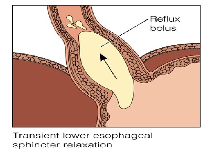
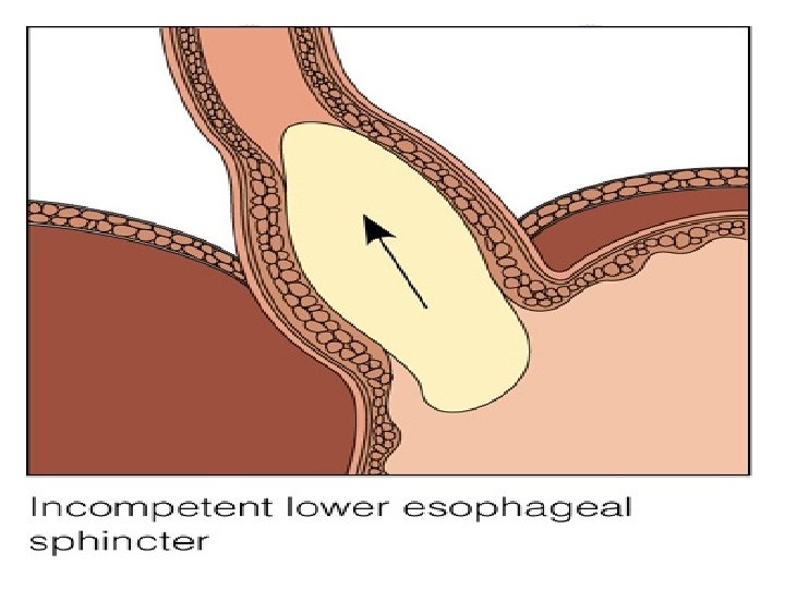
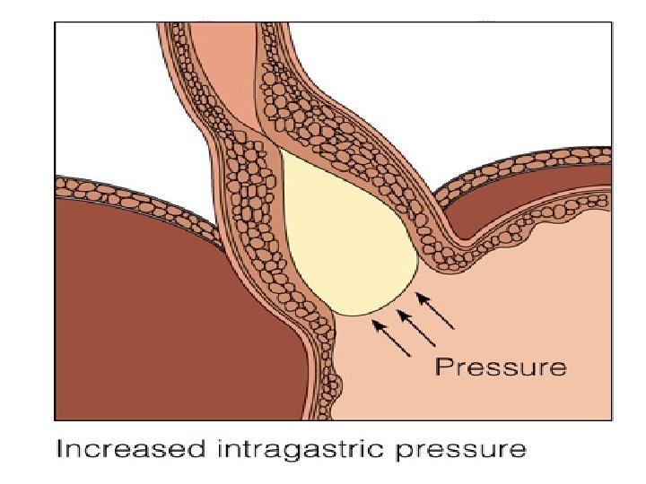
Gastroesophageal Reflux Disease (GERD) 2. Pathophysiology l a. Gastroesophageal reflux results from transient relaxation or incompetence of lower esophageal sphincter, or increased pressure within stomach l b. Factors contributing to gastroesophageal reflux 1. Increased gastric volume (post meals) 2. Position pushing gastric contents close to gastroesophageal juncture (such as bending or lying down) 3. Increased gastric pressure (obesity or tight clothing) 4. Hiatal hernia
Gastroesophageal Reflux Disease (GERD) l c. Normally the peristalsis in esophagus and bicarbonate in salivary secretions neutralize any gastric juices (acidic) that contact the esophagus; during sleep and with gastroesophageal reflux esophageal mucosa is damaged and inflamed; prolonged exposure causes ulceration, friable mucosa, and bleeding; untreated there is scarring and stricture 3. Manifestations l a. Heartburn after meals, while bending over, or recumbent l b. May have regurgitation of sour materials in mouth, pain with swallowing l c. Atypical chest pain l d. Sore throat with hoarseness l e. Bronchospasm and laryngospasm
Gastroesophageal Reflux Disease (GERD) 4. Complications l a. Esophageal strictures, which can progress to dysphagia l b. Barrett’s esophagus: changes in cells lining esophagus with increased risk for esophageal cancer 5. Collaborative Care l a. Diagnosis may be made from history of symptoms and risks l b. Treatment includes 1. Life style changes 2. Diet modifications 3. Medications
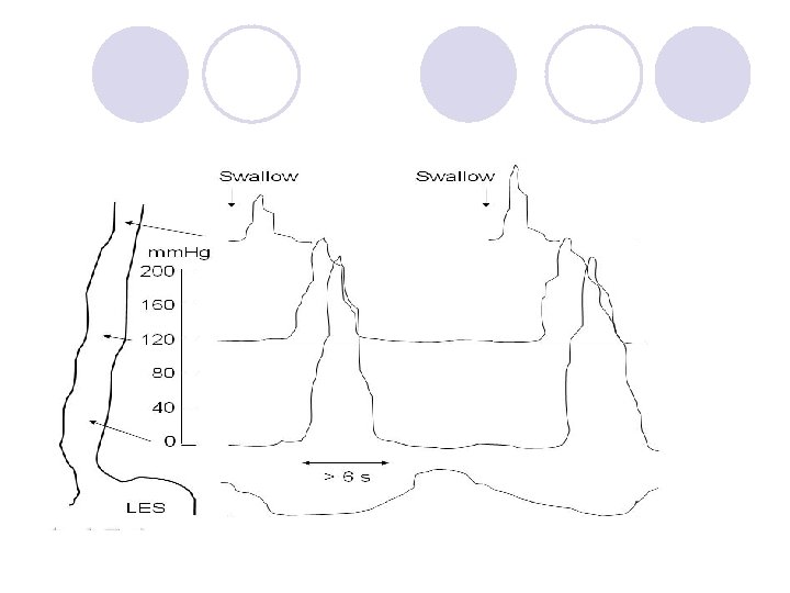
Gastroesophageal Reflux Disease (GERD) 6. Diagnostic Tests l a. Barium swallow (evaluation of esophagus, stomach, small intestine) l b. Upper endoscopy: direct visualization; biopsies may be done l c. 24 -hour ambulatory p. H monitoring l d. Esophageal manometry, which measure pressures of esophageal sphincter and peristalsis l e. Esophageal motility studies
Gastroesophageal Reflux Disease (GERD) 7. Medications l a. Antacids for mild to moderate symptoms, e. g. Maalox, Mylanta, Gaviscon l b. H 2 -receptor blockers: decrease acid production; given BID or more often, e. g. cimetidine, ranitidine, famotidine, nizatidine l c. Proton-pump inhibitors: reduce gastric secretions, promote healing of esophageal erosion and relieve symptoms, e. g. omeprazole (prilosec); lansoprazole (Prevacid) initially for 8 weeks; or 3 to 6 months l d. Promotility agent: enhances esophageal clearance and gastric emptying, e. g. metoclopramide (reglan)
Gastroesophageal Reflux Disease 8. Dietary and Lifestyle Management l a. Elimination of acid foods (tomatoes, spicy, citrus foods, coffee) l b. Avoiding food which relax esophageal sphincter or delay gastric emptying (fatty foods, chocolate, peppermint, alcohol) l c. Maintain ideal body weight l d. Eat small meals and stay upright 2 hours post eating; no eating 3 hours prior to going to bed l e. Elevate head of bed on 6 – 8 blocks to decrease reflux l f. No smoking l g. Avoiding bending and wear loose fitting clothing
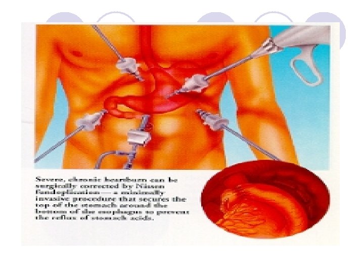
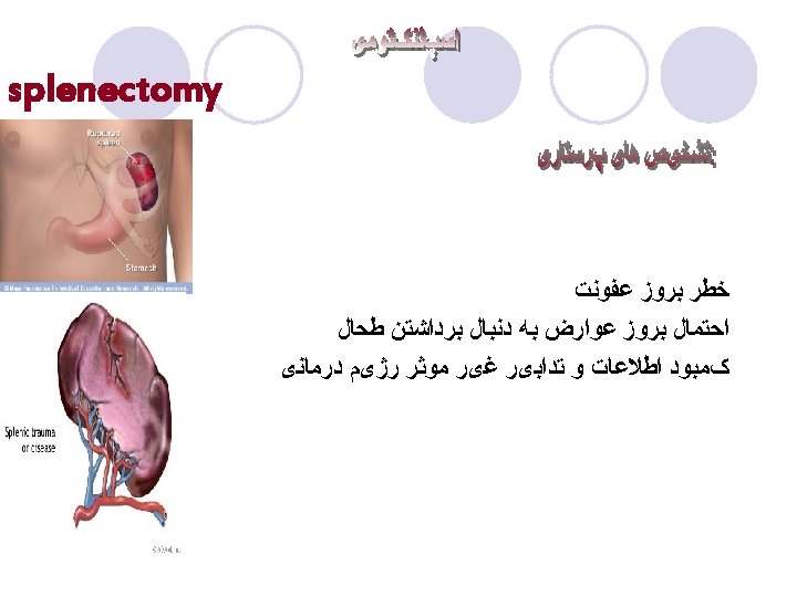
Gastroesophageal Reflux Disease (GERD) 9. Surgery indicated for persons not improved by diet and life style changes l a. Laparoscopic procedures to tighten lower esophageal sphincter l b. Open surgical procedure: Nissen fundoplication 10. Nursing Care l a. Pain usually controlled by treatment l b. Assist client to institute home plan
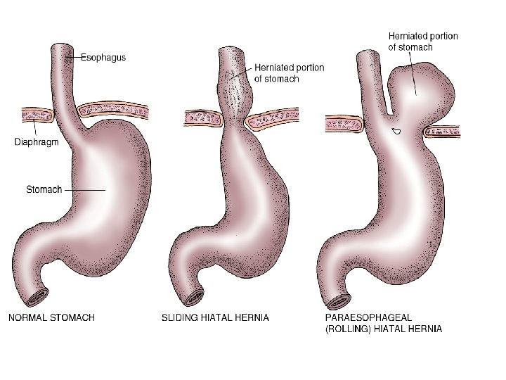
Hiatal Hernia 1. Definition l a. Part of stomach protrudes through the esophageal hiatus of the diaphragm into thoracic cavity l b. Predisposing factors include: ¡Increased intra-abdominal pressure ¡Increased age ¡Trauma ¡Congenital weakness ¡Forced recumbent position
Hiatal Hernia l c. Most cases are asymptomatic; incidence increases with age l d. Sliding hiatal hernia: gastroesophageal junction and fundus of stomach slide through the esophageal hiatus l e. Paraesophageal hiatal hernia: the gastroesophageal junction is in normal place but part of stomach herniates through esophageal hiatus; hernia can become strangulated; client may develop gastritis with bleeding
Hiatal Hernia 2. Manifestations: Similar to GERD 3. Diagnostic Tests l a. Barium swallow l b. Upper endoscopy 4. Treatment l a. Similar to GERD: diet and lifestyle changes, medications l b. If medical treatment is not effective or hernia becomes incarcerated, then surgery; usually Nissen fundoplication by thoracic or abdominal approach ¡ Anchoring the lower esophageal sphincter by wrapping a portion of the stomach around it to anchor it in place
Impaired Esophageal Motility 1. Types l a. Achalasia: characterized by impaired peristalsis of smooth muscle of esophagus and impaired relaxation of lower esophageal sphincter l b. Diffuse esophageal spasm: nonperistaltic contraction of esophageal smooth muscle 2. Manifestations: Dysphagia and/or chest pain 3. Treatment l a. Endoscopically guided injection of botulinum toxin ¡Denervates cholinergic nerves in the distal esophagus to stop spams l b. Balloon dilation of lower esophageal sphincter ¡ May place stents to keep esophagus open
Esophageal Cancer 1. Definition: Relatively uncommon malignancy with high mortality rate, usually diagnosed late 2. Pathophysiology l a. Squamous cell carcinoma 1. Most common affecting middle or distal portion of esophagus 2. More common in African Americans than Caucasians 3. Risk factors cigarette smoking and chronic alcohol use l b. Adenocarcinoma 1. Nearly as common as squamous cell affecting distal portion of esophagus 2. More common in Caucasians 3. Associated with Barrett’s esophagus, complication of chronic GERD and achalasia
Esophageal Cancer 3. Manifestations l a. Progressive dysphagia with pain while swallowing l b. Choking, hoarseness, cough l c. Anorexia, weight loss 4. Collaborative Care: Treatment goals l a. Controlling dysphagia l b. Maintaining nutritional status while treating carcinoma (surgery, radiation therapy, and/or chemotherapy
Esophageal Cancer 5. Diagnostic Tests l a. Barium swallow: identify irregular mucosal patterns or narrowing of lumen l b. Esophagoscopy: allow direct visualization of tumor and biopsy l c. Chest xray, CT scans, MRI: determine tumor metastases l d. Complete Blood Count: identify anemia l e. Serum albumin: low levels indicate malnutrition l f. Liver function tests: elevated with liver metastasis
Esophageal Cancer 6. Treatments: dependent on stage of disease, client’s condition and preference l a. Early (curable) stage: surgical resection of affected portion with anastomosis of stomach to remaining esophagus; may also include radiation therapy and chemotherapy prior to surgery l b. More advanced carcinoma: treatment is palliative and may include surgery, radiation and chemotherapy to control dysphagia and pain l c. Complications of radiation therapy include perforation, hemorrhage, stricture
Esophageal Cancer 7. Nursing Care: Health promotion; education regarding risks associated with smoking and excessive alcohol intake 8. Nursing Diagnoses l a. Imbalanced Nutrition: Less than body requirements (may include enteral tube feeding or parenteral nutrition in hospital and home) l b. Anticipatory Grieving (dealing with cancer diagnosis) l c. Risk for Ineffective Airway Clearance (especially during postoperative period if surgery was done)
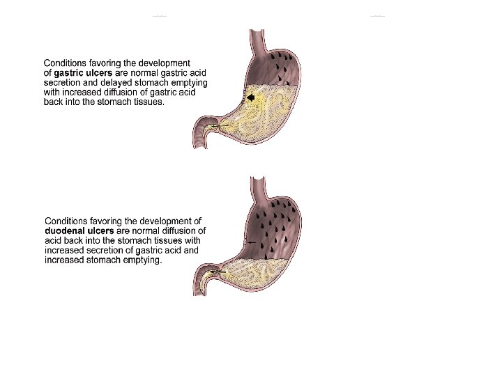
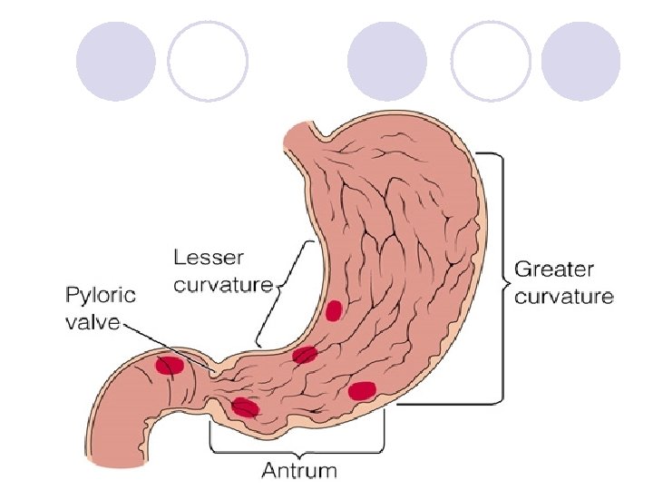
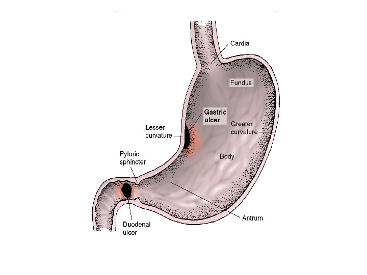
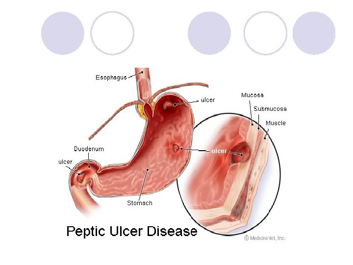
Peptic Ulcer Disease (PUD) Definition and Risk factors l a. Break in mucous lining of GI tract comes into contact with gastric juice; affects 10% of US population l b. Duodenal ulcers: most common; affect mostly males ages 30 – 55; ulcers found near pyloris l c. Gastric ulcers: affect older persons (ages 55 – 70); found on lesser curvature and associated with increased incidence of gastric cancer l d. Common in smokers, users of NSAIDS; familial pattern, ASA, alcohol, cigarettes
Peptic Ulcer Disease (PUD) 2. Pathophysiology l A. Ulcers or breaks in mucosa of GI tract occur with 1. H. pylori infection (spread by oral to oral, fecal-oral routes) damages gastric epithelial cells reducing effectiveness of gastric mucus 2. Use of NSAIDS: interrupts prostaglandin synthesis which maintains mucous barrier of gastric mucosa l b. Chronic with spontaneous remissions and exacerbations associated with trauma, infection, physical or psychological stress
Peptic Ulcer Disease l. Diagnosis ¡Endoscopy with cultures l. Looking for H. Pylori ¡Upper GI barium contrast studies ¡EGD-esophagogastroduodenoscopy ¡Serum and stool studies
Peptic Ulcer Disease (PUD) 3. Manifestations l a. Pain is classic symptom: gnawing, burning, aching hungerlike in epigastric region possibly radiating to back; occurs when stomach is empty and relieved by food (pain: food: relief pattern) l b. Symptoms less clear in older adult; may have poorly localized discomfort, dysphagia, weight loss; presenting symptom may be complication: GI hemorrhage or perforation of stomach or duodenum
Peptic Ulcer Disease l. Treatment ¡Rest and stress reduction ¡Nutritional management ¡Pharmacological management l. Antacids (Mylanta) • Neutralizes acids l. Proton pump inhibitors (omeprazole) • Block gastric acid secretion
Peptic Ulcer Disease l. Pharmacological management ¡Histamine blockers (Tagamet, Zantac) l. Blocks gastric acid secretion ¡Carafate l. Forms protective layer over the site ¡Mucosal barrier enhancers (bismuth subsalicylate , prostoglandins) l. Protect mucosa from injury ¡Antibiotics( ﻣﺘﺮﻭﻧیﺪﺍﺯﻭﻝ ﻭ ، کﻼﺭیﺘﺮﻭﻣﺎیﺴیﻦ ﺗﺘﺮﺍﺳیکﻠیﻦ , ) آﻤﻮکﺴی ﺳیﻠیﻦ l. Treat H. Pylori infection
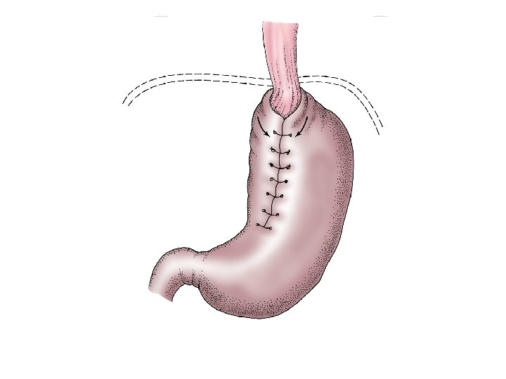
Peptic Ulcer Disease l Surgical intervention ¡Minimally invasive gastrectomy l. Partial gastric removal with laproscopic surgery ¡Bilroth I and II l. Removal of portions of the stomach ¡Vagotomy l. Cutting of the vagus nerve to decrease acid secretion ¡Pyloroplasty l. Widens the pyloric sphincter
Billroth I
Billroth II
Peptic Ulcer Disease (PUD) Complications l a. Hemorrhage: frequent in older adult: hematemesis, melena, hematochezia (blood in stool); weakness, fatigue, dizziness, orthostatic hypotension and anemia; with significant bleed loss may develop hypovolemic shock l b. Obstruction: gastric outlet (pyloric sphincter) obstruction: edema surrounding ulcer blocks GI tract from muscle spasm or scar tissue 1. Gradual process 2. Symptoms: feelings of epigastric fullness, nausea, worsened ulcer symptoms 4.
Peptic Ulcer Disease l c. Perforation: ulcer erodes through mucosal wall and gastric or duodenal contents enter peritoneum leading to peritonitis; chemical at first (inflammatory) and then bacterial in 6 to 12 hours 1. Time of ulceration: severe upper abdominal pain radiating throughout abdomen and possibly to shoulder 2. Abdomen becomes rigid, boardlike with absent bowel sounds; symptoms of shock 3. Older adults may present with mental confusion and non-specific symptoms
Upper GI Bleed l Mortality approx 10% l Predisposing factors include: drugs, esophageal varacies, esophagitis, PUD, gastritis and carcinoma
Upper GI Bleed l Signs and Symptoms ¡Coffee ground vomitus ¡Black, tarry stools ¡Melena ¡Decreased B/P ¡Vertigo ¡Drop in Hct, Hgb ¡Confusion ¡syncope
Upper GI Bleed l Diagnosis ¡History ¡Blood, stool, vomitus studies ¡Endoscopy
Upper GI Bleed l Treatments ¡Volume replacement l. Crystalloids- normal saline l. Blood transfusions ¡NG lavage ¡EGD l. Endoscopic treatment of bleeding ulcer l. Sclerotheraphy-injecting bleeding ulcer with necrotizing agent to stop bleeding( ethanol amin)
Upper GI Bleed l Treatments ¡Sengstaken-Blakemore tube l. Used with bleeding esophageal varacies ¡Surgical intervention l. Removal of part of the stomach
Sengstaken-Blakemore Tube
Cancer of Stomach 1. Incidence l a. Worldwide common cancer, but less common in US l b. Incidence highest among Hispanics, African Americans, Asian Americans, males twice as often as females l c. Older adults of lower socioeconomic groups higher risk 2. Pathophysiology l a. Adenocarcinoma most common form involving mucus-producing cells of stomach in distal portion l b. Begins as localized lesion (in situ) progresses to mucosa; spreads to lymph nodes and metastasizes early in disease to liver, lungs, ovaries, peritoneum
Cancer of Stomach 3. l l l 4. l l l Risk Factors a. H. pylori infection b. Genetic predisposition c. Chronic gastritis, pernicious anemia, gastric polyps d. Achlorhydria (lack of hydrochloric acid) e. Diet high in smoked foods and nitrates Manifestations a. Disease often advanced with metastasis when diagnosed b. Early symptoms are vague: early satiety, anorexia, indigestion, vomiting, pain after meals not responding to antacids c. Later symptoms weight loss, cachexia (wasted away appearance), abdominal mass, stool positive for occult blood
Cancer of Stomach 5. Collaborative Care a. Support client through testing b. Assist client to maintain adequate nutrition 6. Diagnostic Tests a. CBC indicates anemia b. Upper GI series, ultrasound identifies a mass c. Upper endoscopy: visualization and tissue biopsy of lesion
Cancer of Stomach 7. Treatment l a. Surgery, if diagnosis made prior to metastasis 1. Partial gastrectomy with anastomosis to duodenum: Bilroth I or gastroduodenostomy 2. Partial gastrectomy with anastomosis to jejunum: Bilroth II or gastrojejunostomy 3. Total gastrectomy (if cancer diffuse but limited to stomach) with esophagojejunostomy
Cancer of Stomach b. Complications associated with gastric surgery l 1. Dumping Syndrome a. Occurs with partial gastrectomy; hypertonic, undigested chyme bolus rapidly enters small intestine and pulls fluid into intestine causing decrease in circulating blood volume and increased intestinal peristalsis and motility b. Manifestations 5 – 30 minutes after meal: nausea with possible vomiting, epigastric pain and cramping, borborygmi, and diarrhea; client becomes tachycardic, hypotensive, dizzy, flushed, diaphoretic c. Manifestations 2 – 3 hours after meal: symptoms of hypoglycemia in response to excessive release of insulin that occurred from rise in blood glucose when chyme entered intestine
Cancer of Stomach d. Treatment: dietary pattern to delay gastric emptying and allow smaller amounts of chyme to enter intestine l 1. Liquids and solids taken separately l 2. Increased amounts of fat and protein l 3. Carbohydrates, especially simple sugars, reduced l 4. Client to rest recumbent or semi-recumbent 30 – 60 minutes after eating l 5. Anticholinergics(hyosin), sedatives, antispasmodic medications may be added(colofac) l 6. Limit amount of food taken at one time
Cancer of the Stomach l Common post-op complications ¡Pneumonia ¡Anastomotic leak ¡Hemorrhage ¡Relux aspiration ¡Sepsis ¡Reflux gastritis ¡Paralytic ileus ¡Bowel obstruction ¡Wound infection ¡Dumping syndrome
Cancer of Stomach l Nutritional problems related to rapid entry of food into the bowel and the shortage of intrinsic factor l 1 Anemia: iron deficiency and/or pernicious l 2 Folic acid deficiency l 3. Poor absorption of calcium, vitamin D c. Radiation and/or chemotherapy to control metastasic spread d. Palliative treatment including surgery, chemotherapy; client may have gastrostomy or jejunostomy tube inserted 7. Nursing Diagnoses l a. Imbalanced Nutrition: Less than body requirement: consult dietician since client at risk for protein-calorie malnutrition l b. Anticipatory Grieving
Diagnostic Evaluation l Lower GI tract studies: ¡Barium Enema: detect polyps, tumors, lesions of colon ¡Radiopaque substance instilled rectally ¡Gastrografin (water-soluble iodine contrast) used in inflammatory disease or perforated colon l Nursing Interventions: May vary according to MD orders ¡Bowel cleansing 1 -2 days before, clear liquid diet day before, NPO after midnight, enema until clear morning of l Computed Tomography: cross-sectional images l Nursing Interventions: NPO for 6 -8 hrs prior, assess for allergies to contrast dye
Diagnostic Evaluation l Magnetic Resonance Imaging: Noninvasive, uses magnetic fields and radio waves: ¡Useful in evaluating soft tissues, vessels, neoplasm ¡Contraindicated for pts with pacemakers, metal implants l Nursing Interventions: NPO for 6 -8 hrs prior, remove all jewelry, procedure takes 30 -90 minutes, close fitting scanner may cause feelings of claustrophobia l Anoscopy, Proctoscopy, Sigmoidoscopy: ¡Viewing of lower colon through rigid or flexible scope l Nursing Interventions: Minimal bowel cleansing, monitor vital signs during and after procedure
Diagnostic Evaluation l Colonoscopy: Direct visual inspection of colon to cecum ¡Flexible fiberoptic colonoscope, can obtain biopsies and remove polyps ¡Usually takes one hour, pt on left side, legs drawn toward chest l Nursing Interventions: May vary according to MD orders ¡Bowel cleansing : clear liquids day before, Informed consent, NPO night before, IV midazolam (Versed) for sedation. ¡During procedure monitor vital signs, O 2 saturation, color and temp of skin, level of consciousness, vagal response
Gerontologic Considerations l Oral Cavity ¡Tooth loss or decay ¡Atrophy of taste buds l Esophagus ¡Weakened gag reflex l Stomach ¡Decrease gastric secretions ¡Decrease motility l Small Intestine ¡Atrophy of muscle and mucosal surfaces l Large Intestine ¡Decrease mucus production ¡Decrease tone of anal sphincter
Nursing Care of Clients with Bowel Disorders Factors affecting bodily function of elimination A. GI tract l 1. Food intake l 2. Bacterial flora in bowel B. Indirect l 1. Psychologic stress l 2. Voluntary postponement of defecation C. Normal bowel elimination pattern l 1. l 2. Varies with the individual 2 – 3 times daily to 3 stools per week
Irritable Bowel Syndrome (IBS) (spastic bowel, functional colitis) Definition l a. Functional GI tract disorder without identifiable cause characterized by abdominal pain and constipation, diarrhea, or both l b. Affects up to 20% of persons in Western civilization; more common in females
Irritable Bowel Syndrome (IBS) (spastic bowel, functional colitis) Pathophysiology l a. Appears there is altered CNS regulation of motor and sensory functions of bowel 1. Increased bowel activity in response to food intake, hormones, stress 2. Increased sensations of chyme movement through gut 3. Hypersecretion of colonic mucus l b. Lower visceral pain threshold causing abdominal pain and bloating with normal levels of gas l c. Some linkage of depression and anxiety
Irritable Bowel Syndrome (IBS) (spastic bowel, functional colitis) Manifestations l a. Abdominal pain relieved by defecation; may be colicky, occurring in spasms, dull or continuous l b. Altered bowel habits including frequency, hard or watery stool, straining or urgency with stooling, incomplete evacuation, passage of mucus; abdominal bloating, excess gas l c. Nausea, vomiting, anorexia, fatigue, headache, anxiety l d. Tenderness over sigmoid colon upon palpation 4. Collaborative Care l a. Management of distressing symptoms l b. Elimination of precipitating factors, stress reduction
Irritable Bowel Syndrome (IBS) (spastic bowel, functional colitis) 5. l l Diagnostic Tests: to find a cause for client’s abdominal pain, changes in feces elimination a. Stool examination for occult blood, ova and parasites, culture b. CBC with differential, Erythrocyte Sedimentation Rate (ESR): to determine if anemia, bacterial infection, or inflammatory process c. Sigmoidoscopy or colonoscopy 1. Visualize bowel mucosa, measure intraluminal pressures, obtain biopsies if indicated 2. Findings with IBS: normal appearance increased mucus, intraluminal pressures, marked spasms, possible hyperemia without lesions d. Small bowel series (Upper GI series with small bowel-follow through) and barium enema: examination of entire GI tract; IBS: increased motility
Irritable Bowel Syndrome (IBS) (spastic bowel, functional colitis) Medications l a. Purpose: to manage symptoms l b. Bulk-forming laxatives: reduce bowel spasm, normalize bowel movement in number and form l c. Anticholinergic drugs (dicyclomine (Bentyl), hyoscyamine) to inhibit bowel motility and prevent spasms; given before meals l d. Antidiarrheal medications (loperamide (Imodium), diphenoxylate (Lomotil): prevent diarrhea prophylactically l e. Antidepressant medications l f. Research: medications altering serotonin receptors in GI tract to stimulate peristalsis of the GI tract
Irritable Bowel Syndrome (IBS) (spastic bowel, functional colitis) Dietary Management l a. Often benefit from additional dietary fiber: adds bulk and water content to stool reducing diarrhea and constipation l b. Some benefit from elimination of lactose, fructose, sorbitol l c. Limiting intake of gas-forming foods, caffeinated beverages 8. Nursing Care l a. Contact in health environments outside acute care l b. Home care focus on improving symptoms with changes of diet, stress management, medications; seek medical attention if serious changes occur
Peritonitis Definition l a. Inflammation of peritoneum, lining that covers wall (parietal peritoneum) and organs (visceral peritoneum) of abdominal cavity l b. Enteric bacteria enter the peritoneal cavity through a break of intact GI tract (e. g. perforated ulcer, ruptured appendix)
Peritonitis l Causes include: ¡Ruptured appendix ¡Perforated bowel secondary to PUD ¡Diverticulitis ¡Gangrenous gall bladder ¡Ulcerative colitis ¡Trauma ¡Peritoneal dialysis
Peritonitis Pathophysiology l a. Peritonitis results from contamination of normal sterile peritoneal cavity with infections or chemical irritant l b. Release of bile or gastric juices initially causes chemical peritonitis; infection occurs when bacteria enter the space l c. Bacterial peritonitis usually caused by these bacteria (normal bowel flora): Escherichia coli, Klebsiella, Proteus, Pseudomonas l d. Inflammatory process causes fluid shift into peritoneal space (third spacing); leading to hypovolemia, then septicemia
Peritonitis 3. Manifestations l a. Depends on severity and extent of infection, age and health of client l b. Presents with “acute abdomen” 1. Abrupt onset of diffuse, severe abdominal pain 2. Pain may localize near site of infection (may have rebound tenderness) 3. Intensifies with movement l c. Entire abdomen is tender with boardlike guarding or rigidity of abdominal muscle
Peritonitis l d. paralytic ileus; l bowel sounds are diminished or absent l distention; nausea and vomiting l e. Systemically: fever, malaise, tachycardia and tachypnea, restlessness, disorientation, oliguria with dehydration and shock l f. Older or immunosuppressed client may have 1. Few of classic signs 2. Increased confusion and restlessness 3. Decreased urinary output 4. Vague abdominal complaints 5. At risk for delayed diagnosis and higher mortality rates
Peritonitis 4. Complications l a. mortality rate overall 40% l b. Abscess l c. Fibrous adhesions l d. Septicemia, septic shock; hypovolemic shock 5. Collaborative Care l a. Diagnosis and identifying and treating cause l b. Prevention of complications
Peritonitis 6. Diagnostic Tests l a. WBC with differential: elevated WBC to 20, 000; shift to left l b. Blood cultures: identify bacteria in blood l c. Liver and renal function studies, serum electrolytes: evaluate effects of peritonitis l d. Abdominal xrays: detect intestinal distension, air-fluid levels, free air under diaphragm (sign of GI perforation) l e. Diagnostic paracentesis
Medications l a. Antibiotics 1. Broad-spectrum before definitive culture results identifying specific organism(s) causing infection 2. Specific antibiotic(s) treating causative pathogens l b. Analgesics
Peritonitis 8. Surgery l a. Laparotomy to treat cause (close perforation, removed inflamed tissue) l b. Peritoneal Lavage: washing out peritoneal cavity with copious amounts of warm isotonic fluid during surgery to dilute residual bacterial and remove gross contaminants l c. Often have drain in place and/or incision left unsutured to continue drainage
Peritonitis 9. Treatment l a. Intravenous fluids and electrolytes to maintain vascular volume and electrolyte balance l b. Bed rest in Fowler’s position to localize infection and promote lung ventilation l c. Intestinal decompression with nasogastric tube or intestinal tube connected to suction l 1. Relieves abdominal distension secondary to paralytic ileus l 2. NPO with intravenous fluids while having nasogastric suction
Peritonitis 10. Nursing Diagnoses l a. Pain l b. Deficient Fluid Volume: often on hourly output; nasogastric drainage is considered when ordering intravenous fluids l c. Ineffective Protection l d. Anxiety 11. Home Care l a. Client may have prolonged hospitalization l b. Home care often includes l 1. Wound care l 2. Home health referral l 3. Home intravenous antibiotics
Neoplastic Disorders Background l 1. Large intestine and rectum most common GI site affected by cancer l 2. Colon cancer is second leading cause of death from cancer in U. S. B. Client with Polyps 1. Definition l a. Polyp is mass of tissue arising from bowel wall and protruding into lumen l b. Most often occur in sigmoid and rectum l c. 30% of people over 50 have polyps
Neoplastic Disorders Pathophysiology la. Most polyps are adenomas, benign but considered premalignant; < 1% become malignant but all colorectal cancers arise from these polyps lb. Familial polyposis is uncommon autosomal dominant genetic disorder with hundreds of adenomatous polyps throughout large intestine; untreated, near 100% malignancy by age 40
Client with Polyps Manifestations l a. Most asymptomatic l b. Intermittent painless rectal bleeding is most common presenting symptom Collaborative Care l a. Diagnosis is based on colonoscopy l b. Most reliable since allows inspection of entire colon with biopsy or polypectomy if indicated l c. Repeat every 3 years since polyps recur
Client with Polyps Nursing Care la. All clients advised to have screening colonoscopy at age 50 and every 5 years thereafter lb. Bowel preparation ordered prior to colonoscopy with cathartics and/or enemas
Polyps
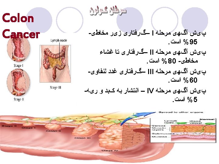
Client with Colorectal Cancer Definition l a. Third most common cancer diagnosed l b. Affects sexes equally l c. Five-year survival rate is 90%, with early diagnosis and treatment Risk Factors l a. Family history l b. Inflammatory bowel disease l c. Diet high in fat, calories, protein
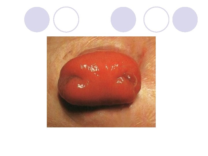
Client with Colorectal Cancer Manifestations la. Often produces no symptoms until it is advanced lb. Presenting manifestation is bleeding; also change in bowel habits (diarrhea or constipation); pain, anorexia, weight loss, palpable abdominal or rectal mass; anemia
Colon Cancer
Client with Colorectal Cancer Complications l a. Bowel obstruction l b. Perforation of bowel by tumor, peritonitis l c. Direct extension of cancer to adjacent organs; reoccurrences within 4 years Screening l a. Digital exam beginning at age 40, annually l b. Fecal occult blood testing beginning at age 50, annually l c. Colonoscopies or sigmoidoscopies beginning at age 50, every 3 – 5 years
Client with Colorectal Cancer Diagnostic Tests l a. CBC: anemia from blood loss, tumor growth l b. Fecal occult blood (guiac or Hemoccult testing): all colorectal cancers bleed intermittently l c. Carcinoembryonic antigen (CEA): not used as screening test, but is a tumor marker and used to estimate prognosis, monitor treatment l d. Colonoscopy or sigmoidoscopy; tissue biopsy of suspicious lesions, polyps l e. Chest xray, CTscans, MRI, ultrasounds: to determine tumor depth, organ involvement, metastasis
Client with Colorectal Cancer l Pre-op care ¡Consult with ET nurse if ostomy is planned ¡Bowel prep with Go. Lytely ¡NPO ¡NG
Client with Colorectal Cancer Surgery l a. Surgical resection of tumor, adjacent colon, and regional lymph nodes is treatment of choice l b. Whenever possible anal sphincter is preserved and colostomy avoided; anastomosis of remaining bowel is performed l c. Tumors of rectum are treated with abdominoperineal resection (A-P resection) in which sigmoid colon, rectum, and anus are removed through abdominal and perineal incisions and permanent colostomy created
Client with Colorectal Cancer Colostomy Named for area of colon is which formed l a. Sigmoid colostomy: used with abdominoperineal resection formed on LLQ l b. Double-barrel colostomy: 2 stomas: proximal for feces diversion; distal is mucous fistula l c. Transverse loop colostomy: emergency procedure; loop suspended over a bridge; temporary l d. Hartman procedure: Distal portion is left in place and oversewn; only proximal colostomy is brought to abdomen as stoma; temporary; colon reconnected at later time when client ready for surgical repair 1.
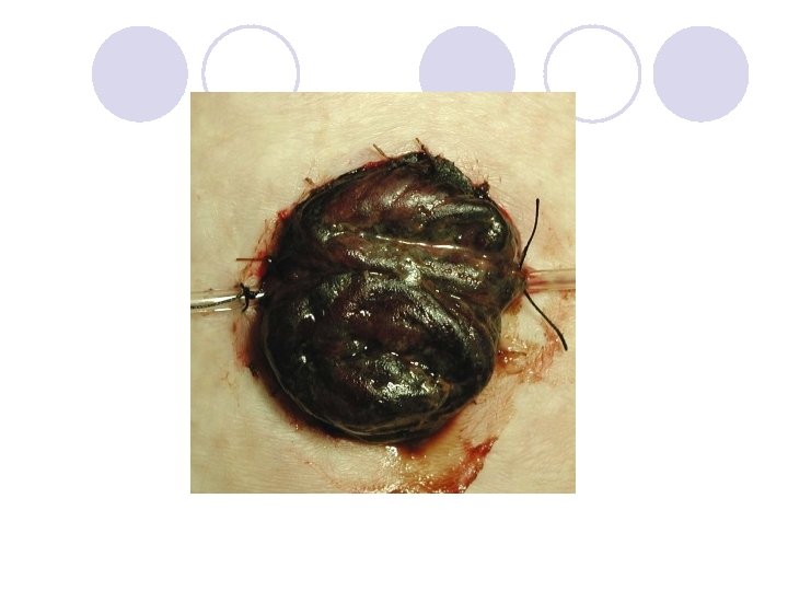
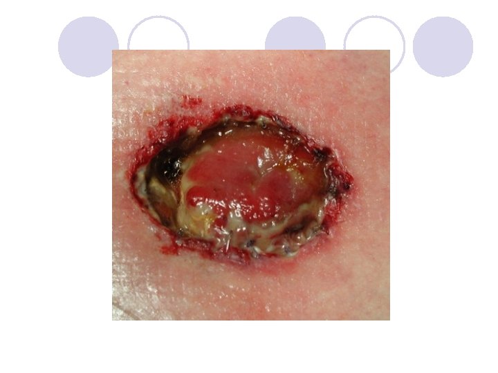
Client with Colorectal Cancer l Post-op care ¡Pain ¡NG tube ¡Wound management l. Stoma • Should be pink and moist • Dark red or black indicates ischemic necrosis • Look for excessive bleeding • Observe for possible separation of suture securing stoma to abdominal wall
Client with Colorectal Cancer l Post-op care ¡Evaluate stool after 2 -4 days postop l. Ascending stoma (right side) • Liquid stool l. Transverse stoma • Pasty l. Descending stoma • Normal, solid stool
End stoma with Hartmann’s pouch
Good stoma Bad stoma
Stomal prolapse
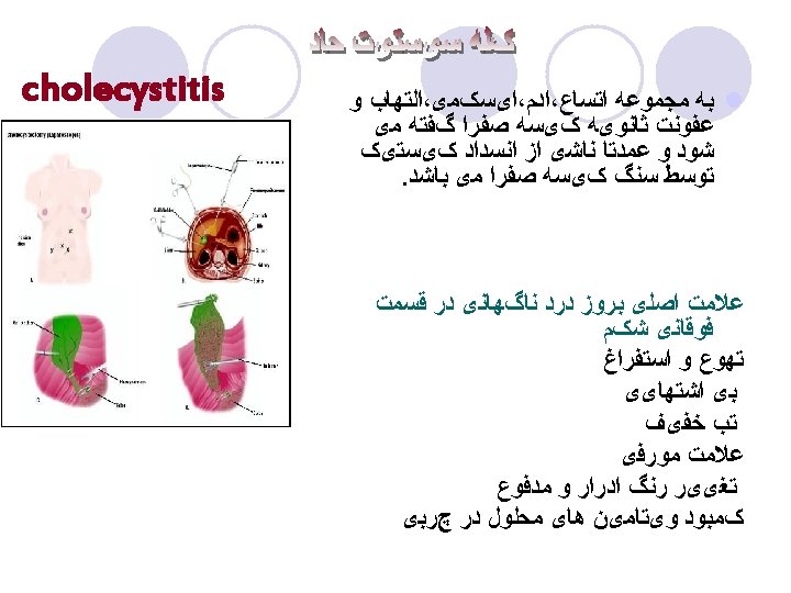
cholecystitis
l. The end
Client with Intestinal Obstruction Definition l a. May be partial or complete obstruction l b. Failure of intestinal contents to move through the bowel lumen; most common site is small intestine l c. With obstruction, gas and fluid accumulate proximal to and within obstructed segment causing bowel distention l d. Bowel distention, vomiting, third-spacing leads to hypovolemia, hypokalemia, renal insufficiency, shock
Client with Intestinal Obstruction Pathophysiology a. Mechanical 1. Problems outside intestines: adhesions (bands of scar tissue), hernias 2. Problems within intestines: tumors, IBD 3. Obstruction of intestinal lumen (partial or complete) l a. Intussusception: telescoping bowel l b. Volvulus: twisted bowel l c. Foreign bodies l d. Strictures
Client with Intestinal Obstruction Functional 1. Failure of peristalsis to move intestinal contents: adynamic ileus (paralytic ileus, ileus) due to neurologic or muscular impairment 2. Accounts for most bowel obstructions 3. Causes include l a. Post gastrointestinal surgery l b. Tissue anoxia or peritoneal irritation from hemorrhage, peritonitis, or perforation l c. Hypokalemia l d. Medications: narcotics, anticholinergic drugs, antidiarrheal medications l e. Spinal cord injuries, uremia, alterations in electrolytes
Client with Intestinal Obstruction a. b. c. l l d. l l e. Manifestations Small Bowel Obstruction Vary depend on level of obstruction and speed of development Cramping or colicky abdominal pain, intermittent, intensifying Vomiting 1. Proximal intestinal distention stimulates vomiting center 2. Distal obstruction vomiting may become feculent Bowel sounds 1. Early in course of mechanical obstruction: borborygmi and high-pitched tinkling, may have visible peristaltic waves 2. Later silent; with paralytic ileus, diminished or absent bowel sounds throughout Signs of dehydration
Client with Intestinal Obstruction Complications l a. Hypovolemia and hypovolemic shock can result in multiple organ dysfunction (acute renal failure, impaired ventilation, death) l b. Strangulated bowel can result in gangrene, perforation, peritonitis, possible septic shock l c. Delay in surgical intervention leads to higher mortality rate
Client with Intestinal Obstruction Large Bowel Obstruction l a. Only accounts for 15% of obstructions l b. Causes include cancer of bowel, volvulus, diverticular disease, inflammatory disorders, fecal impaction l c. Manifestations: deep, cramping pain; severe, continuous pain signals bowel ischemia and possible perforation; localized tenderness or palpable mass may be noted
Client with Intestinal Obstruction Collaborative Care l a. Relieving pressure and obstruction l b. Supportive care Diagnostic Tests a. Abdominal Xrays and CT scans with contrast media l 1. Show distended loops of intestine with fluid and /or gas in small intestine, confirm mechanical obstruction; indicates free air under diaphragm l 2. If CT with contrast media meglumine diatrizoate (Gastrografin), check for allergy to iodine, need BUN and Creatinine to determine renal function b. Laboratory testing to evaluate for presence of infection and electrolyte imbalance: WBC, Serum amylase, osmolality, electrolytes, arterial blood gases c. Barium enema or colonoscopy/sigmoidoscopy to identify large bowel obstruction Gastrointestinal Decompression l a. Treatment with nasogastric or long intestinal tube provides bowel rest and removal of air and fluid l b. Successfully relieves many partial small bowel obstructions
Client with Intestinal Obstruction Surgery l a. Treatment for complete mechanical obstructions, strangulated or incarcerated obstructions of small bowel, persistent incomplete mechanical obstructions l b. Preoperative care l 1. Insertion of nasogastric tube to relieve vomiting, abdominal distention, and to prevent aspiration of intestinal contents l 2. Restore fluid and electrolyte balance; correct acid and alkaline imbalances l 3. Laparotomy: inspection of intestine and removal of infarcted or gangrenous tissue l 4. Removal of cause of obstruction: adhesions, tumors, foreign bodies, gangrenous portion of intestines and anastomosis or creation of colostomy depending on individual case
Client with Intestinal Obstruction Nursing Care l a. Prevention includes healthy diet, fluid intake l b. Exercise, especially in clients with recurrent small bowel obstructions Nursing Diagnoses l a. Deficient Fluid Volume l b. Ineffective Tissue Perfusion, gastrointestinal l c. Ineffective Breathing Pattern Home Care l a. Home care referral as indicated l b. Teaching about signs of recurrent obstruction and seeking medical attention
Client with Diverticular Disease Definition l a. Diverticula are saclike projections of mucosa through muscular layer of colon mainly in sigmoid colon l b. Incidence increases with age; less than a third of persons with diverticulosis develop symptoms Risk Factors l a. Cultural changes in western world with diet of highly refined and fiber-deficient foods l b. Decreased activity levels l c. Postponement of defecation
Client with Diverticular Disease Pathophysiology l a. Diverticulosis is the presence of diverticula which form due to increased pressure within bowel lumen causing bowel mucosa to herniate through defects in colon wall, causing outpouchings l b. Muscle in bowel wall thickens narrowing bowel lumen and increasing intraluminal pressure l c. Complications of diverticulosis include hemorrhage and diverticulitis, the inflammation of the diverticular sac
Clients with Diverticular Disease l d. Diverticulitis: diverticulum in sigmoid colon irritated with undigested food and bacteria forming a hard mass (fecalith) that impairs blood supply leading to perforation l e. With microscopic perforation, inflammation is localized; more extensive perforation may lead to peritonitis or abscess formation
Diverticulits
Diverticulitis
Client with Diverticular Disease Manifestations l a. Pain, left-sided, mild to moderate and cramping or steady l b. Constipation or frequency of defecation l c. May also have nausea, vomiting, low-grade fever, abdominal distention, tenderness and palpable LLQ mass l d. Older adult may have vague abdominal pain Complications l a. Peritonitis l b. Abscess formation l c. Bowel obstruction l d. Fistula formation l e. Hemorrhage
Client with Diverticular Disease Collaborative Care: Focus is on management of symptoms and complications Diagnostic Tests l a. Abdominal Xray: detection of free air with perforation, location of abscess, fistula l b. Barium enema contraindicated in early diverticulitis due to risk of barium leakage into peritoneal cavity, but will confirm diverticulosis l c. Abdominal CT scan, sigmoidoscopy or colonscopy used in diagnosis of diverticulosis l d. WBC count with differential: leukocytosis with shift to left in diverticulitis l e. Hemocult or guiac testing: determine presence of occult blood
Client with Diverticular Disease Medications l a. Broad spectrum antibiotics against gram negative and anaerobic bacteria to treat acute diverticulitis, oral or intravenous route depending on severity of symptoms ¡ Flagyl plus Bactrim or Cipro l b. Analgesics for pain (non-narcotic) l c. Fluids to correct dehydration l d. Stool softener but not cathartic may be prescribed (nothing to increase pressure within bowel) l e. Anticholinergics to decrease intestinal hypermotility
Clients with Diverticular Disease Dietary Management l a. Diet modification may decrease risk of complications l b. High-fiber diet (bran, commercial bulk -forming products such as psyllium seed (Metamucil) or methycelluose) l c. Some clients advised against foods with small seeds which could obstruct diverticula
Client with Diverticular Disease Treatment for acute episode of diverticulitis l a. Client initially NPO with intravenous fluids (possibly TPN) l b. As symptoms subside reintroduce food: clear liquid diet, to soft, low-roughage diet psyillium seed products to soften stool and increase bulk l c. High fiber diet is resumed after full recovery Surgery l a. Surgical intervention indicated for clients with generalized peritonitis or abscess that does not respond to treatment l b. With acute infection, 2 stage Hartman procedure done with temporary colostomy; re-anastomosis performed 2 – 3 months later
Client with Diverticular Disease Nursing Care: Health promotion includes teaching high -fiber foods in diet generally, may be contraindicated for persons with known conditions Nursing Diagnoses l a. Impaired Tissue Integrity, gastrointestinal l b. Pain l c. Anxiety, related to unknown outcome of treatment, possible surgery Home Care l a. Teaching regarding prescribed diet, fluid intake, medications l b. Referral for home health care agency, if new colostomy client