DNA RNA and Protein Synthesis Molecular structure of


 differs from DNA: Usually one polynucleotide strand The sugar is ribose")

—carries copy of a")
 Produced by transcription as a complementary copy of DNA")


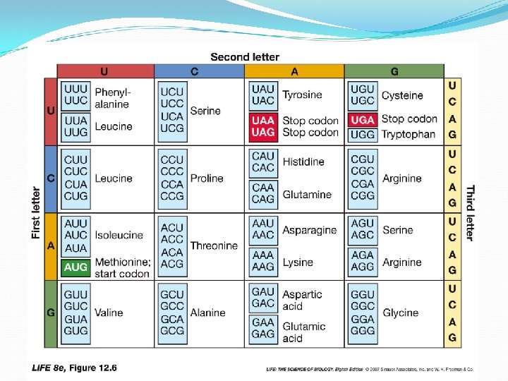
 designates first amino acid (methionine) in all proteins Large")


")


 Substrate of a metabolic pathway")
 Product of a metabolic pathway")

- Slides: 96
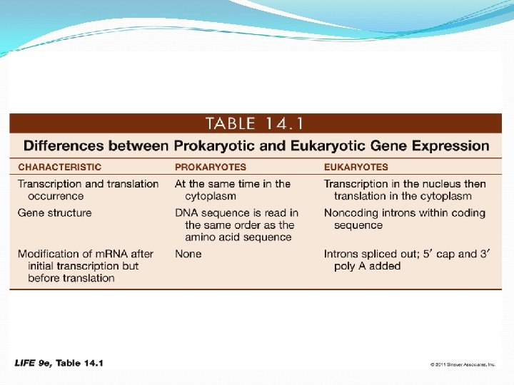
DNA, RNA, and Protein Synthesis
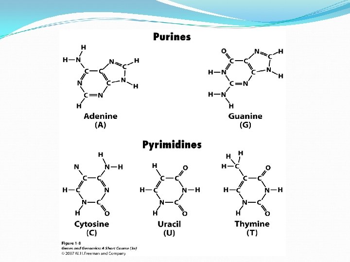
Molecular structure of DNA Chromosomes contain both nucleic acid and protein. There are two types of nucleic acid, DNA and RNA. Nucleic acid is a polymer of nucleotides, each comprised of a ribose sugar, a phosphate group and a nitrogen containing base. There are two types of bases, Purines and Pyrimidines.
In the 1920 s, Frederick Griffith performed experiments with two strains of the bacterium Streptococcus pneumoniae. A biological assay implicates DNA as the primary genetic molecule.
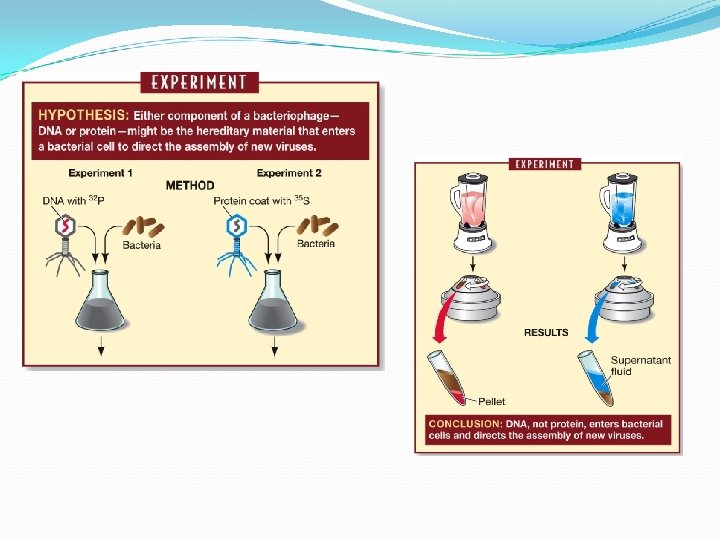
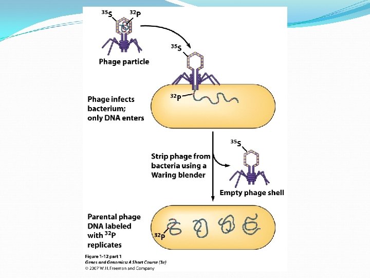
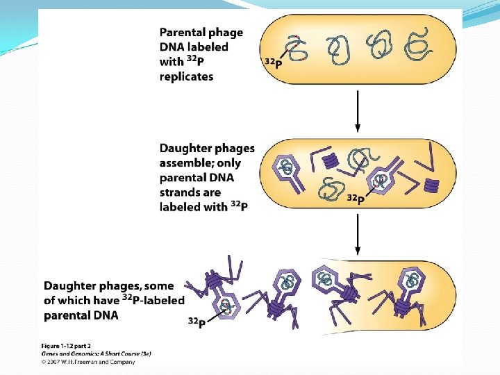
In 1952, Alfred D. Hershey and Martha Chase confirmed DNA is the genetic material T 2 bacteriophage (virus that attacks bacteria) consists of DNA core packed in protein coat Phage DNA alone carries genetic information
A DNA nucleotide
Individual nucleotides of DNA are linked by phosphodiester bonds. This sugar-phosphate chain forms the “backbone” of the DNA molecule.
In the 1950 s, chemists Rosalind Franklin and Maurice Wilkins provided key information about DNA structure using X-ray crystallography
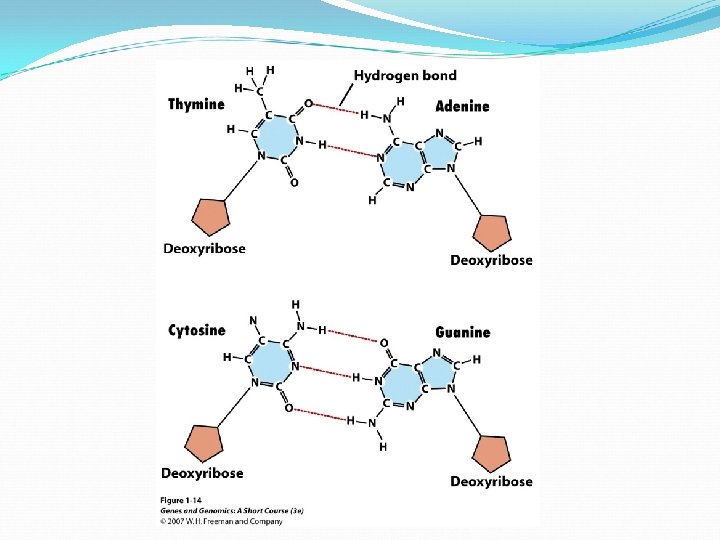
Erwin Chargaff noted that A = T, and G = C
James D. Watson and Francis Crick established the general structure of DNA in 1953 X-ray crystallography convinced them that the DNA molecule was helical, with certain dimensions. Other evidence for two polynucleotide chains running antiparallel to each other
Key features of DNA: – double-stranded helix – diameter is uniform – the twist is right-handed – antiparallel strands – two strands run in different directions – Semiconservative replication uses each parent strand as template for new strand
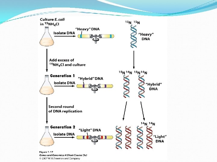
DNA Replication The Meselson – Stahl experiment demonstrating replication is semi-conservative using density labeling.
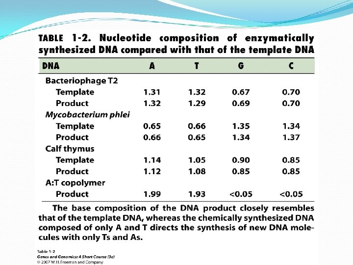
DNA replication is carried out by a highly complex assembly of proteins DNA Polymerase III Helicase Single-strand binding proteins Primase
When DNA polymerase III reaches previous Okazaki fragment, it is released DNA polymerase I replaces the primer of previous Okazaki fragment with DNA, but leaving a small “nick” Finally, DNA ligase catalyzes formation of the phosphodiester linkage that joins the two Okazaki fragments
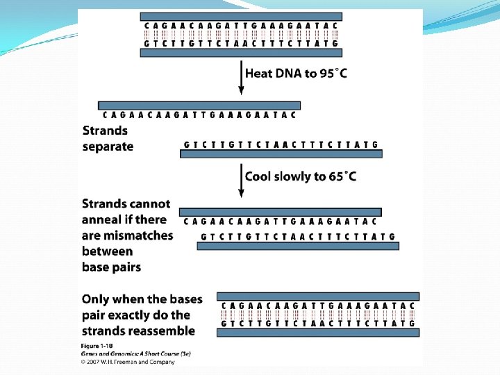
Denature and Renature of DNA double helix is stabilized by numerous hydrogen bonds between complimentary base pairs. DNA is denatured or melt if exposed to high temperature or extreme of p. H. The melting temperature (Tm) is the temperature at which half of the DNA molecules in a sample have been denatured. Double helices with an access of G: C base pairs are more stable and have higher Tm than helices in which A: T base pairs predominate.
Annealing or hybridization is an essential and powerful tool Labeled DNA fragment can be used as a probe to find its complement, even in a whole genome. The gene of interest can be identified and isolate from DNA library. Specific primers can be designed to amplify the gene of interest in polymerase chain reaction. By observing the extent of annealing between DNA strands in solution, researchers can determine the degree of similarity between DNA molecules from different species.
Decoding Genetic Information: DNA to RNA to Protein Gene expression takes place in two steps: Transcription – makes single- stranded RNA copy of a DNA segment Translation – uses information encoded in RNA to make a polypeptide http: //www. anselm. edu/homepage/jpitocch/genbio/transcrtransl. JPG
Transcription In normal prokaryotic and eukaryotic cells, transcription requires: DNA template for complementary base pairing appropriate ribonucleoside triphosphates (ATP, GTP, CTP, and UTP) to act as substrates RNA polymerase enzyme http: //www. brooklyn. cuny. edu/bc/ahp/Bio. Info/graphics/Transcription. 01. GIF
In prokaryotes, translation of m. RNA often begins before transcription is complete http: //www. phschool. com/science/biology_place/biocoach/images/transcription
Figure 14. 2 From Gene to Protein
Each eukaryote gene has one promoter to which RNA polymerase binds, with the help of other molecules. At the other end of the gene there is a terminator sequence to signal end of transcription.
Transcription
In the nucleus, pre-m. RNA is modified at both ends: G cap is added at the 5′ end (modified guanosine triphosphate)—facilitates m. RNA binding to ribosome. G cap protects m. RNA from being digested by ribonucleases.
Poly A tail added at 3′ end. AAUAAA sequence after last codon is a signal for an enzyme to cut the pre-m. RNA; then another enzyme adds 100 to 300 adenines—the “tail. ” May assist in export from nucleus; important for stability of m. RNA.
Eukaryotic genes may have noncoding sequences—introns. The coding sequences are exons. Introns and exons appear in the primary m. RNA transcript—pre-m. RNA; introns are removed from the final m. RNA.
Introns interrupt, but do not scramble, the DNA sequence that encodes a polypeptide. Sometimes, the separated exons code for different domains (functional regions) of the protein.
Intron Splicing
Alternative Splicing
Decoding Genetic Information: DNA to RNA to Protein Francis Crick’s central dogma stated that information flow is: DNA codes for RNA, and RNA codes for protein Replication Transcription Translation
Exception to the central dogma: Viruses: Non-cellular particles that reproduce inside cells; many have RNA instead of DNA. Viruses can replicate by transcribing from RNA to RNA, and then making multiple copies by transcription.
Other viruses such as HIV are retroviruses. After infecting a host cell a copy of the viral genome is incorporated into the host’s genome to make more RNA. Synthesis of DNA from RNA is reverse transcription. http: //www. brown. edu/Courses/Bio_160/Projects 1999/hiv/images/Virion 2. jpg HIV, a retrovirus
Translation Concurrent transcription and translation in prokaryotes.
Translation Transcription and translation are spatially separated in eukaryotes.
RNA (ribonucleic acid) differs from DNA: Usually one polynucleotide strand The sugar is ribose Contains uracil (U) instead of thymine (T)
Bases in RNA can pair with a single strand of DNA, except that adenine pairs with uracil instead of thymine. Single-strand RNA can fold into complex shapes by internal base pairing.
Three kinds of RNA in protein synthesis: Messenger RNA (m. RNA)—carries copy of a DNA sequence to site of protein synthesis at the ribosome Transfer RNA (t. RNA)—carries amino acids for polypeptide assembly Ribosomal RNA (r. RNA)—catalyzes peptide bonds and provides structure
Messenger RNA (m. RNA) Produced by transcription as a complementary copy of DNA moves from nucleus of eukaryotic cells into cytoplasm serves as template for protein synthesis
t. RNA – the “adapter” molecule required to assure specificity in translation of m. RNA into proteins t. RNAs must read m. RNA correctly Specific t. RNAs must carry correct amino acids
t. RNAs are “adapters” link a specific codon with specific amino acid t. RNA has three functions: carries an amino acid associates with m. RNA molecules interacts with ribosomes
Amino acid binding site at 3’ end of every t. RNA (for covalently binding a specific amino acid) Anticodon On other side of the molecule three bases which are complementary to appropriate codon The codon and anticodon unite by complementary base pairing. http: //www. wiley. com/legacy/college/boyer/0470003790/structure/t. RNA/trna_diagram. gif
Wobble Specificity for base at codon’s third position is not always observed Ex: codons for alanine — GCA, GCC, and GCU — are recognized by the same t. RNA Allows cells to produce fewer t. RNA species; but not in all cases — the genetic code remains unambiguous http: //www. wiley. com/legacy/college/boyer/0470003790/structure/t. RNA/trna_diagram. gif
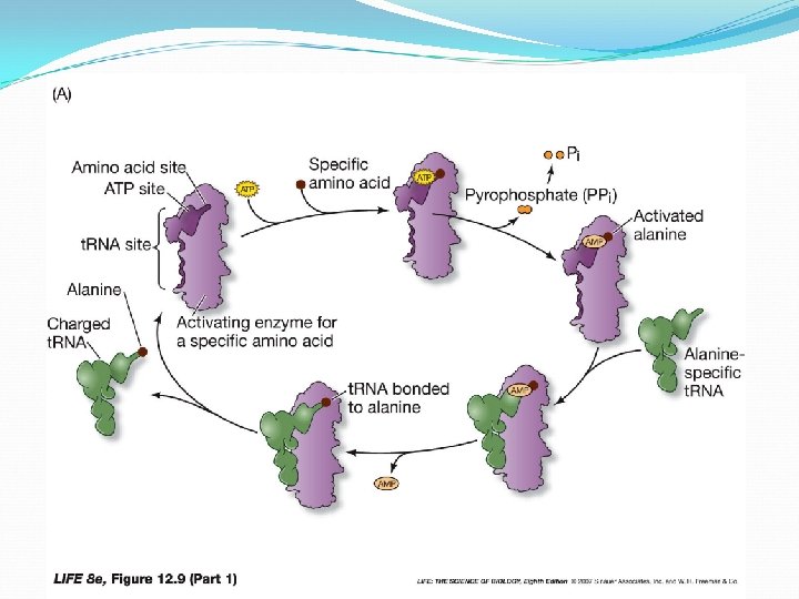
Aminoacyl-t. RNA synthetases Attaches amino acids to t. RNAs Their three-part active sites bind: a specific amino acid ATP a specific t. RNA, charged with a high-energy bond High-energy bond provides energy for making a peptide bond
Translation also occurs in three steps: Initiation Elongation Termination http: //srv 2. lycoming. edu/~newman/courses/bio 43704/ribosome 1. jpg
Initiation Start codon (AUG) designates first amino acid (methionine) in all proteins Large subunit then joins complex with t. RNA at P-site, all guided by initiation factors
Initiation The m. RNA binds to the small ribosomal subunit. The Shine-Dalgarno sequence near the 5’ end of the m. RNA base pairs with a sequence near the 3’ end of the r. RNA of the small ribosomal subunit.
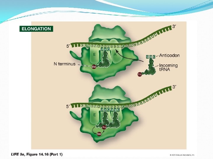
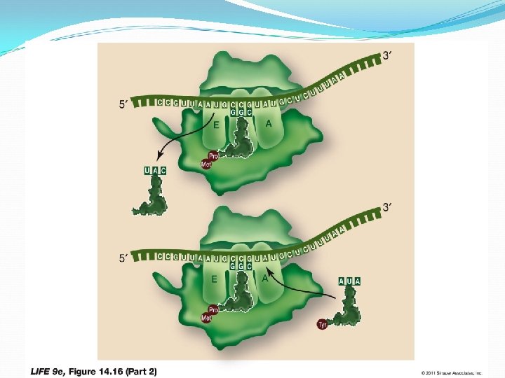
Elongation: The second charged t. RNA enters the A site. Large subunit catalyzes two reactions: It breaks bond between t. RNA in P site and its amino acid Peptide bond forms between that amino acid and the amino acid on t. RNA in the A site When the first t. RNA has released its methionine, it moves to the E site and dissociates from the ribosome—can then become charged again. Elongation occurs as the steps are repeated, assisted by proteins called elongation factors.
Termination When stop codon is reached… Release factor and a water molecule enter A site, instead of an amino acid Newly completed protein separates from ribosome, and subunits separate
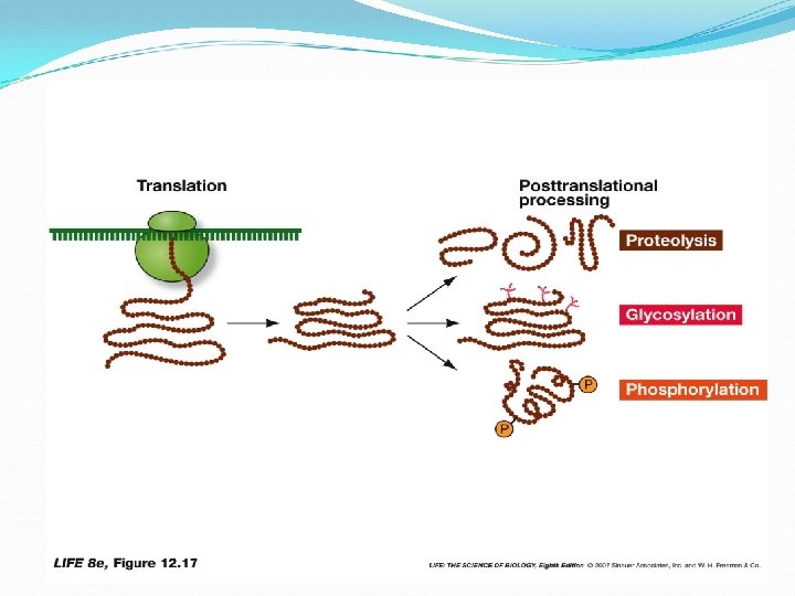
Protein modifications: Proteolysis: Cutting of a long polypeptide chain into final products, by proteases Glycosylation: Addition of sugars to form glycoproteins Phosphorylation: Addition of phosphate groups catalyzed by protein kinases— charged phosphate groups change the conformation
High accuracy of protein synthesis
Mutation Results in Mutant Proteins The genetic disease, sickle-cell anemia, caused by mutant proteins with single amino acid replacement. A single amino acid change in such a large complex hemoglobin could produce profound changes in activity.
Control of Gene Expression Cells conserve energy and resources by making proteins only when needed. Intricate molecular mechanisms must exist in cells to control the numbers of their many proteins. There are three ways that might achieve this differential synthesis: transcriptional control, translational control, and post-transcriptional control.
Control of Gene Expression Cells regulate protein synthesis at any of four points Controlling the rate of m. RNA synthesis The stability of m. RNAs Changing the rate of protein synthesis The stability of proteins
Control of Gene Expression Cells regulate protein synthesis by several methods: Block transcription of the gene Hydrolyze the m. RNA after it is made Prevent translation of m. RNA at the ribosome Hydrolyze the protein after it is made Inhibit the function of the protein
Regulation of m. RNA Transcription Positive regulation Gene is not normally transcribed (usually “off”) Activator protein binds to stimulate transcription
Regulation of m. RNA Transcription One of two types of regulatory protein binds to promoter : Negative regulation Gene is normally transcribed (usually “on”) Binding of a repressor protein prevents transcription
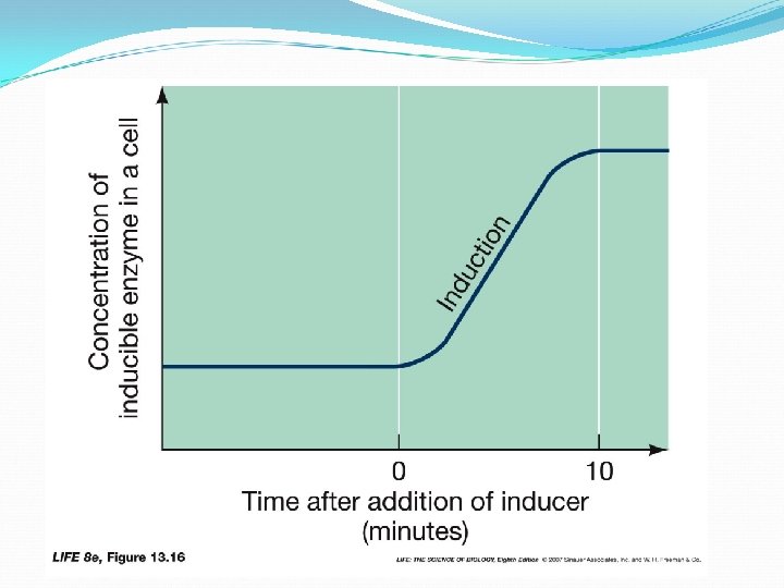
Regulation of m. RNA Transcription E. coli prefers glucose as an energy source, but can use lactose if glucose is low. 3 enzymes are required for lactose metabolism presence of lactose stimulates production of these enzymes lactose is an inducer E. coli http: //spacebio. net/modules
Regulation of m. RNA Transcription • enzymes that are produced in the presence of an inducer are said to be inducible E. coli • enzymes that are made all the time are said to be constitutive http: //spacebio. net/modules
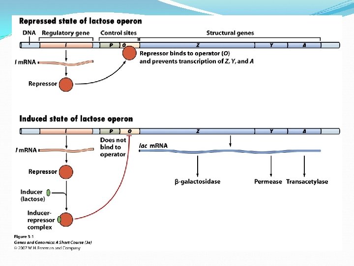
Regulation of m. RNA Transcription Operon is the whole unit promoter, operator, and one or more structural genes Operon containing genes for lactose metabolism: lac operon
Regulation of m. RNA Transcription Structural genes specify primary protein structure — the amino acid sequence The three structural genes for lactose enzymes are adjacent on chromosome They share a promoter, and are transcribed together
Regulation of m. RNA Transcription Prokaryotes shut down transcription by placing obstacle between promoter and structural gene a protein called a repressor bind to operator— blocks transcription of m. RNA
Regulation of lac Operon
Regulation of lac Operon A repressor protein is coded by a regulatory gene. The regulatory gene that codes for the lac repressor is the i (inducibility) gene is near lac structural genes, but not all regulatory genes are near their operons
Regulation of lac Operon Regulatory genes like i have their own promoter, called pi The i gene is expressed constitutively (expression is constant).
Regulation of lac Operon The repressor protein has two binding sites one for the operator one for inducer (lactose) Operator binding site
Regulation of lac Operon Binding inducer to repressor, changes repressor shape allows promoter to bind RNA polymerase When lactose concentration drops, inducers separate from repressors — repressor again binds operator, transcriptions stops
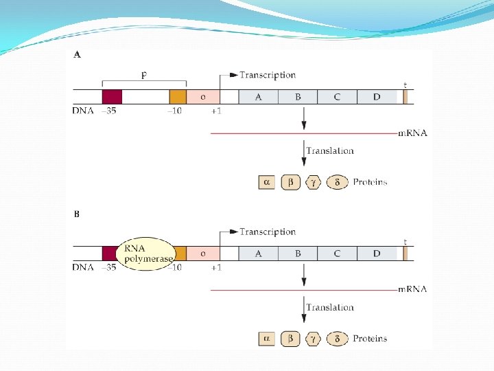
• Promoter is the specific region of the DNA that bound to RNA polymerase for efficient transcription. • Two highly conserved, separate nucleotide blocks make up the promoter of the E. coli lac operon. • These blocked are designated -10 and -35 upstream of the m. RNA start site +1. • Mutations in these regions have mild and severe effects on transcription.
The sequences required for RNA polymerase binding and initiation are contained within less than 40 nucleotides. The initial step is the recognition and binding of an RNA polymerase to the -35 region. Subsequently, the -10 region melts into its component single strands allowing transcription to begin at +1 position.
Bacterial RNA polymerase core complexes are bound by a single initiation factor σ (sigma). E coli can control gene expression by activation of σ factor in response to stimuli. σ is the most common initiation factor in E. coli genome that binds to many other promoters besides lac promoter. σ 32 is activated in response to “heat shock” and directs polymerase to transcribe specific genes to respond to this stress. σ 54 is activated when cells are starved for nitrogen, turning on genes that are responsible for uptake of nitrogen. 70
Sigma factors produced by E. coli
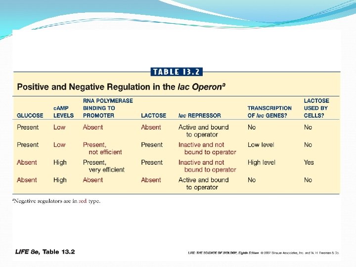
Genes are activated when transcription factors bind specific DNA sequences near promoter If both glucose and lactose are present, E. coli will first use glucose. Presence of glucose lowers concentration of c. AMP thus less CRP binding to promoter, resulting in less efficient transcription of lactose -metabolizing enzymes
When glucose is used up, starvation signals accumulate in the cell, and trigger an activation of lac operon can increase efficiency of the promoter A regulatory protein CRP binds c. AMP This complex binds to DNA just upstream of promoter. Allows more efficient binding of RNA polymerase to promoter
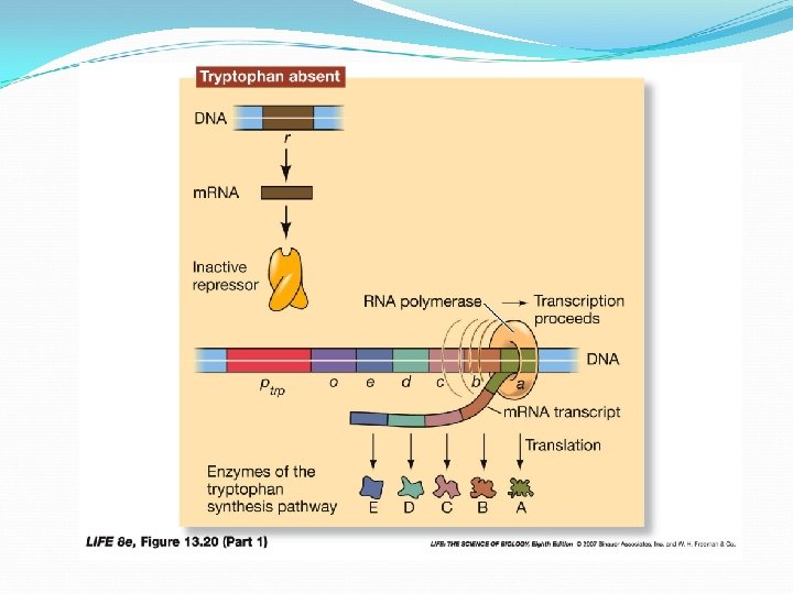
Regulation of trp Operon A protein is repressible if synthesis can be turned off by a biochemical cue (e. g. , ample supply of that protein) The trp operon controls synthesis of tryptophan — it is a repressible system Gene is normally “on”, transcribing m. RNA and synthesizing structural proteins
Regulation of trp Operon The repressor must first bind with a corepressor, in this case tryptophan (when its abundant), activating repressor Active repressor turns gene “off”, blocking transcription
lac Operon VS trp Operon Inducible systems (lac operon) Substrate of a metabolic pathway (inducer) interacts with a regulatory protein (repressor) — repressor cannot bind to operator —allowing transcription Control catabolic pathways (turned on when substrate, lactose, is present)
lac Operon VS trp Operon Repressible system (trp operon) Product of a metabolic pathway (corepressor) interacts with a regulatory protein (repressor) allowing it to bind to operator, blocking transcription Control anabolic pathways (turned on when product, tryptophan, is not present).
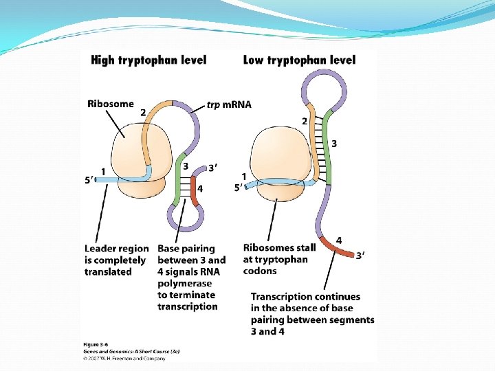
The Attenuation in trp operon • Attenuation is a method of regulating transcription by preventing complete m. RNA synthesis. – The 5’ end of trp operon m. RNA, the leader region (region 1) is rich in tryptophan codon. – When tryptophan is available, the translation of this region occurs. – As this happens, the trp m. RNA forms a stem-loop structure between region 3 and 4, transcription is attenuated. – When the tryptophan level drops, the attenuation is relieved. – The ribosome is stalled at the leader sequence, different stem-loop forms (2 and 3), allows the transcription to continue.