CHAPTER 16 Viral Diversity Viruses of Prokaryotes RNA


")


.")


 – Small, irregular, rod shaped")


- Slides: 55
CHAPTER 16 Viral Diversity
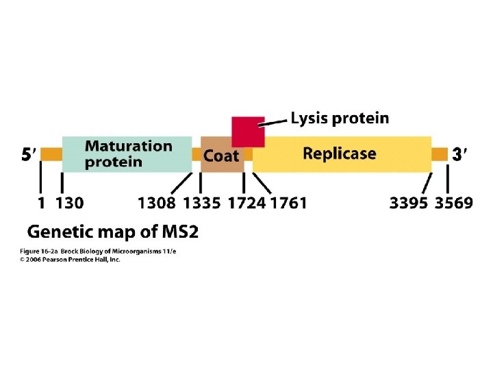
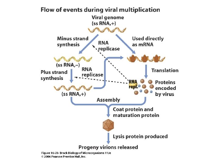
Viruses of Prokaryotes RNA Bacteriophages • A variety of RNA viruses that infect bacteria are known. The small RNA genome of these bacterial viruses is translated directly and encodes only a few proteins. • Figure 16. 2 a shows the genetic map of RNA bacteriophage MS 2, and Figure 16. 2 b shows the flow of events of MS 2 multiplication.
• The small genome encodes only four proteins. • These are the maturation protein (present in the mature virus particle as a single copy), coat protein, lysis protein (involved in the lysis process that results in release of mature virus particles), and a subunit of RNA replicase, the enzyme that brings about replication of the viral RNA.
Icosahedral Single-Stranded DNA Bacteriophages • M 13 and X 174 are ss. DNA viruses - ~25 nm • The single-stranded DNA genome (5356 nt)of the virus X 174 is so small that overlapping genes are required to encode all its essential proteins. This virus provided the first example of overlapping genes.
• Because cellular DNA always replicates in the double-stranded configuration, the replication process of the single-stranded genome of X 174 is of interest. • On infection, the plus-sense viral DNA becomes separated from the protein coat. Entrance into the cell is accompanied by the conversion of this singlestranded DNA into a double-stranded molecule called the replicative form (RF). • Figure 16. 3 a shows the genetic map of phage X 174, and Figure 16. 3 b shows the flow of events during X 174 replication.
A m. RNA is read twice by ribosomes, once for A and second for A*
• The production of progeny viral DNA involves rolling circle replication. Figure 16. 4 shows the mechanism in phage X 174. A cleaves the plus strand of the RF
Filamentous Single-Stranded DNA Bacteriophages • Some single-stranded DNA viruses, such as M 13, have filamentous virions (Figure 16. 5) related to f 1 and fd phages. • These viruses are very useful tools for DNA sequencing and genetic engineering. They are released without actually killing the host. • M 13 is only 6 nm in diameter but 860 nm long. • Filamentous phages are released without killing the host cell. • All phages with protein A (including X 174) do not kill host cells.
Release of phages
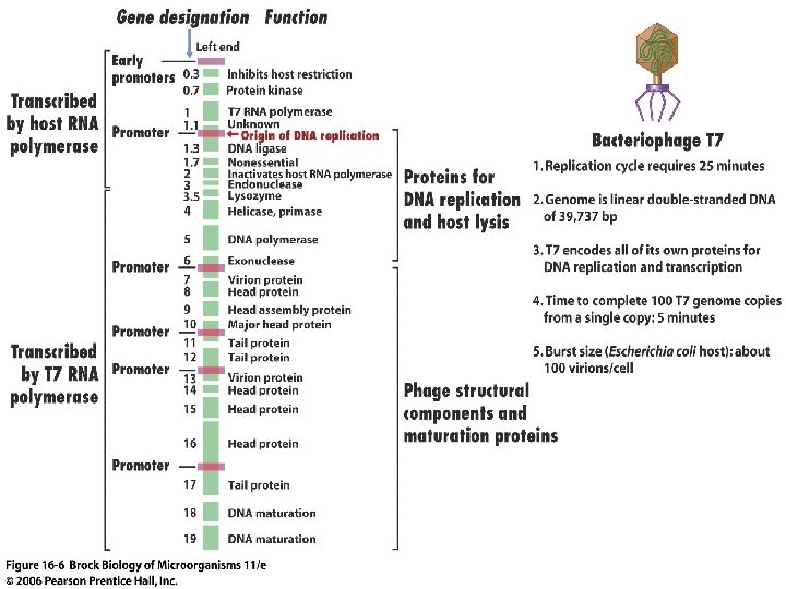
Double-Stranded DNA Bacteriophages: T 7 • The bacteriophage T 7 double-stranded DNA genome always enters the host cell in the same orientation. Figure 16. 6 shows the genetic map for T 7.
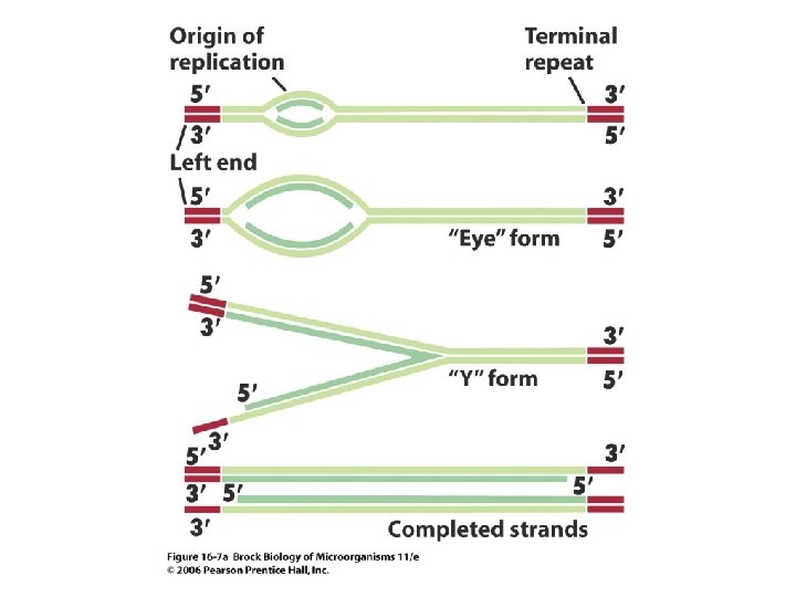
• The late genes in T 7 are transcribed by a virus-encoded RNA polymerase. • The replication strategy for the T 7 genome employs T 7 DNA polymerase and involves terminal repeats and the formation of concatemers (Figure 16. 7).
Formation of concatamers by joining DNA at the unreplicated terminal ends
Production of mature viral DNA
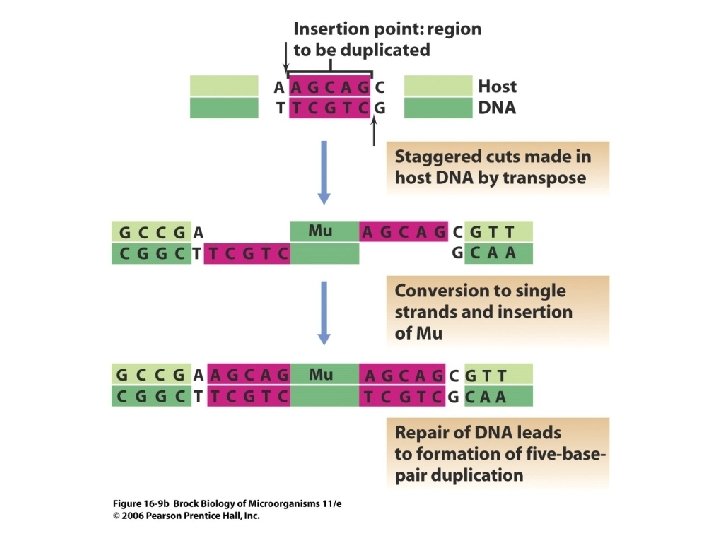
Mu: A Double-Stranded Transposable DNA Bacteriophage
• Bacteriophage Mu is a temperate virus that is also a transposable element. In either the lytic or lysogenic pathway, its genome is integrated into the host chromosome by the activity of a transposase. • Even in the lytic pathway, its genome is replicated as part of a larger DNA molecule. The genome is packaged into the virion in such a way that there are short sequences of host DNA at either end.
• Figure 16. 9 illustrates replication of bacteriophage Mu. Genome – 39 kb (37. 2 kb viral DNA and 1. 8 kb host DNA) 50 -150 bp 1 -2 kb Lambda. T 4, T 7, and Mu have linear ds. DNA
Viruses of Eukaryotes Plant Viruses • Most plant viruses have positive-strand RNA genomes. One example is tobacco mosaic virus (TMV), the first virus discovered (Figure 16. 11).
• The genomes of these viruses can move within the plant through intercellular connections that span the cell walls. • Other types of plant viruses are also known, including the Chlorella viruses, which have very large double-stranded DNA genomes.
Positive-Strand RNA Viruses of Animals: Poliovirus and Coronaviruses
• In small RNA viruses such as poliovirus, the viral RNA is translated directly, causing the production of a long polyprotein that is broken down by enzymes into the many small proteins necessary for nucleic acid multiplication and virus assembly (Figure 16. 13).
Poliovirus
RNA linked 22 a. a. protein serve as a primer
• Coronavirus is a large single-stranded RNA virus that resembles poliovirus in some but not all of its replication features (Figure 16. 14).
Coronavirus – Flow of information
Negative-Strand RNA Viruses of Animals: Rabies, Influenza, and Related Viruses • In negative-strand viruses, the virus RNA is not the m. RNA but is copied into m. RNA by an enzyme present in the virion. • Figure 16. 16 illustrates the flow of events during multiplication of a negative-strand RNA virus.
• Vesicular stomatitis virus (VSV)
• Translation of viral m. RNAs leads to the synthesis of viral coat proteins. • Assembly of an enveloped virus is considerably more complex than assembly of a naked virion.
• Two kinds of coat proteins are formed, nucleocapsid proteins and envelope proteins. The nucleocapsid is formed first by association of the nucleocapsid protein molecules around the viral RNA.
• Important negative-strand viruses include rabies virus and influenza virus (Figure 6. 17).
influenza virus
Influenza virus
Double-Stranded RNA Viruses: Reoviruses • Reoviruses contain segmented doublestranded RNA genomes. • Like negative-strand RNA viruses, reoviruses contain an RNA-dependent RNA polymerase within the virion.
Replication of Double-Stranded DNA Viruses of Animals • Most double-stranded DNA animal viruses, such as SV 40, replicate in the nucleus. SV 40 has a tiny genome and employs the strategy of overlapping genes to boost its genetic-coding potential. Some of these viruses cause cancer.
• Figure 16. 20 shows the genetic map of polyomavirus SV 40. 5. 2 kb
• Figure 16. 21 shows the general scheme of molecular events involved in cell transformation by a polyomavirus such as SV 40.
• Herpesviruses cause a variety of disease syndromes and can maintain themselves in a latent state in the host indefinitely, initiating viral replication periodically.
Double-Stranded DNA Viruses: Herpesviruses • Herpesviruses are large, double-stranded DNA viruses. The viral DNA circularizes and is replicated by a rolling circle mechanism.
• Figure 16. 22 illustrates the flow of events in multiplication of herpes simplex virus.
Double-Stranded DNA Viruses: Pox Viruses • The pox viruses, unlike the other DNA viruses discussed so far, are very large viruses that replicate entirely in the cytoplasm. These viruses are responsible for several human diseases, but a vaccination campaign has eradicated the smallpox virus in the wild.
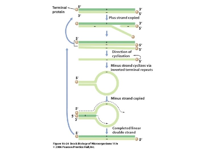
Double-Stranded DNA Viruses: Adenoviruses • Different double-stranded DNA animal viruses have different genome replication strategies.
• The strategy of the adenoviruses involves protein primers and a mode of replication that avoids the synthesis of a lagging strand occurs within the nucleus (Figure 16. 24).
Viruses Using Reverse Transcriptase: Retroviruses and Hepadnavirus
• The retroviruses contain RNA genomes and use reverse transcriptase to make a DNA copy during their life cycle (Figure 16. 25). Lys 3
• Figure 16. 26 illustrates translation of retrovirus m. RNA and processing of the proteins.
• The hepadnaviruses contain DNA genomes and use reverse transcriptase to make genomic DNA from an RNA copy. Figure 16. 27 shows the genome of hepatitis B, a hepadnavirus.
Hepadnaviruses (Liver infecting) – Small, irregular, rod shaped
Hepatitis B virus – serious bloodborne pathogen 3 -4 kb DNA genome is partially ds Replicated through RNA intermediate
• These viruses have complex patterns of gene expression.