Reproductive system Reproductive System Gonads Ducts Accessory organs


 and hormones •")


 • Produce secretions")


 Secretory phase (postovulatory)")


- Slides: 66
Reproductive system
Reproductive System • • Gonads Ducts Accessory organs and glands External genitalia
Functions of the reproductive system • Production of gametes (reproductive cells) and hormones • Fertilization • Male – Sperm production, transport of sperm to the uterus • Female – Ova production, fusion of gametes, development of the zygote
Fig 27. 1 The scrotum is a pouch of skin Inside the scrotum there are two scrotal cavities that each contain a testis Male reproductive system
Fig 27. 1
• Diagrammatical sectional view at representative stages of the descent of the testes.
Fig 27. 7 Sperm is produced in the seminiferous tubules of the testis
Interstitial cell
Fig 27. 4
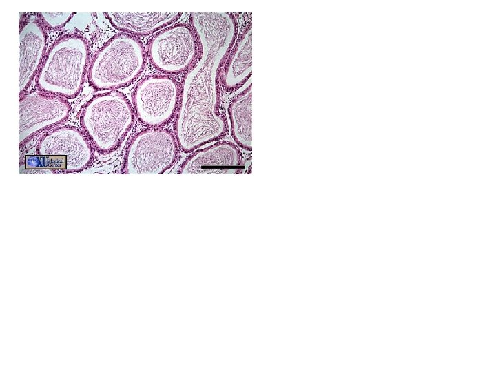
Fig 27. 5 There is about 700 feet of seminiferous tubule in men/teste This allows men to produce about 200, 000 per day
lumen Fig 27. 5
Sperm production • Spermatogonia-stem cells that develop into spermatids • Become active at puberty • Spermatogenesisproduction of spermatids • Occurs in the seminiferous tubules of the testis Fig 27. 5
Spermiogenesis – first phase of development • Anatomical maturation of sperm cells • A single Spermatid develops into a Spermatozoon • Occurs in the seminiferous tubules of the testis Fig 27. 6
Sustentacular cells in seminiferous tubules • Maintain the blood testis barrier – Allows the sperm to develop is an environment that is different from the general circulation • Support of spermatogenesis & spermiogenesis • Release the hormone Inhibin • Inhibin-stops production of FSH in the anterior pituitary
Hormones • Anterior pituitary-FSH & LH • Follicle-stimulating hormone-supports sperm production • Luteinizing hormone-stimulates interstitial cells of the testis to release testosterone
• Interstitial cells of the testis release testosterone • Testosterone • Promotes production of mature sperm • Maintain accessory organs (glands) of the reproductive tract • Influences secondary sexual characteristics – Facial hair, muscle mass, body fat
Sperm development • FSH, LH, & Testosterone, promote sperm production
Anatomy of Sperm • Head – Acrosomal cap • Neck • Middle piece • Tail – Flagellum
lumen Fig 27. 5
Male reproductive tract • The pathway the sperm travel to exit the body • Along the way sperm mix with secretions from the accessory glands • The epididymis ~ 16’ of tube – Storage and maturation of sperm
Fig 27. 3 Regulate the temperature of the testissperm develop 1. 1°C below body temp
Fig 27. 8
Fig 27. 9
Fig 27. 9
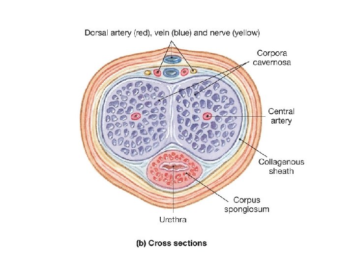
penis Corpora caveronsa Corpus spongiosum urethra
Accessory glands • Seminal vesicles, Prostate gland, Bulbourethral glands (Cowper’s gland) • Produce secretions that make up the majority of semen volume • Sympathetic nervous systems controls release
• These secretions: • Activate the sperm-the flagella become functional • Provide nutrients for the sperm cells • Proving p. H buffers for the semen – The urethra and the vagina are acidic environments
Seminal vesicles • Makes up 60% of semen volume • Fluid contains high levels of sugar to provide nutrients to sperms cells • Secretions are released into the ejaculatory duct
Prostate gland • Makes up 30% of semen volume • Fluid is a milky solution that contains several enzymes (prostaglandins) • Secretions are released into the urethra
Bulbourethral glands • Makes up 5% of semen volume • Fluid is a alkaline mucus that is a p. H buffer • Secretions are released into the urethra preejaculation
Fig 27. 10 Female reproductive system ovaries The ovaries & uterus are held in place by ligaments in the pelvic cavity
Fig 27. 10
Fig 27. 20
Fig 27. 11
Fig Oogenesis: 27. 13 Oocytes are produced in the ovaries before birth. By puberty there are 400, 000 oocytes total During the ovarian cycle the oocyte mature Only about 500 will mature and ovulate during life
Ovarian cycle • Follicule cells provide nutrients to the oocytes • Oocytes +follicule cells = follicle • Development of oocytes occurs within the follicles
Fig 27. 12 Mature follice Development of the follicle cells that surround the oocytes Few primary follicles develop into secondary follicles Thecal cells produce estrogens The ovaries usually contain a single secondary follicle destined for further development
Mature follicle 2 -primary follicle 5 -oocyte
Step 4, 5, & 6 • Step 4 -Ovulation-The oocyte is released from the ovary into the uterine tubes • Step 5 -Formation of the corpus luteum • The remaining follicle cells for the corpus luteum which produces progesterone & estrogen • Step 6 -Degradion of the corpus luteum (unless pregnancy occurs) • The corpus luteum degrades into scar tissue and a new ovarian cycle begins
Fig 27. 11
Fig 27. 20
Fig 27. 14
Fig 27. 11
Fig 27. 15
Fig 27. 17
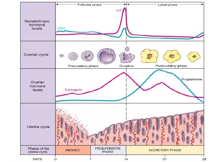
Uterine cycle • • • Three phases. Menes Proliferative phase (preovulatory) Secretory phase (postovulatory) Menes-Arteries constrict reducing, blood flow to the endometrium (inner layer of the uterus) • The tissue of the endometrium and blood from ruptured vessels in to the lumen of the uterus
• Proliferative phase-Repair and growth of the endometrium tissue and blood vessels • Secretory phase-Further development of the endometrium and increased glandular activity
Fig 27. 17
Hormones • Anterior pituitary-FSH, LH, & Prolactin • Follicle stimulating hormone-supports oocyte maturation, stimulates the follicle cells to release estrogen • Luteinizing hormone-stimulates ovulation and formation of the corpus luteum • Prolactin-stimulates production of breast milk
• Thecal of the ovaries release estrogen • Estrogen • Stimulates growth of the uterine of the endometrium • Maintain accessory organs & glands of the reproductive tract • Influences secondary sexual characteristics
• The corpus luteum releases Progesterone & Estrogen • Progesterone • Prepares the uterus for development of the embryo
Fig 27. 21
FYI • If no method of birth control is used there is a 85% chance of a pregnancy within in a year • • Total abstinence-the only 100% effective method Surgical sterilization-vasectomy & tubal ligation, 99. 6% Hormonal methods-inhibits ovulation, 99. 6% Methods to block implantation- Intrauterine device, 99. 2% Barrier methods-block entry of sperm to the uterus, 80% Spermatocides-kills sperm, 74% Periodic abstinence, 77% • Abortion-induced miscarriage, surgical procedure
If fertilization does occur
FYI
FYI Sperm count range from 20 – 100 million sperm per cubic m. L
Chorion - villi Amnion Chorionic gonadaltropin HCG
FYI
FYI
FYI 772 in text book
FYI
FYI
FYI Epidural shot