Thoracic Wall Lecture Objectives Describe the shape and

Thoracic Wall

Lecture Objectives • Describe the shape and outline of the thoracic cage including inlet and outlet. • Describe the anatomical landmarks of the anterior chest wall. • List various structures making the thoracic wall. • Make a list of muscles of the thoracic wall including their nerve and blood supply and their actions. • List various parts of the thoracic vertebrae and name its characteristic features. • Describe the sternum with its joints. • Classify ribs, name their various parts and compare them with each other. • Define intercostal spaces and discuss their various components including intercostal muscles. • Describe the diaphragm, its origin, insertion, function, nerve and blood supply. Study openings in the diaphragm and structures that pass through.
 • Ribs")
Thoracic Cage Bony cage flattened from front to back • Sternum (breastbone) • Ribs – 1‐ 7 are true ribs (vertebrosternal) – 8‐ 10 are false ribs (vertebrochondral) – 11‐ 12 are floating • Costal cartilages • Bodies of the thoracic vertebrae

Skeleton of Thoracic Wall

Thoracic Cage: Functions • Enclose and protect the organs in the thoracic and abdominal cavities • Provide support for the bones of the upper limbs • Play a role in breathing

• Manubrium Sternum – 1 st & 2 nd ribs – Clavicular notch (sternoclavicular joint) – Sternal angle – T 3‐T 4 • Body – Costal cartilages of 2‐ 7 ribs • Xiphoid – – Ossifies by 40 CPR position Abdominal mm. T 9

Sternum

Ribs • Increase in length from ribs 1‐ 7, thereafter decreasing • Head and tubercle articulate with facets • Body with costal groove containing nerve & blood vessels • Intercostal spaces contain intercostal muscles

• Typical ribs Ribs – Long & twisted – Rounded superior edge – Grooved inferior edge (costal groove) – Head (2 facets 2‐ 9, 1 facet 1, 10‐ 12), neck, tubercle, shaft & angle

Ribs • Atypical ribs – 1 st rib • Widest, shortest, most curved true rib • Articulate with T 1 • Surface marking … – 11 th and 12 th • No neck • No tubercle • Floating
 –")
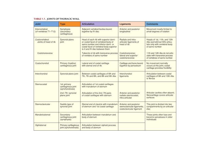
Thoracic Wall: Joints Cartilaginous Joints • Joints of sternum – Manubriosternal joint (2°) – Xiphisternal joint • Costochondral joints • 1 st sternocostal joint Synovial Joints (plane joints) • Joints between ribs and thoracic vertebrae • 2 nd ‐ 7 th sternocostal joints • 6 th – 10 th interchondral joints

Joints of the Heads of Ribs • 2‐ 9 ribs – 2 synovial joints – With the corresponding vertebra and one above – Intra‐articular ligament • Between head and IVD • 1, 10‐ 12 ribs – 1 synovial joint with the corresponding vertebra
 – With the transverse")
Costotransverse Joint • Joints of the tubercles (1‐ 10 ribs) – With the transverse process of the corresponding vertebra

Thoracic Wall: Joints

Thoracic Apertures

Superior Thoracic Aperture • Between thoracic cavity and the root of the neck • Boundaries. . • Orientation. . • Content – – Trachea Esophagus Nerves & BVs Lungs & pleurae • Suprapleural membrane – Close the sides of the opening above the parietal pleurae

Superior Thoracic Aperture

Inferior Thoracic Aperture

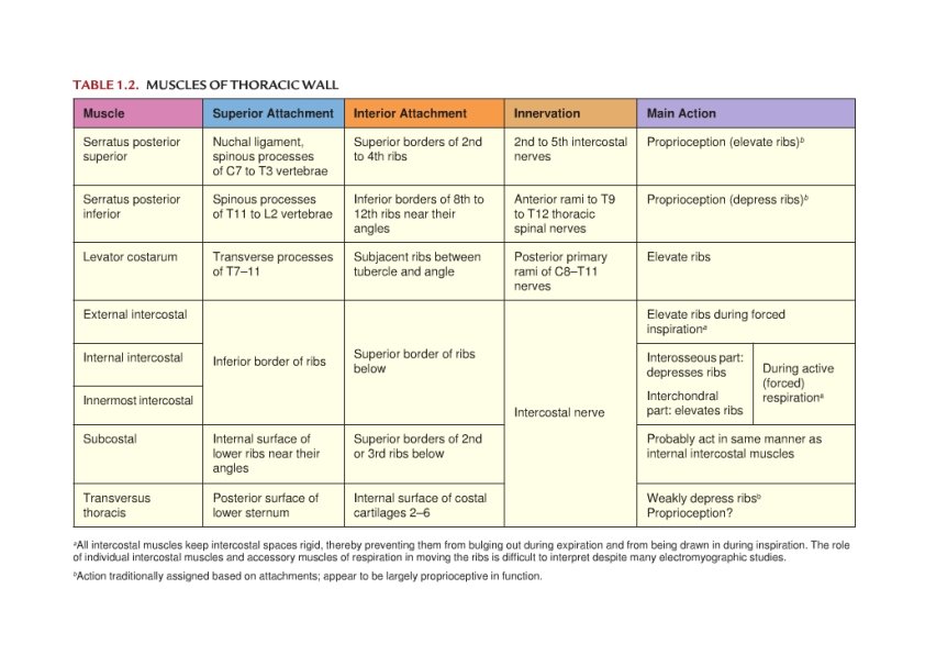
Intercostal Spaces • Between successive ribs • Contain the intercostal mm. – External , internal, and innermost intercostal mm. • Neurovascular bundle run superficial to the innermost intercostal m. – Arranged from superior to inferior as vein, artery , and nerve

Intercostal Muscles Nerve supply: intercostal nerves • Three layers – External intercostal • Orientation • Anterior (external) intercostal membrane • Helps in inspiration – Internal intercostal • Orientation • Posterior (internal) intercostal membrane • Helps in expiration – Innermost intercostal • Cross more than one intercostal spaces • Attached to the endothoracic fascia internally – Attached to parietal pleura • Divided into three parts • Works with the internal intercostal

Accessory Muscles of Respiration • Transversus thoracis – Help in expiration • Pectoralis major, pectoralis minor, serratus anterior, scalene mm. – May help in inspiration

Accessory Muscles of Respiration • Levator costarum – Between the transverse processes and the ribs – Nerve supply: posterior rami of thoracic spinal nerves – Help in inspiration • Serratus posterior superior m. – Deep to rhomboids – Nerve supply: 1‐ 4 intercostal nerves – Help in inspiration • Serratus posterior inferior m. – Deep to latissmus dorsi – Nerve supply: last 4 intercostal nerves – Help in expiration

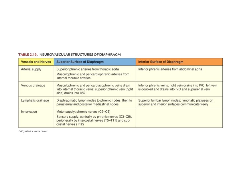
Diaphragm • Physical barrier between thoracic and abdominal cavities • Functions – Inspiration – Abdominal pressure – Abdominothoracic pump • Dome like shape – Peripheral muscular • Reach up to the 5 th rib – Central tendon • At level of xiphisternal joint

Origin of the diaphragm • Sternal‐ posterior surface of xiphoid process • Costal‐ the lower six ribs and their costal cartilages • Vertebral‐ – Right crus‐ bodies of L 1‐L 3 – Left crus‐ bodies of L 1‐L 2 • Arcuate ligaments – Medial‐ L 2 (body) to L 1 (transverse process) – Lateral‐ L 1 (transverse process) to 12 th rib – Median‐ connects crura anterior to aorta

Openings in the Diaphragm • Aortic opening‐ T 12 – Between crura – Content • Aorta, thoracic duct, & azygos vein • Esophageal opening‐ T 10 – In right crus – Content • Esophagus, vagi, BVs & lymphatic vessels • Caval opening‐ T 8 – Content • IVC, branches of right phrenic nerve

Openings in the Diaphragm • Other structures pass the diaphragm – Splanchnic nerves – through crura – Sympathetic trunk – medial arcuate lig. – Subcostal nerve – lateral arcuate lig. – Superior epigastric vessels – between sternal & costal origins
 • Sensory – Centrally")
Diaphragm: Innervation • Motor – Phrenic nerves (C 3‐C 5) • Sensory – Centrally – phrenic nerves – Peripherally – intercostal nerves (T 7‐ T 12)

Diaphragm: Blood Supply

Arteries of Thoracic Wall • Posterior intercostal aa. – 1‐ 2 ← superior intercostal ← costocervical trunk ← 2 nd part of supclavian – 3‐ 12 ←descending thoracic aorta • Anterior intercostal aa. – 1‐ 6 ← internal thoracic ← 1 st part of subclavian – 7‐ 12 ← musculophrenic ← internal thoracic
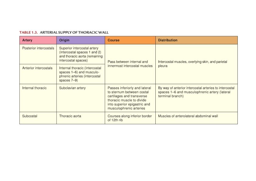

Veins of Thoracic Wall • Posterior intercostal veins – Drain into azygos and hemiazygos veins • Anterior intercostal veins – Follow the corresponding aa. (internal intercostal and musculophrenic vv. )

Nerves of Thoracic Wall • Anterior rami of thoracic spinal nerves – 1‐ 11 intercostal nerves • 1‐ 6 end within the intercostal spaces • 7‐ 9 pass anterior deep to the costal cartilage to reach the abdominal wall • 10‐ 11 continue anteriorly to the abdominal wall – 12 subcostal nerve • In the abdominal wall

Branches of the Intercostal Nerves • Rami communicants • Collateral branch • Lateral cutaneous branch – 1 st ‐ part of the brachial plexus – Intercostobrachial nerve (2 nd) • • • Referral pain in coronary artery disease Anterior cutaneous branch Muscular branches Pleural sensory branches Peritoneal sensory branches (6‐ 11)

Surface Anatomy
- Slides: 37