Vagus accessory and hypoglossal nerves CN 10 11
 Mark Kozsurek, M. D. ,")
")


")


")


")


- Slides: 28
Vagus, accessory and hypoglossal nerves (CN 10, 11, 12) Mark Kozsurek, M. D. , Ph. D. mark@kozsurek. hu ED II. , 04/11/2019.
Vagus nerve (CN 10)
Why do we need the vagus nerve? (What are the symptomes in vagus insufficiency? ) n swallowing: Levator veli palatini muscle, lower half of pharyngeal constrictors, esophageal muscles n phonation: laryngeal muscles n controlling viscera: lungs, heart, esophagus, stomach, liver, biliary system, pancreas, small and large intestine, kidney Clinical symptomes: dysphagia, aspiration, dysphonia, deviation of uvula.
ambiguus nucleus: branchialmotor dorsal vagal nucleus: general visceromotor spinal trigeminal nucleus: general somatosensory lateral nucleus of ala cinerea: general viscerosensory solitary nucleus: special viscerosensory – taste sensing
Ala cinerea is a synonim for the vagal trigone seen in the rhomboid fossa. Within that two nuclei are found: one dorsomedially and one ventrolaterally. https: //web. duke. edu/brain/lab 04. html For this reason: dorsal vagal nucl. = medial nucl. of ala cinerea ventral vagal nucl. = lateral nucl. of ala cinerea
https: //www. researchgate. net/figure/General-organization-of-the-nucleus-of-the-solitary-tract-NTS_fig 2_313783771 Term „solitary nucleus” (or nucleus of the solitary tract) can be used for the entire viscerosensory nucleus of the vagus nerve including its rostral special viscerosensory (taste-sensing) division and for the caudal general somatosensory portion - in many textbooks. „Solitary nucleus” sometimes exclusively refers to the taste-sensing nucleus shared by the facial, glossopharyngeal and vagal nerves - in German anatomy and our department.
Brainstem, dural and cranial exit of the vagus nerve
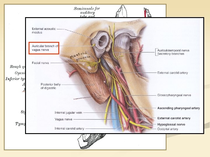
I. Branches on the head and neck superior or jugular ggl. inferior or nodose ggl. 1. Posterior meningeal branch 2. Auricular branch of vagus nerve inferoposterior wall of the external auditory meatus, posterior surface of the auricle (mastoid canaliculus: jugular fossa → tympanomastoid fissure) Both of these ganglia are sensory and contain pseudounipolar cells! Both of these branches contain exclusively general somatosensory fibres!
1. Post. meningeal branch 2. Auricular branch 3. Pharyngeal branches PHARYNGEAL PLEXUS Pharyngeal plexus is formed by the pharyngeal branches of glossopharyngeal and vagus nerves and sympathetic fibres from the superior cervical ganglion.
1. Posterior meningeal branch 2. Auricular branch 3. Pharyngeal branches 4. Superior laryngeal branch - external br. (for Cricothyroid) - internal br. 5. Inf. and recurrent laryngeal nerve
6. CARDIAC PLEXUS - superior cervical cardiac branches - inferior cervical cardiac branches - (thoracic cardiac branches) Note that the cardiac plexus is in the thorax but is mainly formed by vagal branches descending from the cervical levels!
II. Thoracic branches - thoracic cardiac branches - bronchial/pulmonary branches - pericardiac branches - esophageal branches
III. Abdominal branches * Cannon-Böhm’s point - gastric plexus - hepatic plexus The most distal branches follow the celiac trunk and the superior mesenteric artery to reach their targets. - celiac plexus - renal plexus
poterior meningeal br. : general somatosensory GSS r. auricularis n. vagi: general somatosensory GSS, BM, GVM superior laryngeal nerve: external br. – branchialmotor; iternal br. – general somatosensory (mucosa), special viscerosensory (taste sensing), general visceromotor (glands) fibres head, neck rr. pharyngei: branchialmotor (striated muscles), general visceromotor (glands), general somatosensory (mucosa) inferior and recurrent laryngeal nerve: branchialmotor, general somatosensory (mucosa), general visceromotor (glands) abdomen thorax cardiac plexus: general visceromotor and viscerosensory (blood pressure, heart rate) GSS, GVM bronchial, pulmonary, pericardiac and oesophageal plexus: general visceromotor and viscerosensory gastric, coeliac, hepatic, renal plexus: general visceromotor and viscerosensory
General viscerosensory and visceromotor functions of the vagus nerve n Vagus controls the autonomic functions of the thoracic and abdominal viscera. lateral nucleus of ala cinerea inferior ganglion (pseudounipolar neurons) viscera dorsal vagus nucleus parasympathetic (intramural) ganglia submucous and myenteric plexus multipolar neurons
Accessory nerve (CN 11)
What does the accessory nerve do? n phonation: fibres joining the vagus mainly innervate the interarythenoid muscles. n innervation of trapezius and sternocleidomastoid muscles Clinical symptoms: abnormal, asymmetrical position of the head or neck („wry neck”), abduction of the upper limb over the horizontal plane is difficult.
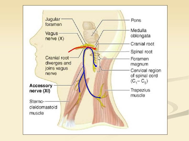
ambiguus nucleus: branchialmotor (fibres arising from this nucleus constitute the cranial roots which exits in the lateral parolivary sulcus and than join the vagus nerve as internal branches) spinal accessory nucleus: general somatomotor nucleus in the lateral portion of the ventral horn of C 1 -C 6 spinal segments. (fibers coming from the nucleus give the spinal roots of the accessory nerve which enter the skull through the foramen magnum exit again through the pars nervosa of the jugular foramen and target sternocleidomastoid and trapezius muscles as external branches)
glossopharyngeus nerve vagus nerve
Hypoglossal nerve (CN 12)
Functions: 1. Somatomotor innervation of extrinsic and intrinsic muscles of the tongue. 2. Contributes to the innervation of infrahyoid muscles (and the geniohyoid) by taking guest fibres from the cervical plexus. hypoglossal nucleus: general somatomotor
Clinical consideration: Peripheral injury of the hypoglossal nerve results in deviation of the protruded tongue toward the side of injury. Tongue might be atrophic. Chewing, swallowing, talking is difficult.
deep cervical ansa: a branch of C 1 spinal nerve joins the hypoglossus as a guest fibre and later leaves that as the superior root of cervical ansa. This will form a loop with the inferior root of ansa cervicalis arrising from the ventral branches of C 2 -3 spinal nerves. Deep cervical ansa innervates infrahyoid muscles and the Geniohyoid.
Pirogov’s triangle is formed by the intermediate tendon of the digastric muscle, the posterior border of the mylohyoid muscle and the hypoglossal nerve. Béclard's triangle is an area whose boundaries are the posterior border of the hyoglossus, the posterior belly of the digastric/stylohyoid muscles and the greater horn of the hyoid bone.
Hypoglossus nerve is found in the lateral sulcus of the tongue bellow the lingual nerve and the submandibular duct.
Thank you for your attention!