Structures of the Respiratory System Continued Bronchi and
 Bronchi and Lungs Pages 440 -445")
Structures of the Respiratory System (Continued) Bronchi and Lungs Pages 440 -445
 Bronchi Formed by division of the trachea Each bronchus enters the lung")
Main (Primary) Bronchi Formed by division of the trachea Each bronchus enters the lung at the hilum (medial depression) ◦ then subdivide into smaller and smaller branches © 2015 Pearson Education, Inc.

Figure 13. 1 The major respiratory organs shown in relation to surrounding structures. Nasal cavity Nostril Oral cavity Pharynx Larynx Trachea Right main (primary) bronchus Left lung Right lung Diaphragm

Lungs Occupy most of the thoracic cavity Each lung is divided into lobes by fissures Left lung—two lobes Right lung—three lobes ◦ Connective tissue lines the fissures © 2015 Pearson Education, Inc.
 covers the outer surface of")
Membranous layers of the Lungs • Serous membrane (“serosa”) covers the outer surface of the lungs; 2 -layer membrane: – Pulmonary (visceral) pleura covers the lung surface – Parietal pleura lines the walls of the thoracic cavity • Pleural (serous) fluid fills the area between layers – Allows gliding and decreases friction during breathing • Pleural space lies between the layers © 2015 Pearson Education, Inc.

Figure 13. 4 a Anatomical relationships of organs in the thoracic cavity. Intercostal muscle Rib Trachea Parietal pleura Pleural cavity Visceral pleura Lung Thymus Apex of lung Right superior lobe Horizontal fissure Right middle lobe Oblique fissure Right inferior lobe Heart (in pericardial cavity of mediastinum) Diaphragm Base of lung Left superior lobe Oblique fissure Left inferior lobe (a) Anterior view. The lungs flank mediastinal structures laterally.

Figure 13. 4 b Anatomical relationships of organs in the thoracic cavity. Vertebra Posterior Esophagus (in posterior mediastinum) Root of lung at hilum • Left main bronchus • Left pulmonary artery • Left pulmonary vein Right lung Parietal pleura Visceral pleura Left lung Pleural cavity Thoracic wall Pulmonary trunk Pericardial membranes Sternum Heart (in mediastinum) Anterior mediastinum Anterior (b) Transverse section through the thorax, viewed from above.
 Tree Divisions All have reinforcing cartilage in their walls ◦ Exception are")
Bronchial (Respiratory) Tree Divisions All have reinforcing cartilage in their walls ◦ Exception are the smallest branches Hierarchy of branches: ◦ Bronchi Primary (largest) Secondary Tertiary ◦ Bronchioles ◦ Terminal bronchioles (smallest) © 2015 Pearson Education, Inc.
 Alveolar")
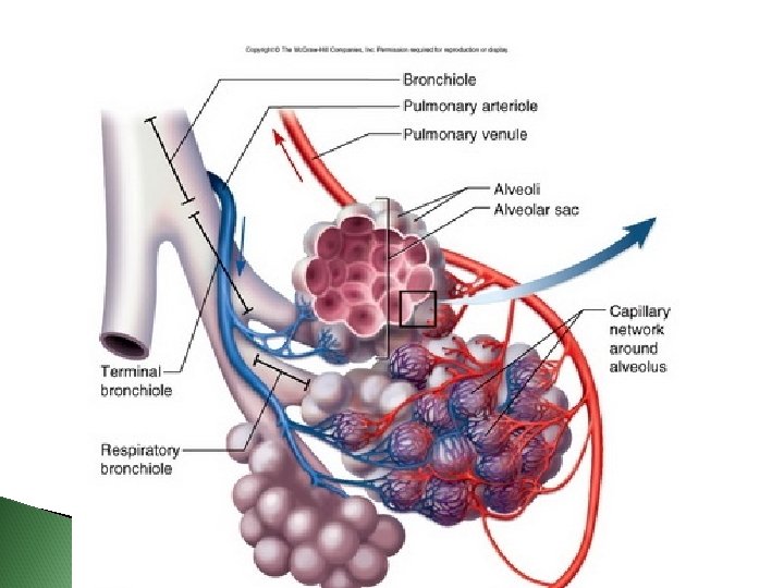
The Respiratory Zone includes: Respiratory bronchioles Alveolar ducts> Alveolar sacs> Alveoli (air sacs) Alveolar duct Respiratory bronchioles Terminal bronchiole (a) Diagrammatic view of respiratory bronchioles, alveolar ducts, and alveoli This is the only site of gas exchange Alveoli Alveolar duct Alveolar sac

The Respiratory Membrane The lungs are mostly air spaces; the rest is the stroma, mostly elastic connective tissue Alveoli: Respiratory membrane (air-blood barrier) Alveolar pores connect neighboring air sacs ◦ Inner walls lined with a squamous epithelial layer ◦ Pulmonary capillaries cover external surfaces ◦ one side is air, and the other side is flowing blood ◦ Formed by alveolar and capillary walls © 2015 Pearson Education, Inc.
. Red blood cell Capillary")
Figure 13. 6 Anatomy of the respiratory membrane (air-blood barrier). Red blood cell Capillary Endothelial cell nucleus Alveolar pores Capillary Macrophage Nucleus of squamous epithelial cell Respiratory membrane Alveoli (gasfilled air spaces) Red blood cell in capillary Surfactantsecreting cell Squamous epithelial cell of alveolar wall O 2 CO 2 Alveolus Alveolar epithelium Fused basement membranes Capillary endothelium
- Slides: 12