
Chapter 25 The Urinary System Urinary System Organs
- Slides: 109
Chapter 25 The Urinary System
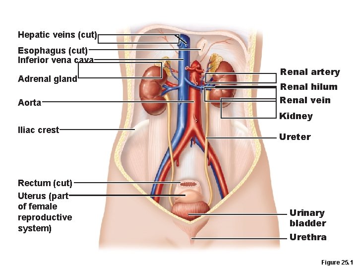
Urinary System Organs • Kidneys are major excretory organs • Urinary bladder is the temporary storage reservoir for urine • Ureters transport urine from the kidneys to the bladder • Urethra transports urine out of the body
Hepatic veins (cut) Esophagus (cut) Inferior vena cava Adrenal gland Aorta Renal artery Renal hilum Renal vein Kidney Iliac crest Rectum (cut) Uterus (part of female reproductive system) Ureter Urinary bladder Urethra Figure 25. 1
Kidney Functions • Removal of toxins, metabolic wastes, and excess ions from the blood • Regulation of blood volume, chemical composition, and p. H • Gluconeogenesis during prolonged fasting • Endocrine functions –Renin: regulation of blood pressure and kidney function –Erythropoietin: regulation of RBC production • Activation of vitamin D
Kidney Anatomy • Retroperitoneal, in the superior lumbar region • Right kidney is lower than the left • Convex lateral surface, concave medial surface • Renal hilum leads to the renal sinus • Ureters, renal blood vessels, lymphatics, and nerves enter and exit at the hilum
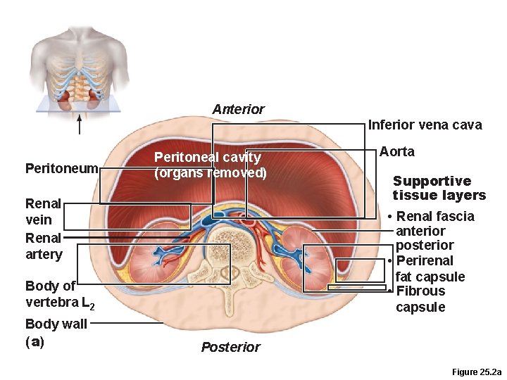
Kidney Anatomy • Layers of supportive tissue 1. Renal fascia • The anchoring outer layer of dense fibrous connective tissue 2. Perirenal fat capsule • A fatty cushion 3. Fibrous capsule • Prevents spread of infection to kidney
Anterior Inferior vena cava Peritoneum Peritoneal cavity (organs removed) Renal vein Renal artery Supportive tissue layers • Renal fascia anterior posterior • Perirenal fat capsule • Fibrous capsule Body of vertebra L 2 Body wall (a) Aorta Posterior Figure 25. 2 a
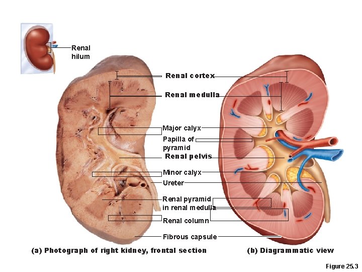
Internal Anatomy • Renal cortex – Granular superficial region • Renal medulla – Cone-shaped medullary (renal) pyramids separated by renal columns • Lobe – Medullary pyramid & surrounding cortical tissue • Papilla - Tip of pyramid; releases urine into minor calyx • Renal pelvis – Funnel-shaped tube within the renal sinus • Major calyces – Branching channels of the renal pelvis that collect urine from minor calyces & empty urine into the pelvis
Renal hilum Renal cortex Renal medulla Major calyx Papilla of pyramid Renal pelvis Minor calyx Ureter Renal pyramid in renal medulla Renal column Fibrous capsule (a) Photograph of right kidney, frontal section (b) Diagrammatic view Figure 25. 3
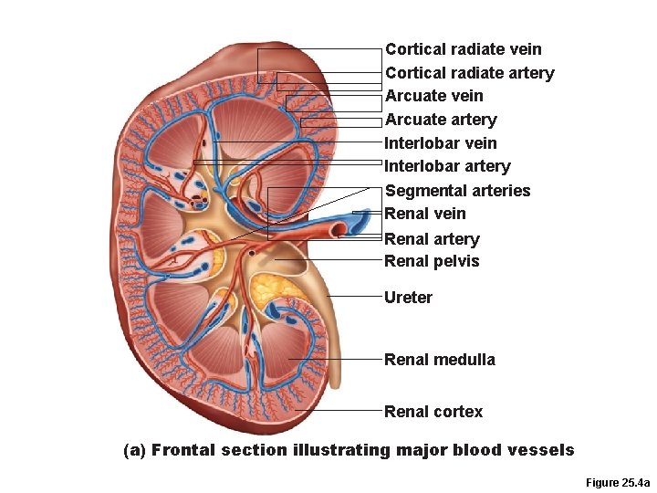
Cortical radiate vein Cortical radiate artery Arcuate vein Arcuate artery Interlobar vein Interlobar artery Segmental arteries Renal vein Renal artery Renal pelvis Ureter Renal medulla Renal cortex (a) Frontal section illustrating major blood vessels Figure 25. 4 a
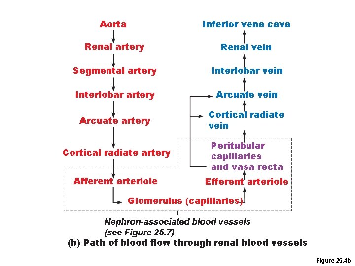
Aorta Inferior vena cava Renal artery Renal vein Segmental artery Interlobar vein Interlobar artery Arcuate vein Arcuate artery Cortical radiate vein Cortical radiate artery Peritubular capillaries and vasa recta Afferent arteriole Efferent arteriole Glomerulus (capillaries) Nephron-associated blood vessels (see Figure 25. 7) (b) Path of blood flow through renal blood vessels Figure 25. 4 b
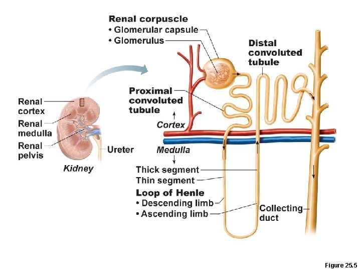
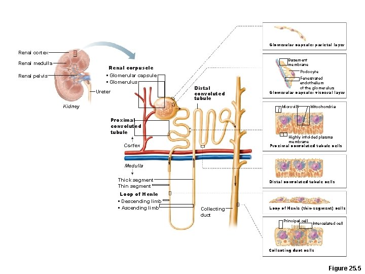
Nephrons • Structural and functional units that form urine • ~1 million per kidney • Two main parts 1. Glomerulus: a tuft of capillaries 2. Renal tubule: begins as cup-shaped glomerular (Bowman’s) capsule surrounding the glomerulus
Figure 25. 5
Ureters • Convey urine from kidneys to bladder • Retroperitoneal • Enter the base of the bladder through the posterior wall –As bladder pressure increases, distal ends of the ureters close, preventing backflow of urine
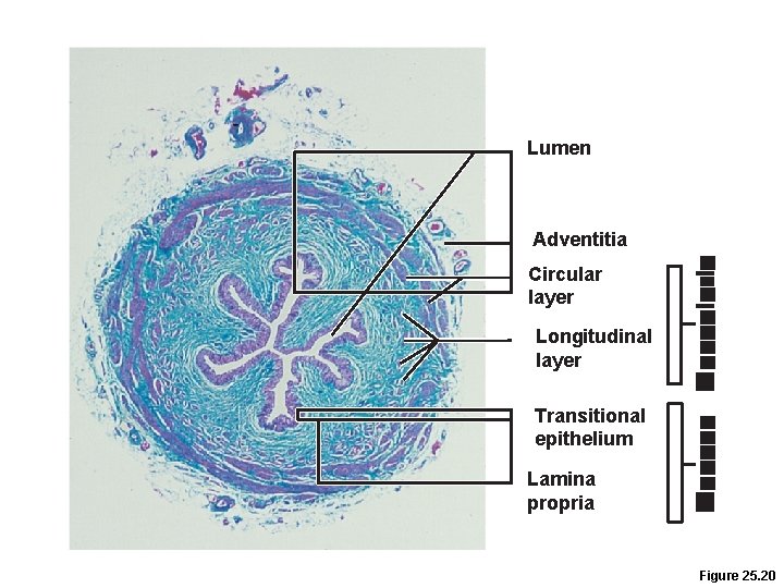
Ureters • Three layers of wall of ureter 1. Lining of transitional epithelium 2. Smooth muscle muscularis • Contracts in response to stretch 3. Outer adventitia of fibrous connective tissue
Lumen Adventitia Circular layer Longitudinal layer Transitional epithelium Lamina propria Figure 25. 20
Renal Calculi • Kidney stones form in renal pelvis –Crystallized calcium, magnesium, or uric acid salts • Larger stones block ureter, cause pressure and pain in kidneys • May be due to chronic bacterial infection, urine retention, Ca 2+ in blood, p. H of urine
Urinary Bladder • Muscular sac for temporary storage of urine • Retroperitoneal, on pelvic floor posterior to pubic symphysis –Males—prostate gland surrounds the neck inferiorly –Females—anterior to the vagina and uterus
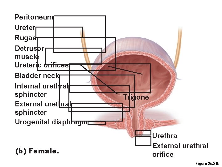
Urinary Bladder • Trigone –Smooth triangular area outlined by the openings for the ureters and the urethra –Infections tend to persist in this region
Urinary Bladder • Layers of the bladder wall 1. Transitional epithelial mucosa 2. Thick detrusor muscle (three layers of smooth muscle) 3. Fibrous adventitia (peritoneum on superior surface only)
Urinary Bladder • Collapses when empty; rugae appear • Expands and rises superiorly during filling without significant rise in internal pressure
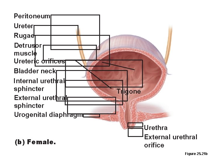
Peritoneum Ureter Rugae Detrusor muscle Ureteric orifices Bladder neck Internal urethral sphincter External urethral sphincter Urogenital diaphragm (b) Female. Trigone Urethra External urethral orifice Figure 25. 21 b
Urethra • Muscular tube –Lining epithelium • Mostly pseudostratified columnar epithelium, except –Transitional epithelium near bladder –Stratified squamous epithelium near external urethral orifice
Urethra • Sphincters –Internal urethral sphincter • Involuntary (smooth muscle) at bladder-urethra junction • Contracts to open –External urethral sphincter • Voluntary (skeletal) muscle surrounding the urethra as it passes through the pelvic floor
Urethra • Female urethra (3– 4 cm): –Tightly bound to the anterior vaginal wall –External urethral orifice is anterior to the vaginal opening, posterior to the clitoris
Peritoneum Ureter Rugae Detrusor muscle Ureteric orifices Bladder neck Internal urethral sphincter External urethral sphincter Urogenital diaphragm (b) Female. Trigone Urethra External urethral orifice Figure 25. 21 b
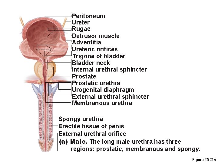
Urethra • Male urethra –Carries semen and urine –Three named regions 1. Prostatic urethra (2. 5 cm)—within prostate gland 2. Membranous urethra (2 cm)—passes through the urogenital diaphragm 3. Spongy urethra (15 cm)—passes through the penis and opens via the external urethral orifice
Peritoneum Ureter Rugae Detrusor muscle Adventitia Ureteric orifices Trigone of bladder Bladder neck Internal urethral sphincter Prostate Prostatic urethra Urogenital diaphragm External urethral sphincter Membranous urethra Spongy urethra Erectile tissue of penis External urethral orifice (a) Male. The long male urethra has three regions: prostatic, membranous and spongy. Figure 25. 21 a
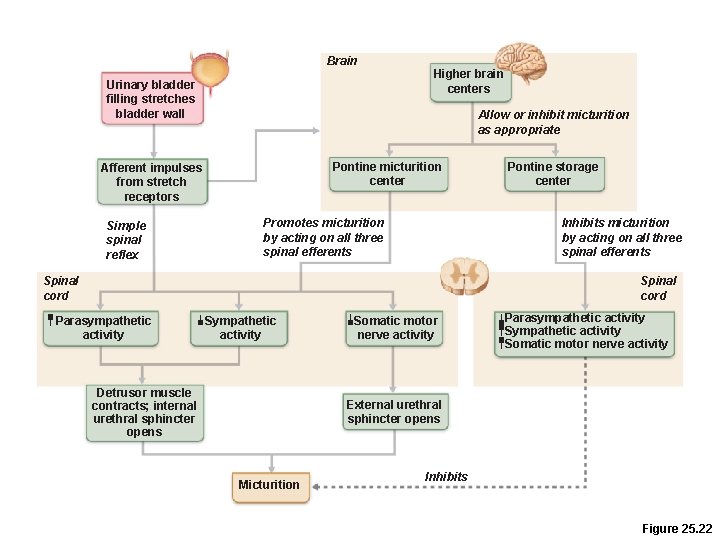
Micturition • Urination or voiding • Three simultaneous events 1. Contraction of detrusor muscle by ANS 2. Opening of internal urethral sphincter by ANS 3. Opening of external urethral sphincter by somatic nervous system
Micturition • Reflexive urination (urination in infants) –Distension of bladder activates stretch receptors –Excitation of parasympathetic neurons in reflex center in sacral region of spinal cord –Contraction of the detrusor muscle –Contraction (opening) of internal sphincter –Inhibition of somatic pathways to external sphincter, allowing its relaxation (opening)
Micturition • Pontine control centers mature between ages 2 and 3 1. Pontine storage center inhibits micturition: • Inhibits parasympathetic pathways • Excites sympathetic and somatic efferent pathways 2. Pontine micturition center promotes micturition: • Excites parasympathetic pathways • Inhibits sympathetic and somatic efferent pathways
Brain Urinary bladder filling stretches bladder wall Allow or inhibit micturition as appropriate Pontine micturition center Afferent impulses from stretch receptors Simple spinal reflex Higher brain centers Promotes micturition by acting on all three spinal efferents Pontine storage center Inhibits micturition by acting on all three spinal efferents Spinal cord Parasympathetic activity Sympathetic activity Detrusor muscle contracts; internal urethral sphincter opens Somatic motor nerve activity Parasympathetic activity Somatic motor nerve activity External urethral sphincter opens Micturition Inhibits Figure 25. 22
Three Basic Processes Of The Nephron Filtration – separating fluid from large particles in the blood to make urine l. Reabsorption – drawing nutrients and other substances out of the urine, to keep in the body l. Secretion – adding substances to the urine for removal from the body l
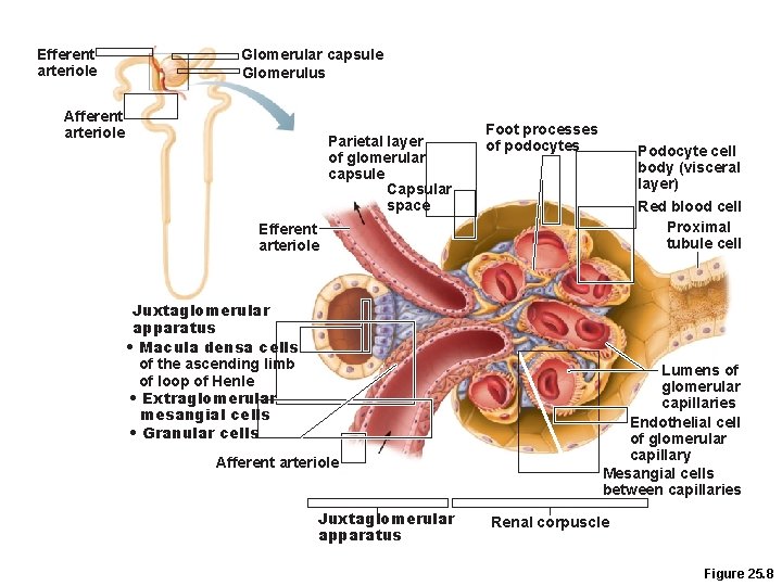
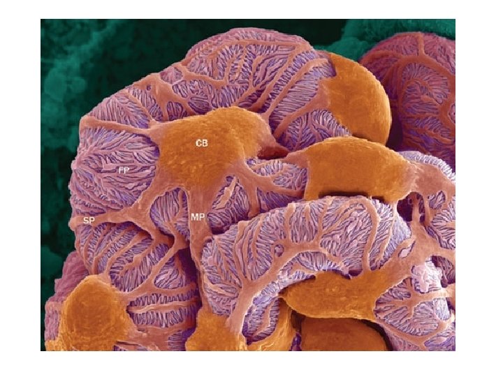
Nephrons Renal corpuscle –Glomerulus + its glomerular capsule • Fenestrated glomerular endothelium –Allows filtrate to pass from plasma into the glomerular capsule • Glomerular capsule • Parietal layer: simple squamous epithelium • Visceral layer: branching epithelial podocytes –Extensions terminate in foot processes that cling to basement membrane –Filtration slits allow filtrate to pass into the capsular space
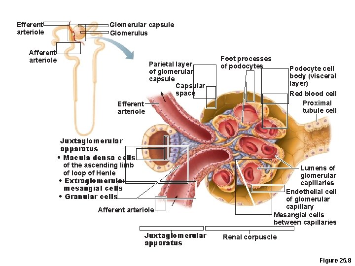
Efferent arteriole Glomerular capsule Glomerulus Afferent arteriole Parietal layer of glomerular capsule Capsular space Foot processes of podocytes Podocyte cell body (visceral layer) Red blood cell Proximal tubule cell Efferent arteriole Juxtaglomerular apparatus • Macula densa cells of the ascending limb of loop of Henle • Extraglomerular mesangial cells • Granular cells Afferent arteriole Juxtaglomerular apparatus Lumens of glomerular capillaries Endothelial cell of glomerular capillary Mesangial cells between capillaries Renal corpuscle Figure 25. 8
Renal Tubule • Proximal convoluted tubule (PCT) –Cuboidal cells with dense microvilli and large mitochondria –Functions in reabsorption and secretion –Confined to the cortex
Renal Tubule • Loop of Henle with descending and ascending limbs –Thin segment usually in descending limb –Simple squamous epithelium –Freely permeable to water –Thick segment of ascending limb • Cuboidal to columnar cells
Renal Tubule • Distal convoluted tubule (DCT) –Cuboidal cells with very few microvilli –Function more in secretion than reabsorption –Confined to the cortex
Collecting Ducts • Receive filtrate from many nephrons • Fuse together to deliver urine through papillae into minor calyces • Cuboidal to columnar epithelium
Glomerular capsule: parietal layer Renal cortex Renal medulla Basement membrane Renal corpuscle • Glomerular capsule • Glomerulus Renal pelvis Ureter Podocyte Distal convoluted tubule Kidney Fenestrated endothelium of the glomerulus Glomerular capsule: visceral layer Microvilli Proximal convoluted tubule Mitochondria Highly infolded plasma membrane Proximal convoluted tubule cells Cortex Medulla Thick segment Thin segment Loop of Henle • Descending limb • Ascending limb Distal convoluted tubule cells Collecting duct Loop of Henle (thin-segment) cells Principal cell Intercalated cell Collecting duct cells Figure 25. 5
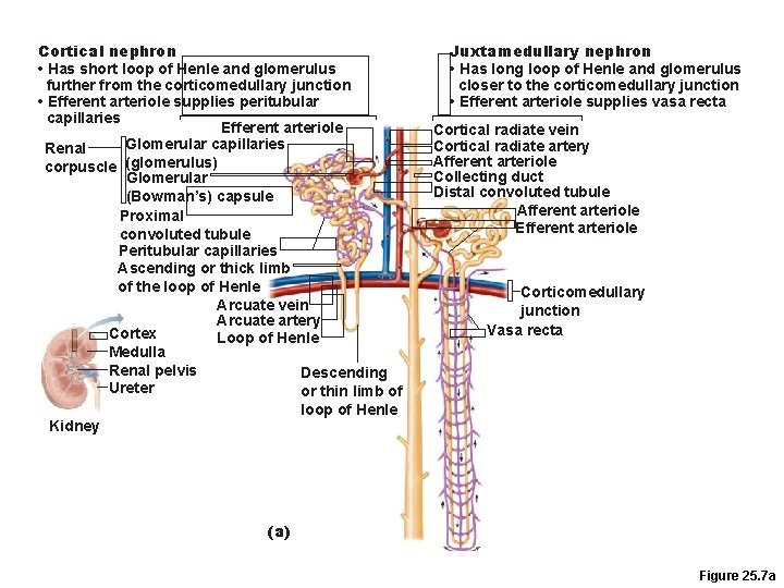
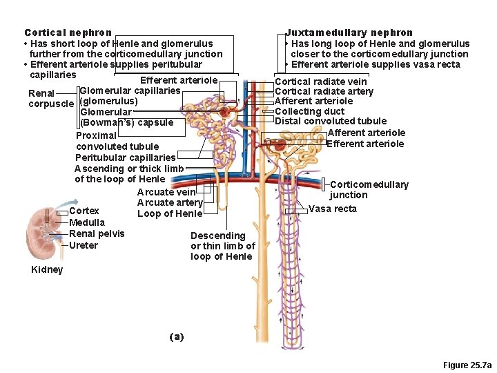
Nephrons Two Types: • Cortical nephrons— 85% of nephrons; almost entirely in the cortex • Juxtamedullary nephrons –Long loops of Henle deeply invade the medulla –Extensive thin segments –Important in the production of concentrated urine
Cortical nephron • Has short loop of Henle and glomerulus further from the corticomedullary junction • Efferent arteriole supplies peritubular capillaries Efferent arteriole Glomerular capillaries Renal corpuscle (glomerulus) Glomerular (Bowman’s) capsule Proximal convoluted tubule Peritubular capillaries Ascending or thick limb of the loop of Henle Arcuate vein Arcuate artery Cortex Loop of Henle Medulla Renal pelvis Descending Ureter or thin limb of loop of Henle Kidney Juxtamedullary nephron • Has long loop of Henle and glomerulus closer to the corticomedullary junction • Efferent arteriole supplies vasa recta Cortical radiate vein Cortical radiate artery Afferent arteriole Collecting duct Distal convoluted tubule Afferent arteriole Efferent arteriole Corticomedullary junction Vasa recta (a) Figure 25. 7 a
Nephron Capillary Beds 1. Glomerulus –Afferent arteriole glomerulus efferent arteriole –Specialized for filtration –Blood pressure is high because • Afferent arterioles are larger in diameter than efferent arterioles • Arterioles are high-resistance vessels
Nephron Capillary Beds 1. Peritubular capillaries –Low-pressure, porous capillaries adapted for absorption –Arise from efferent arterioles –Cling to adjacent renal tubules in cortex –Empty into venules
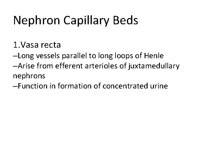
Nephron Capillary Beds 1. Vasa recta –Long vessels parallel to long loops of Henle –Arise from efferent arterioles of juxtamedullary nephrons –Function in formation of concentrated urine
Cortical nephron • Has short loop of Henle and glomerulus further from the corticomedullary junction • Efferent arteriole supplies peritubular capillaries Efferent arteriole Glomerular capillaries Renal corpuscle (glomerulus) Glomerular (Bowman’s) capsule Proximal convoluted tubule Peritubular capillaries Ascending or thick limb of the loop of Henle Arcuate vein Arcuate artery Cortex Loop of Henle Medulla Renal pelvis Descending Ureter or thin limb of loop of Henle Kidney Juxtamedullary nephron • Has long loop of Henle and glomerulus closer to the corticomedullary junction • Efferent arteriole supplies vasa recta Cortical radiate vein Cortical radiate artery Afferent arteriole Collecting duct Distal convoluted tubule Afferent arteriole Efferent arteriole Corticomedullary junction Vasa recta (a) Figure 25. 7 a
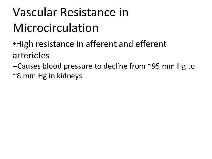
Vascular Resistance in Microcirculation • High resistance in afferent and efferent arterioles –Causes blood pressure to decline from ~95 mm Hg to ~8 mm Hg in kidneys
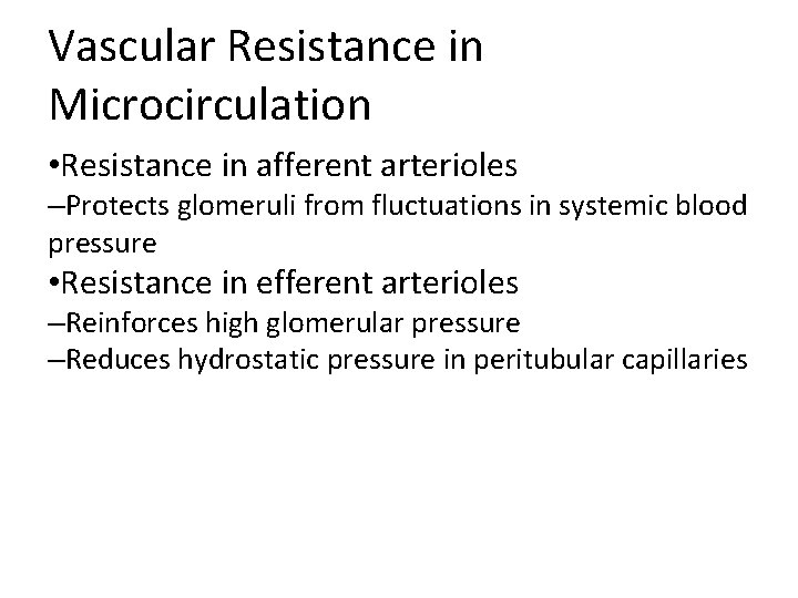
Vascular Resistance in Microcirculation • Resistance in afferent arterioles –Protects glomeruli from fluctuations in systemic blood pressure • Resistance in efferent arterioles –Reinforces high glomerular pressure –Reduces hydrostatic pressure in peritubular capillaries
Juxtaglomerular Apparatus (JGA) • One per nephron • Important in regulation of filtrate formation and blood pressure • Involves modified portions of the –Distal portion of the ascending limb of the loop of Henle –Afferent arteriole
Juxtaglomerular Apparatus (JGA) • Granular cells (juxtaglomerular, or JG cells) –Enlarged, smooth muscle cells of arteriole –Secretory granules contain renin –Act as mechanoreceptors that sense blood pressure
Juxtaglomerular Apparatus (JGA) • Macula densa –Tall, closely packed cells of the ascending limb –Act as chemoreceptors that sense Na. Cl content of filtrate • Extraglomerular mesangial cells –Interconnected with gap junctions –May pass signals between macula densa and granular cells
Efferent arteriole Glomerular capsule Glomerulus Afferent arteriole Parietal layer of glomerular capsule Capsular space Foot processes of podocytes Podocyte cell body (visceral layer) Red blood cell Proximal tubule cell Efferent arteriole Juxtaglomerular apparatus • Macula densa cells of the ascending limb of loop of Henle • Extraglomerular mesangial cells • Granular cells Afferent arteriole Juxtaglomerular apparatus Lumens of glomerular capillaries Endothelial cell of glomerular capillary Mesangial cells between capillaries Renal corpuscle Figure 25. 8
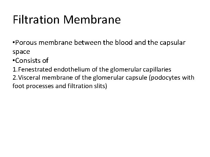
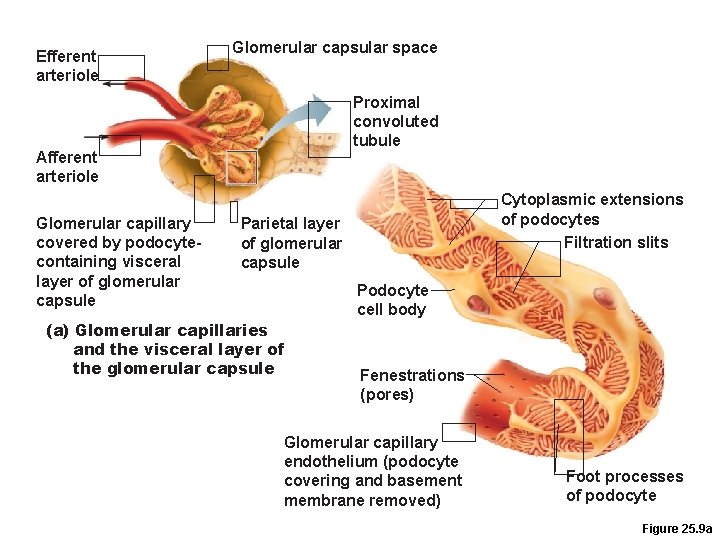
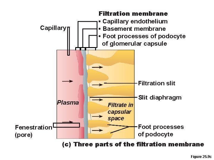
Filtration Membrane • Porous membrane between the blood and the capsular space • Consists of 1. Fenestrated endothelium of the glomerular capillaries 2. Visceral membrane of the glomerular capsule (podocytes with foot processes and filtration slits)
Efferent arteriole Glomerular capsular space Proximal convoluted tubule Afferent arteriole Glomerular capillary covered by podocytecontaining visceral layer of glomerular capsule Cytoplasmic extensions of podocytes Filtration slits Parietal layer of glomerular capsule (a) Glomerular capillaries and the visceral layer of the glomerular capsule Podocyte cell body Fenestrations (pores) Glomerular capillary endothelium (podocyte covering and basement membrane removed) Foot processes of podocyte Figure 25. 9 a
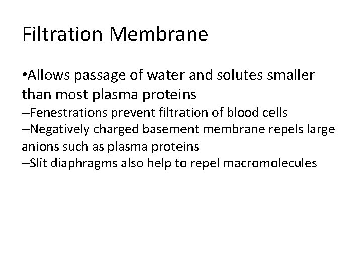
Filtration Membrane • Allows passage of water and solutes smaller than most plasma proteins –Fenestrations prevent filtration of blood cells –Negatively charged basement membrane repels large anions such as plasma proteins –Slit diaphragms also help to repel macromolecules
Capillary Filtration membrane • Capillary endothelium • Basement membrane • Foot processes of podocyte of glomerular capsule Filtration slit Plasma Fenestration (pore) Slit diaphragm Filtrate in capsular space Foot processes of podocyte (c) Three parts of the filtration membrane Figure 25. 9 c
Kidney Physiology: Mechanisms of Urine Formation • The kidneys filter the body’s entire plasma volume 60 times each day • Filtrate –Blood plasma minus proteins • Urine –<1% of total filtrate –Contains metabolic wastes and unneeded substances
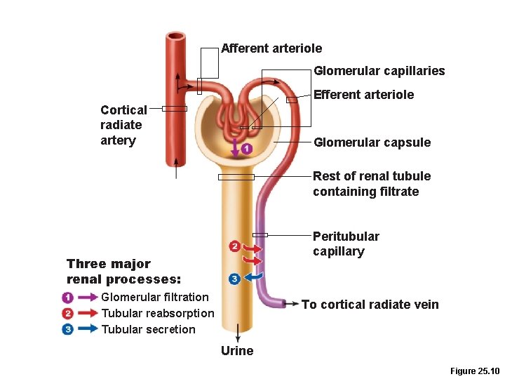
Mechanisms of Urine Formation 1. Glomerular filtration 2. Tubular reabsorption –Returns all glucose and amino acids, 99% of water, salt, and other components to the blood 3. Tubular secretion –Reverse of reabsoprtion: selective addition to urine
Afferent arteriole Glomerular capillaries Efferent arteriole Cortical radiate artery Glomerular capsule Rest of renal tubule containing filtrate Peritubular capillary Three major renal processes: Glomerular filtration Tubular reabsorption Tubular secretion To cortical radiate vein Urine Figure 25. 10
Glomerular Filtration • Passive mechanical process driven by hydrostatic pressure in the capillaries of the glomerulus. • The glomerulus is a very efficient filter because –Its filtration membrane is very permeable and it has a large surface area –Glomerular blood pressure is higher (55 mm Hg) than other capillaries • Molecules >5 nm are not filtered (e. g. , plasma proteins) and function to maintain colloid osmotic pressure of the blood
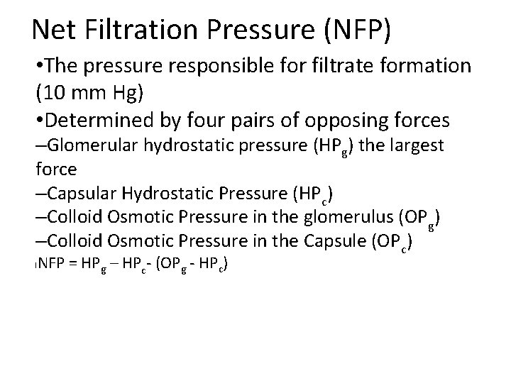
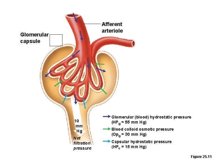
Net Filtration Pressure (NFP) • The pressure responsible for filtrate formation (10 mm Hg) • Determined by four pairs of opposing forces –Glomerular hydrostatic pressure (HPg) the largest force –Capsular Hydrostatic Pressure (HPc) –Colloid Osmotic Pressure in the glomerulus (OPg) –Colloid Osmotic Pressure in the Capsule (OPc) NFP = HPg – HPc- (OPg - HPc) l
Afferent arteriole Glomerular capsule 10 mm Hg Net filtration pressure Glomerular (blood) hydrostatic pressure (HPg = 55 mm Hg) Blood colloid osmotic pressure (Opg = 30 mm Hg) Capsular hydrostatic pressure (HPc = 15 mm Hg) Figure 25. 11
Glomerular Filtration Rate (GFR) • Volume of filtrate formed per minute by the kidneys (120– 125 ml/min) • Governed by (and directly proportional to) –Total surface area available for filtration –Filtration membrane permeability –NFP
Tubular Reabsorption • A selective transepithelial process –All organic nutrients are reabsorbed –Water and ion reabsorption are hormonally regulated • Includes active and passive process
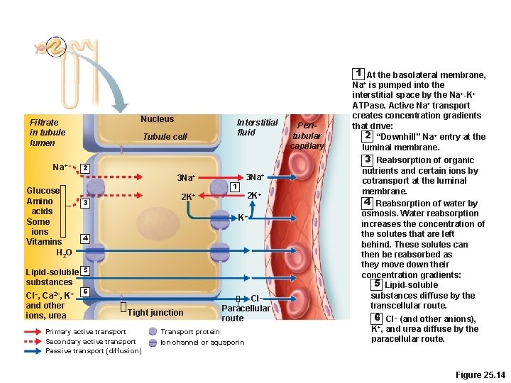
Sodium Reabsorption • Na+ (most abundant cation in filtrate) • Primary active transport out of the tubule cell by – Na+-K+ ATPase in the basolateral membrane
Sodium Reabsorption in Peritubular Capillaries • Low hydrostatic pressure and high osmotic pressure in the peritubular capillaries • Promotes bulk flow of water and solutes (including Na+)
Reabsorption of Nutrients, Water, and Ions • Na+ reabsorption provides the energy and the means for reabsorbing most other substances • Organic nutrients are reabsorbed by secondary active transport –When the carriers are saturated, excess of that substance remains in tubule and is excreted
Reabsorption of Nutrients, Water, and Ions • Water is reabsorbed by osmosis (obligatory water reabsorption), aided by water-filled pores called aquaporins (“Water follows sodium”) • Cations and fat-soluble substances follow by diffusion
1 At the basolateral membrane, Nucleus Filtrate in tubule lumen Na+ Tubule cell 3 Na+ 1 2 K+ 3 K+ 4 Lipid-soluble 5 substances Cl–, Ca 2+, K+ and other ions, urea Peritubular capillary 3 Reabsorption of organic 2 3 Na+ Glucose Amino acids Some ions Vitamins H 2 O Interstitial fluid Na+ is pumped into the interstitial space by the Na+-K+ ATPase. Active Na+ transport creates concentration gradients that drive: 2 “Downhill” Na + entry at the luminal membrane. 6 Tight junction Primary active transport Secondary active transport Passive transport (diffusion) Cl– Paracellular route Transport protein Ion channel or aquaporin nutrients and certain ions by cotransport at the luminal membrane. 4 Reabsorption of water by osmosis. Water reabsorption increases the concentration of the solutes that are left behind. These solutes can then be reabsorbed as they move down their concentration gradients: 5 Lipid-soluble substances diffuse by the transcellular route. 6 Cl– (and other anions), K+, and urea diffuse by the paracellular route. Figure 25. 14
Reabsorptive Capabilities of Renal Tubules and Collecting Ducts • PCT –Site of most reabsorption • 65% of Na+ and water • All nutrients • Ions • Small proteins
Reabsorptive Capabilities of Renal Tubules and Collecting Ducts • Technically, the Loop of Henle is involved in reabsorption –Descending limb: H 2 O –Ascending limb: Na+, K+, Cl • However, the function of this reabsorption is actually to create a counter-current multiplier (see below).
Reabsorptive Capabilities of Renal Tubules and Collecting Ducts • DCT and collecting duct –Reabsorption is hormonally regulated • Ca 2+ (PTH) • Water (ADH) • Na+ (aldosterone and ANP)
Tubular Secretion • Reabsorption in reverse –K+, H+, NH 4+ and organic acids move from peritubular capillaries or tubule cells into filtrate • Disposes of substances that are bound to plasma proteins
Tubular Secretion • Eliminates undesirable substances that have been passively reabsorbed (e. g. , urea and uric acid) • Rids the body of excess K+ • Controls blood p. H by altering amounts of H+ or HCO 3– in urine
Regulation of Urine Concentration and Volume • Osmolality –Number of solute particles in 1 kg of H 2 O –Reflects ability to cause osmosis
Regulation of Urine Concentration and Volume • Osmolality of body fluids –Expressed in milliosmols (m. Osm) –The kidneys maintain osmolality of plasma at ~300 m. Osm, using countercurrent mechanisms
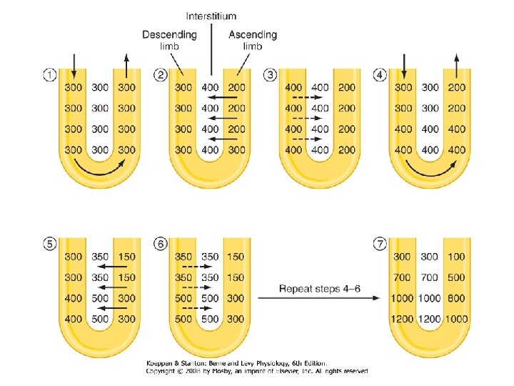
Countercurrent Mechanism • Occurs when fluid flows in opposite directions in two adjacent segments of the same tube –Filtrate flow in the loop of Henle (countercurrent multiplier) –Blood flow in the vasa recta (countercurrent exchanger)
Countercurrent Mechanism • Role of countercurrent mechanisms –Establish and maintain an osmotic gradient (300 m. Osm to 1200 m. Osm) from renal cortex through the medulla –Allow the kidneys to vary urine concentration
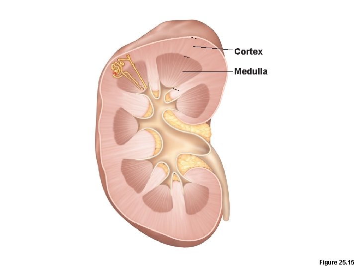
Cortex Medulla Figure 25. 15
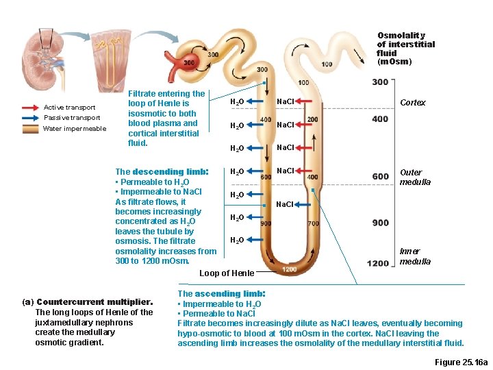
Countercurrent Multiplier: Loop of Henle • Descending limb –Freely permeable to H 2 O, which passes out of the filtrate into the hyperosmotic medullary interstitial fluid –Filtrate osmolality increases to ~1200 m. Osm
Countercurrent Multiplier: Loop of Henle • Ascending limb –Impermeable to H 2 O –Selectively permeable to solutes • Na+ and Cl– are passively reabsorbed in the thin segment, actively reabsorbed in the thick segment • Active transport of Na. Cl in the ascending limb powers the whole system. –Filtrate osmolality decreases to 100 m. Osm
Osmolality of interstitial fluid (m. Osm) Active transport Passive transport Water impermeable Filtrate entering the loop of Henle is isosmotic to both blood plasma and cortical interstitial fluid. H 2 O Na. CI H 2 O The descending limb: • Permeable to H 2 O • Impermeable to Na. Cl H 2 O As filtrate flows, it becomes increasingly H 2 O concentrated as H 2 O leaves the tubule by H 2 O osmosis. The filtrate osmolality increases from 300 to 1200 m. Osm. Loop of Henle (a) Countercurrent multiplier. The long loops of Henle of the juxtamedullary nephrons create the medullary osmotic gradient. Na. CI Cortex Outer medulla Na. CI Inner medulla The ascending limb: • Impermeable to H 2 O • Permeable to Na. Cl Filtrate becomes increasingly dilute as Na. Cl leaves, eventually becoming hypo-osmotic to blood at 100 m. Osm in the cortex. Na. Cl leaving the ascending limb increases the osmolality of the medullary interstitial fluid. Figure 25. 16 a
Urea Recycling • Urea moves between the collecting ducts and the loop of Henle –Secreted into filtrate by facilitated diffusion in the ascending thin segment –Reabsorbed by facilitated diffusion in the collecting ducts deep in the medulla • Contributes to the high osmolality in the medulla
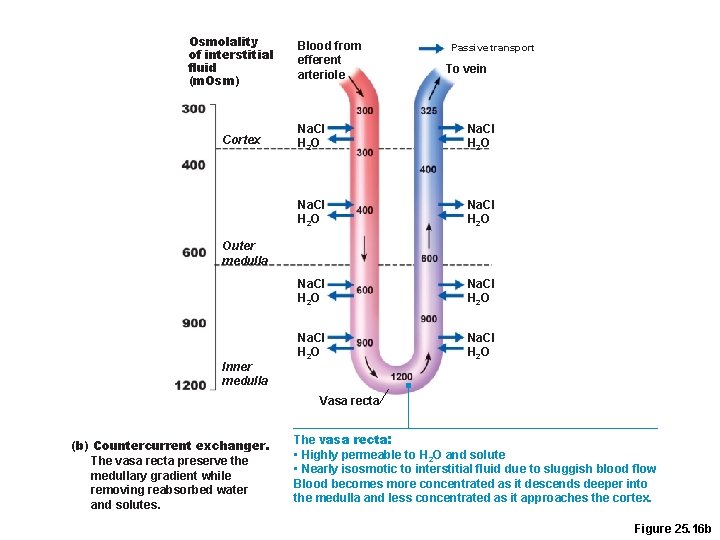
Countercurrent Exchanger: Vasa Recta • The vasa recta –Maintain the osmotic gradient –Deliver blood to the medullary tissues –Protect the medullary osmotic gradient by preventing rapid removal of salt, and by removing reabsorbed H 2 O
Osmolality of interstitial fluid (m. Osm) Cortex Blood from efferent arteriole Passive transport To vein Na. CI H 2 O Na. CI H 2 O Outer medulla Inner medulla Vasa recta (b) Countercurrent exchanger. The vasa recta preserve the medullary gradient while removing reabsorbed water and solutes. The vasa recta: • Highly permeable to H 2 O and solute • Nearly isosmotic to interstitial fluid due to sluggish blood flow Blood becomes more concentrated as it descends deeper into the medulla and less concentrated as it approaches the cortex. Figure 25. 16 b
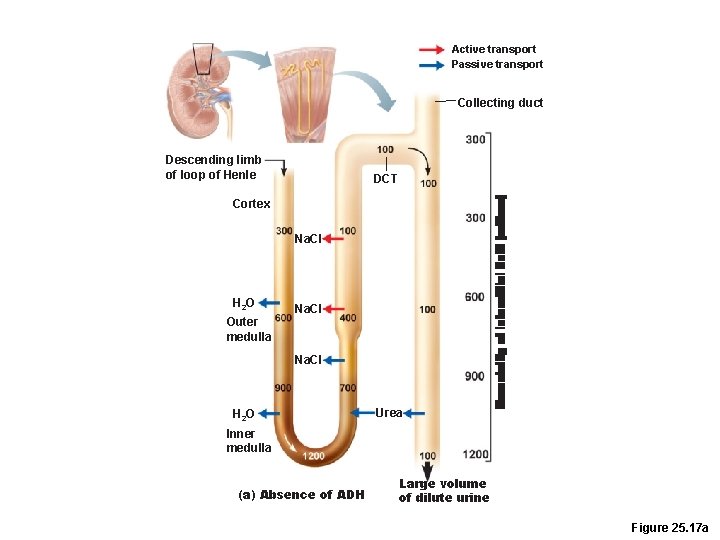
Formation of Dilute Urine • Filtrate is diluted in the ascending loop of Henle • In the absence of ADH, dilute filtrate continues into the renal pelvis as dilute urine • Na+ and other ions may be selectively removed in the DCT and collecting duct, decreasing osmolality to as low as 50 m. Osm
Active transport Passive transport Collecting duct Descending limb of loop of Henle DCT Cortex Na. CI H 2 O Outer medulla Na. CI H 2 O Urea Inner medulla (a) Absence of ADH Large volume of dilute urine Figure 25. 17 a
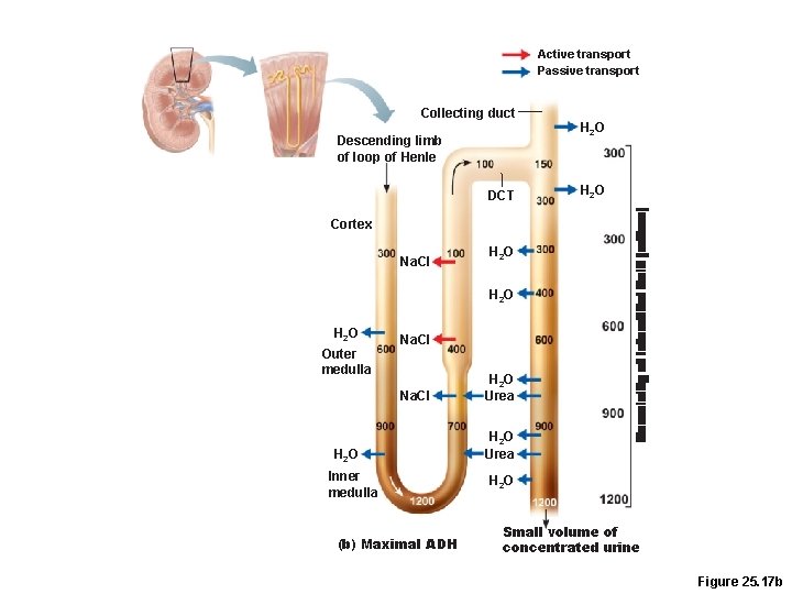
Formation of Concentrated Urine • Depends on the medullary osmotic gradient and ADH • ADH triggers reabsorption of H 2 O in the collecting ducts • Facultative water reabsorption occurs in the presence of ADH so that 99% of H 2 O in filtrate is reabsorbed
Active transport Passive transport Collecting duct Descending limb of loop of Henle DCT H 2 O Cortex Na. CI H 2 O Outer medulla Na. CI H 2 O Inner medulla (b) Maximal ADH H 2 O Urea H 2 O Small volume of concentrated urine Figure 25. 17 b
Physical Characteristics of Urine • Color and transparency –Clear, pale to deep yellow (due to urochrome) –Drugs, vitamin supplements, and diet can alter the color –Cloudy urine may indicate a urinary tract infection
Physical Characteristics of Urine • Odor –Slightly aromatic when fresh –Develops ammonia odor upon standing –May be altered by some drugs and vegetables
Physical Characteristics of Urine • p. H –Slightly acidic (~p. H 6, with a range of 4. 5 to 8. 0) –Diet, prolonged vomiting, or urinary tract infections may alter p. H • Specific gravity – 1. 001 to 1. 035, dependent on solute concentration
Chemical Composition of Urine • 95% water and 5% solutes • Nitrogenous wastes: urea, uric acid, and creatinine • Other normal solutes –Na+, K+, PO 43–, and SO 42–, –Ca 2+, Mg 2+ and HCO 3– • Abnormally high concentrations of any constituent may indicate pathology
Regulation of Glomerular Filtration • GFR is tightly controlled by two types of mechanisms • Intrinsic controls (renal autoregulation) –Act locally within the kidney • Extrinsic controls –Nervous and endocrine mechanisms that maintain blood pressure, but affect kidney function
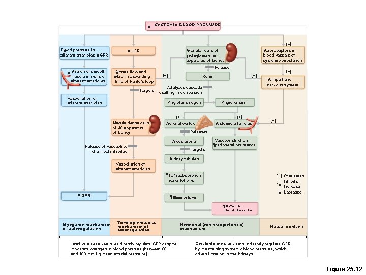
Intrinsic Controls • Maintains a nearly constant GFR when Mean Arterial Pressure is in the range of 80– 180 mm Hg • Two types of renal autoregulation –Myogenic mechanism (Chapter 19) –Tubuloglomerular feedback mechanism, which senses changes in the juxtaglomerular apparatus
Intrinsic Controls: Myogenic Mechanism • BP constriction of afferent arterioles –Helps maintain normal GFR –Protects glomeruli from damaging high BP • BP dilation of afferent arterioles –Helps maintain normal GFR
Intrinsic Controls: Tubuloglomerular Feedback Mechanism • Flow-dependent mechanism directed by the macula densa cells • If GFR increases, filtrate flow rate increases in the tubule • Filtrate Na. Cl concentration will be high because of insufficient time for reabsorption
Intrinsic Controls: Tubuloglomerular Feedback Mechanism • Flow-dependent mechanism directed by the macula densa cells • If GFR increases, filtrate flow rate increases in the tubule • Filtrate Na. Cl concentration will be high because of insufficient time for reabsorption
Extrinsic Controls: Sympathetic Nervous System • Under normal conditions at rest –Renal blood vessels are dilated –Renal autoregulation mechanisms prevail
Extrinsic Controls: Sympathetic Nervous System • Under normal conditions at rest –Renal blood vessels are dilated –Renal autoregulation mechanisms prevail
Extrinsic Controls: Renin. Angiotensin Mechanism • Triggered when the granular cells of the JGA detect decreased GFR • Granular cells release renin. • Renin is an enzyme that activates the hormone Angiotensinogen.
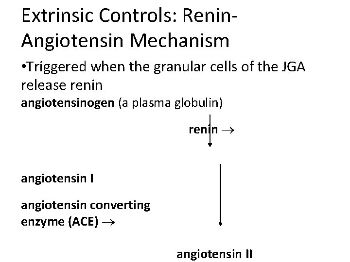
Extrinsic Controls: Renin. Angiotensin Mechanism • Triggered when the granular cells of the JGA release renin angiotensinogen (a plasma globulin) renin angiotensin I angiotensin converting enzyme (ACE) angiotensin II
Effects of Angiotensin II 1. Constricts arteriolar smooth muscle, causing Mean Arterial Pressure to rise 2. Stimulates the reabsorption of Na+ –Acts directly on the renal tubules –Triggers adrenal cortex to release aldosterone 3. Stimulates the hypothalamus to release ADH and activates the thirst center 4. Constricts efferent arterioles, decreasing peritubular capillary hydrostatic pressure and increasing fluid reabsorption 5. Causes glomerular mesangial cells to contract, decreasing the surface area available for filtration
SYSTEMIC BLOOD PRESSURE (–) Blood pressure in afferent arterioles; GFR Stretch of smooth muscle in walls of afferent arterioles Baroreceptors in blood vessels of systemic circulation Granular cells of juxtaglomerular apparatus of kidney GFR Filtrate flow and Na. Cl in ascending limb of Henle’s loop Release (+) Renin Catalyzes cascade Targets resulting in conversion Vasodilation of afferent arterioles Angiotensinogen (+) Macula densa cells of JG apparatus of kidney Sympathetic nervous system Angiotensin II (+) Adrenal cortex (+) Systemic arterioles (+) Releases Aldosterone Release of vasoactive chemical inhibited Targets Vasoconstriction; peripheral resistance Kidney tubules Vasodilation of afferent arterioles Na+ reabsorption; water follows GFR (+) Stimulates (–) Inhibits Increase Decrease Blood volume Systemic blood pressure Myogenic mechanism of autoregulation Tubuloglomerular mechanism of autoregulation Intrinsic mechanisms directly regulate GFR despite moderate changes in blood pressure (between 80 and 180 mm Hg mean arterial pressure). Hormonal (renin-angiotensin) mechanism Neural controls Extrinsic mechanisms indirectly regulate GFR by maintaining systemic blood pressure, which drives filtration in the kidneys. Figure 25. 12
Reabsorptive Capabilities of Renal Tubules and Collecting Ducts • Mechanism of aldosterone –Targets collecting ducts (principal cells) and distal DCT –Promotes synthesis of luminal Na+ and K+ channels –Promotes synthesis of basolateral Na+-K+ ATPases
Summary of Urinary Hormones l ADH l. Released when plasma sodium concentration is high l. Increases reabsorption of water from the collecting duct l Renin l. Released when GFR and/or blood pressure is low l. Activates Angiotensinogen
Summary of Urinary Hormones l Angiotensin II l. Raises MAP by Increasing sodium reabsorption l. Causes release of ADH l. Vasoconstriction l l Aldosterone l. Released when blood pressure is low or sodium conc is low. l. Increases sodium reabsorption