Benign Breast Disease Alireza Mohammadzadeh MD Thoracic Surgeon

Benign Breast Disease Alireza Mohammadzadeh, MD Thoracic Surgeon

Benign breast disorders & diseases encompass a wide range of clinical and pathologic entities

Understanding of these for : clear explanation to affected women appropriate treatment instituted unnecessary follow up

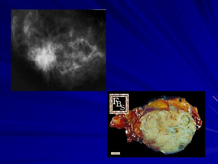
Fibroadenoma Predominantly in younger women aged 15 to 25 years Usually grow to 1 or 2 cm and then are stable Small f. (<1 cm) are considered normal Larger f. (<3 cm) are disorders Giant f. (>3 cm) are disease Multiple f. (more than 5 in one breast) are disease
 –")
Ultrasound Benign – Pure and intensely hyperechoic – Elliptical shape (wider than tall) – Lobulated – Complete tine capsule Malignant – – Hypoechoic, spiculated Taller than wide Duct extension microlobulation

Fibroadenoma

Core-needle biopsy

Treatment Surgical removal Cryoablation observation

Sclerosing adenosis Prevalent during childbearing & perimenopausal years No malignant potential Occasionally presents as a palpable mass Benign calcification Lesions up to 1 cm are called radial scar Larger lesions are called complex sclerosing

Sclerosing adenosis Mimic of cancer On physical examination, by mammography, at gross pathology Wire localized excisionl biopsy

Benign Breast Diseases Glandular breast parenchyma – Mass – Asymmetric nodularity – Pain Nipple-Areolar Complex – Discharge – Rash – Retraction Surrounding breast skin – Dimpling

Management History Clinical Breast Exam Breast imaging Tissue sampling Therapy

History Age – Menarche – Pregnancy Breast feeding – Menopause Family History Prior biopsies Hormone therapy

Clinical Exam Inspection – Skin – Symmetry – Masses Palpable – Gland – Axilla, Supraclavicular spaces – Nipple-areola complex

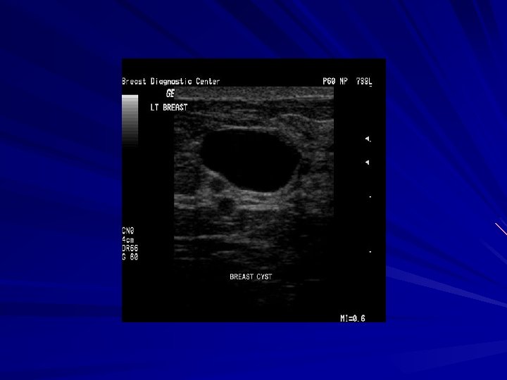
Breast Mass Breast Cysts – Fluid-filled – 1 out of every 14 women 50% multiple and recurrent – Hormonally influenced – Needle aspirated

Breast Cyst

Breast Mass Phyllodes Tumor – Proliferation of connective tissue with ductal elements Whorled and cellular stroma – Firm, lobulated – 2 to 40 cm in size – 10% malignant – Treatment Wide excision

Fibrocystic Disease Clinical, mammographic and histologic findings Exaggerated response from hormones and growth factors – Cyclical pain – Nodularity – upper outer quadrants

Fibrocystic Disease Histology – Adenosis – Apocrine metaplasia – Fibrosis – Duct ectasia – Mild ductal hyperplasia

Fibrocystic Disease Risk Factors – Dense breast – Sclerosing adenosis – Atypical ductal, papillary, or lobular hyperplasia

Breast Pain Cyclical pain – hormonal – Dull, diffuse and bilateral – Luteal phase – Treatment Reassurance NSAIDS Evening primrose oil Non-cyclical pain – Non-breast vs breast – Imaging – Treatment Reassurance NSAIDS Evening primrose oil

Breast Infections Mastitis – Generalized cellulitis of the breast – Ascending infection subareolar ducts commonly occurs during lactation – Staph. aureus – Erythema, pain, tenderness

Mastitis Treatment – Abx – Continue to breast feed – Close follow-up

Breast Abscess – Breast tissue – Treatment Abx Needle aspiration Incision and drainage
 – Non-spontaneous")
Nipple Discharge Physiologic – Bilateral – Involves multiple ducts – Heme (-) – Non-spontaneous
 Most common cause intraductal")
Nipple Discharge Pathologic – Unilateral – Spontaneous – Heme (+) Most common cause intraductal papilloma

Bloody Nipple Discharge

Intraductal Papilloma Single duct Benign 4% of intraductal ca

Imaging Mammography Ultrasound MRI

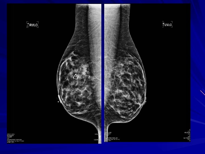
Mammography Screening tool – Age of 40 Estimated reduction in mortality 15 -25% 10% false positive rate Densities & calcifications
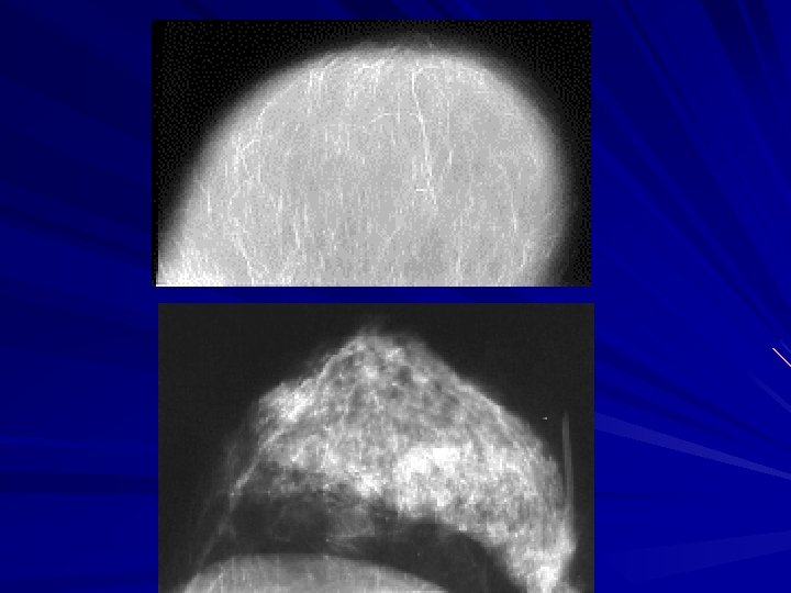
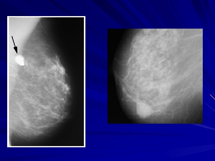

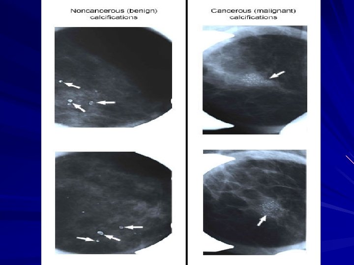
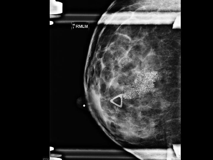
Calcification Macrocalcifications – Large white dots – Almost always noncancerous and require no further follow-up. Microcalcifications – Very fine white specks – Usually noncancerous but can sometimes be a sign of cancer. – Size, shape and pattern

BI-RADS Classification Features 0 1 2 3 4 Need additional imaging Negative – routine in 1 yr Benign finding – routine in 1 yr Probably benign, 6 mo follow-up Suspicious abnormality, biopsy recommended Highly suggestive of malignancy; appropriate action should be taken 5

Ultrasound Not a screening tool Palpable vs cystic Mammographic detected lesion

Ultrasound

Malignant or Benign

Malignant vs Benign

MRI High risk patients – Personal history of breast ca – LCIS, atypia – 1 st degree relative with breast cancer – Very dense breast High sensitivity (95 -100%) – 10 -20% will have a biopsy

MRI Pre Gad Post Gad Color Overlay

Diagnosis Fine needle aspiration – Cytology Core biopsy – Image guided – Stereotactic Excisional biopsy – Needle localization

FNA Fast, inexpensive 96% accuracy Institution dependent Unable to differentiate b/w in situ vs CA

Core Needle Biopsy 14 -18 gauge spring loaded needle Tissue Multiple
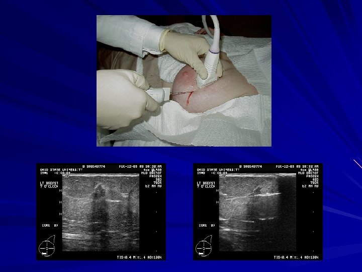

Large Core Biopsy 6 -14 gauge core Large samples Single insertion

Core biopsy Vacuum Assisted

Excisional Biopsy Atypical lesions LCIS Radial scar Atypical papillary lesions Radiologic-pathologic discordance Phyllodes Inadequate tissue harvesting
- Slides: 52