GENITAL SYSTEM The development of the genital n

- Slides: 45
GENITAL SYSTEM The development of the genital n system comprises the development of: (1) Gonads (primitive sex cells). n (2) Genital ducts. n (3) External genitalia. n All three components pass through an indifferent stage in which they may develop into either a male or a female. n
GENITAL SYSTEM The Y chromosome causes n differentiation of the gonad into testis. The indifferent duct system and n external genitalia develop under the influence of hormones. Male and Female morphologic n characteristic do not begin to develop until the 7 TH week
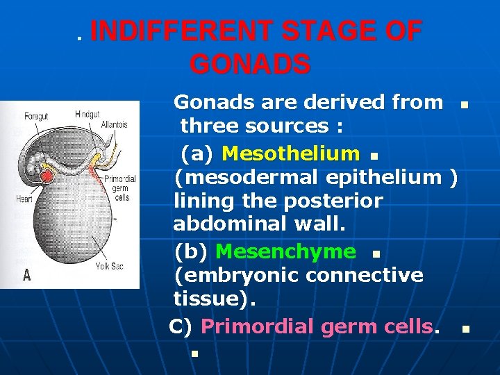
. INDIFFERENT STAGE OF GONADS Gonads are derived from n three sources : (a) Mesothelium n (mesodermal epithelium ) lining the posterior abdominal wall. (b) Mesenchyme n (embryonic connective tissue). C) Primordial germ cells. n n
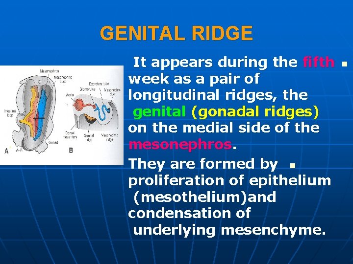
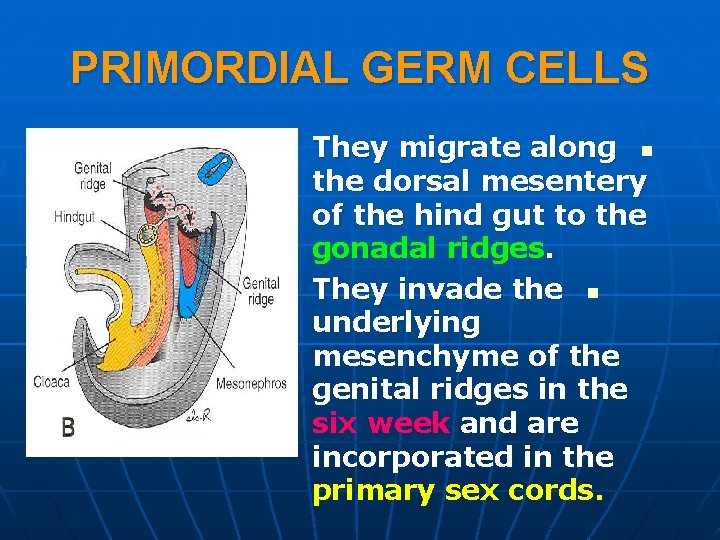
GENITAL RIDGE It appears during the fifth week as a pair of longitudinal ridges, the genital (gonadal ridges) on the medial side of the mesonephros. They are formed by n proliferation of epithelium (mesothelium)and condensation of underlying mesenchyme. n
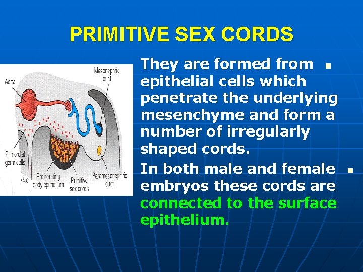
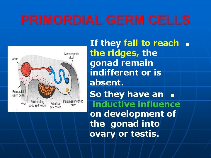
PRIMITIVE SEX CORDS They are formed from n epithelial cells which penetrate the underlying mesenchyme and form a number of irregularly shaped cords. In both male and female embryos these cords are connected to the surface epithelium. n
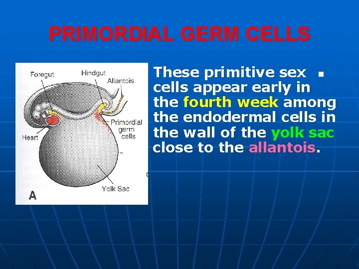
PRIMORDIAL GERM CELLS These primitive sex n cells appear early in the fourth week among the endodermal cells in the wall of the yolk sac close to the allantois.
PRIMORDIAL GERM CELLS They migrate along n the dorsal mesentery of the hind gut to the gonadal ridges. They invade the n underlying mesenchyme of the genital ridges in the six week and are incorporated in the primary sex cords.
PRIMORDIAL GERM CELLS If they fail to reach the ridges, the gonad remain indifferent or is absent. So they have an n inductive influence on development of the gonad into ovary or testis. n
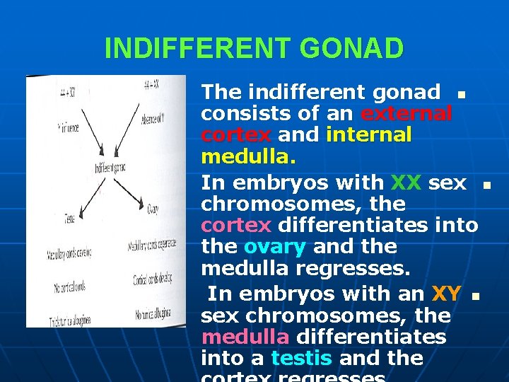
INDIFFERENT GONAD The indifferent gonad n consists of an external cortex and internal medulla. In embryos with XX sex n chromosomes, the cortex differentiates into the ovary and the medulla regresses. In embryos with an XY n sex chromosomes, the medulla differentiates into a testis and the
SEX DETERMINATION 1. The sex of the embryo is n determined genetically at the time of fertilization depending upon the sperm (X or Y ) fertilizing the X ovum. 2. Gonads acquire male or female n morphological characteristics about the seventh week of development.
SEX DETERMINATION The key to this sexual differentiation n is the Y chromosome. The short arm of this chromosome n contains SRY (sex determining region on y ) gene. The protein product of this gene is the TDF (testis determining factor ) that acts on the medulla of the indifferent gonad. n
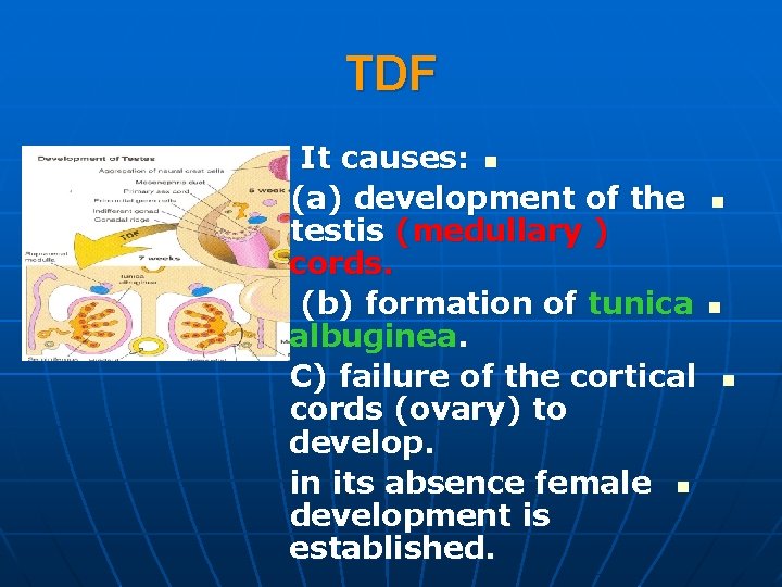
TDF It causes: n (a) development of the testis (medullary ) cords. (b) formation of tunica albuginea. C) failure of the cortical cords (ovary) to develop. in its absence female n development is established. n n n
SEX DIFFERENTIATION (3) The differentiated gonad will n determine the type of sexual differentiation in the genital ducts and external genitalia through the secretion of hormones.
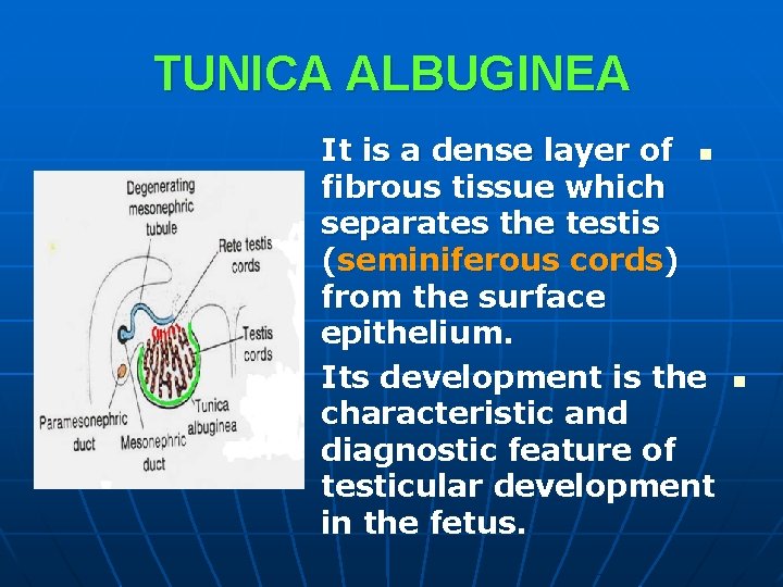
TUNICA ALBUGINEA It is a dense layer of n fibrous tissue which separates the testis (seminiferous cords) from the surface epithelium. Its development is the characteristic and diagnostic feature of testicular development in the fetus. n
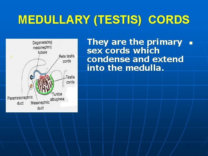
MEDULLARY (TESTIS) CORDS They are the primary sex cords which condense and extend into the medulla. n
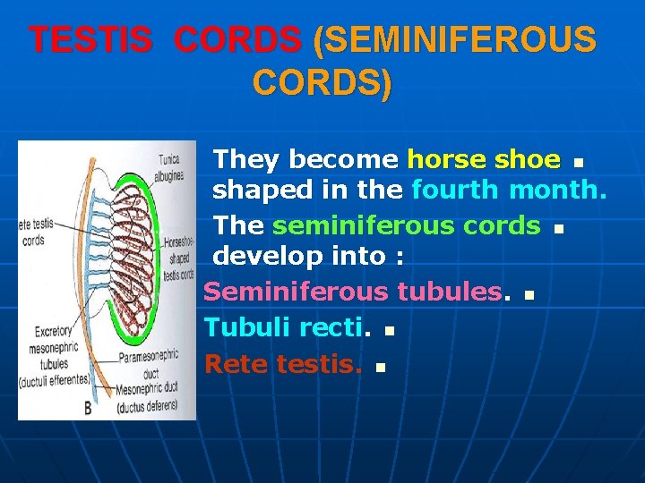
TESTIS CORDS (SEMINIFEROUS CORDS) They become horse shoe n shaped in the fourth month. The seminiferous cords n develop into : Seminiferous tubules. n Tubuli recti. n Rete testis. n
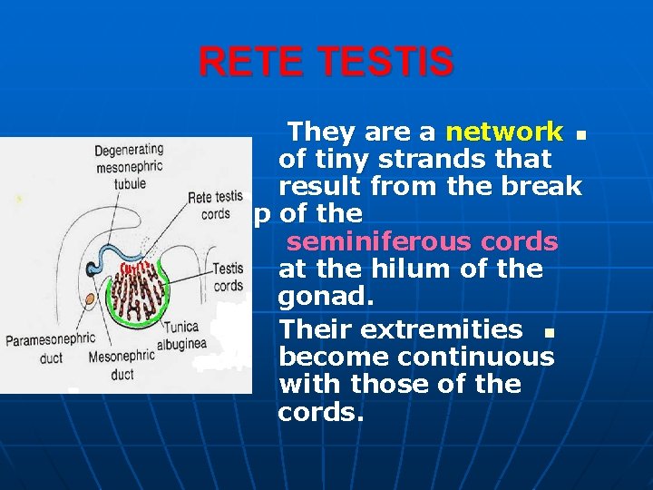
RETE TESTIS They are a network n of tiny strands that result from the break up of the seminiferous cords at the hilum of the gonad. Their extremities n become continuous with those of the cords.
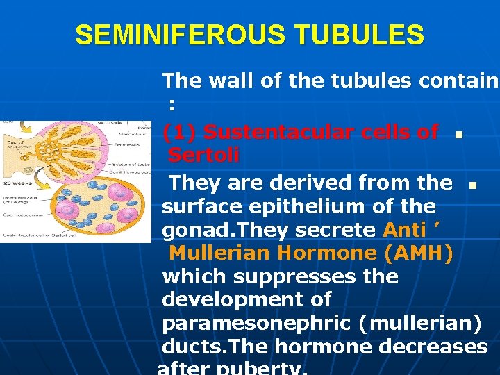
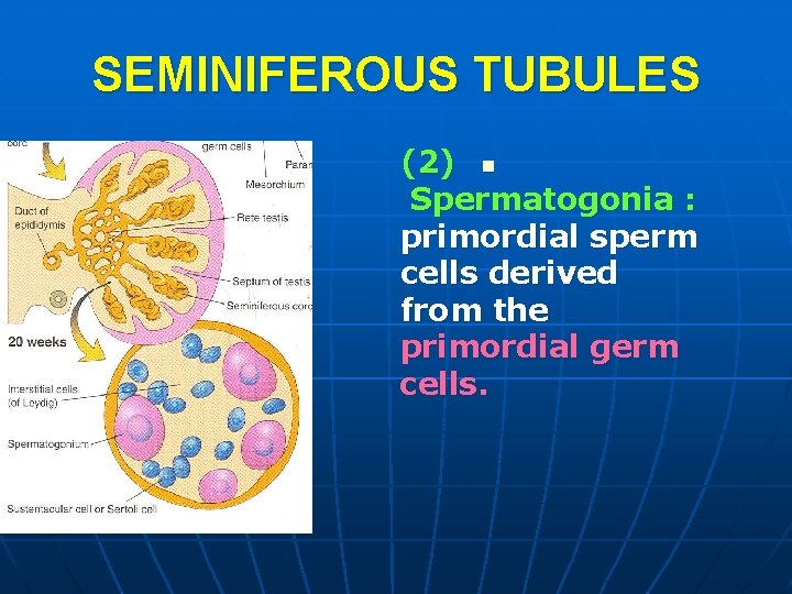
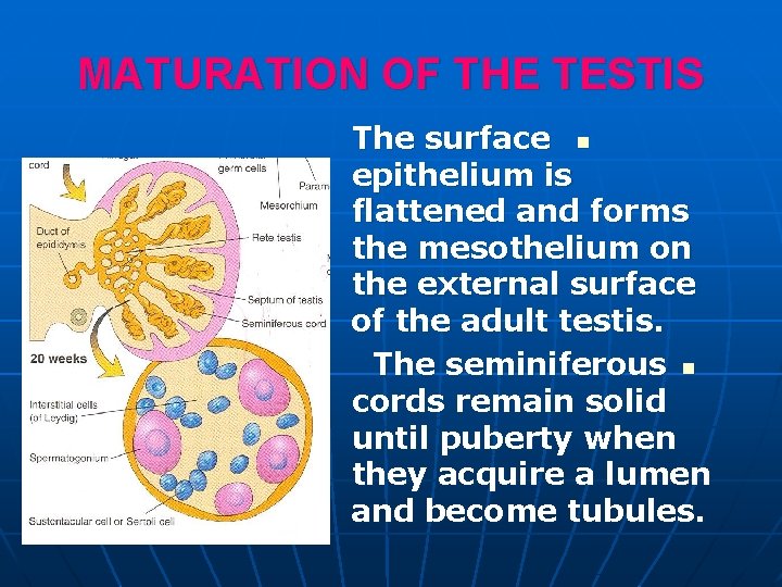
SEMINIFEROUS TUBULES The wall of the tubules contain : (1) Sustentacular cells of n Sertoli They are derived from the n surface epithelium of the gonad. They secrete Anti ’ Mullerian Hormone (AMH) which suppresses the development of paramesonephric (mullerian) ducts. The hormone decreases
SEMINIFEROUS TUBULES (2) n Spermatogonia : primordial sperm cells derived from the primordial germ cells.
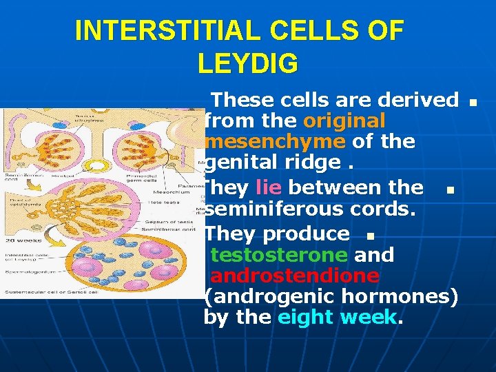
INTERSTITIAL CELLS OF LEYDIG These cells are derived from the original mesenchyme of the genital ridge. They lie between the n seminiferous cords. They produce n testosterone androstendione (androgenic hormones) by the eight week. n
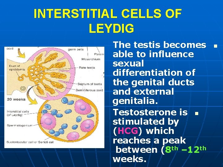
INTERSTITIAL CELLS OF LEYDIG The testis becomes able to influence sexual differentiation of the genital ducts and external genitalia. Testosterone is n stimulated by (HCG) which reaches a peak between (8 th – 12 th weeks. n
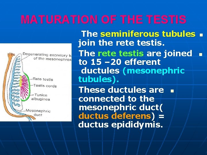
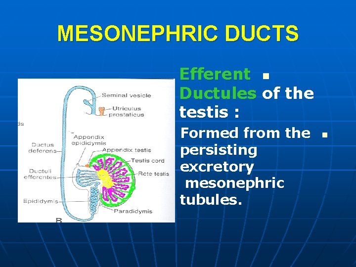
MATURATION OF THE TESTIS The seminiferous tubules join the rete testis. The rete testis are joined to 15 – 20 efferent ductules (mesonephric tubules). These ductules are n connected to the mesonephric duct( ductus deferens) = ductus epididymis. n n
MATURATION OF THE TESTIS The surface n epithelium is flattened and forms the mesothelium on the external surface of the adult testis. The seminiferous n cords remain solid until puberty when they acquire a lumen and become tubules.
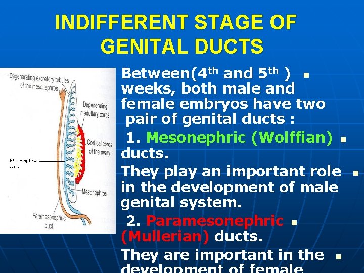
INDIFFERENT STAGE OF GENITAL DUCTS Between(4 th and 5 th ) n weeks, both male and female embryos have two pair of genital ducts : 1. Mesonephric (Wolffian) n ducts. They play an important role in the development of male genital system. 2. Paramesonephric n (Mullerian) ducts. They are important in the n n
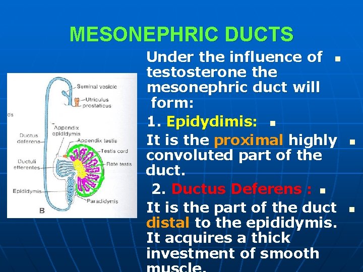
MESONEPHRIC DUCTS Under the influence of n testosterone the mesonephric duct will form: 1. Epidydimis: n It is the proximal highly convoluted part of the duct. 2. Ductus Deferens : n It is the part of the duct distal to the epididymis. It acquires a thick investment of smooth n n
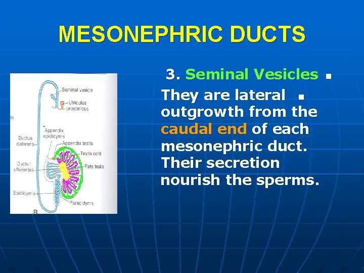
MESONEPHRIC DUCTS 3. Seminal Vesicles They are lateral n outgrowth from the caudal end of each mesonephric duct. Their secretion nourish the sperms. n
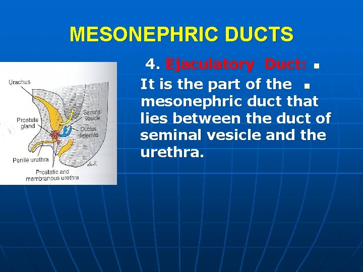
MESONEPHRIC DUCTS 4. Ejaculatory Duct: n It is the part of the n mesonephric duct that lies between the duct of seminal vesicle and the urethra.
MESONEPHRIC DUCTS Efferent n Ductules of the testis : Formed from the persisting excretory mesonephric tubules. n
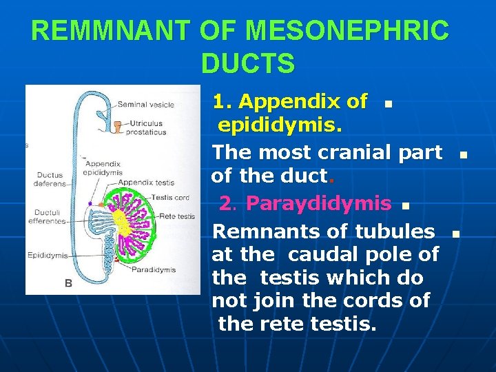
REMMNANT OF MESONEPHRIC DUCTS 1. Appendix of n epididymis. The most cranial part of the duct. 2. Paraydidymis n Remnants of tubules at the caudal pole of the testis which do not join the cords of the rete testis. n n
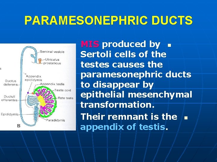
PARAMESONEPHRIC DUCTS MIS produced by n Sertoli cells of the testes causes the paramesonephric ducts to disappear by epithelial mesenchymal transformation. Their remnant is the n appendix of testis.
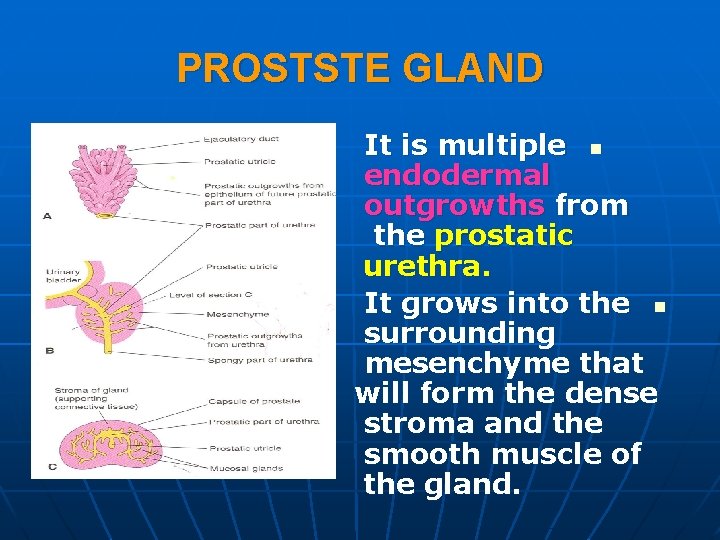
PROSTSTE GLAND It is multiple n endodermal outgrowths from the prostatic urethra. It grows into the n surrounding mesenchyme that will form the dense stroma and the smooth muscle of the gland.
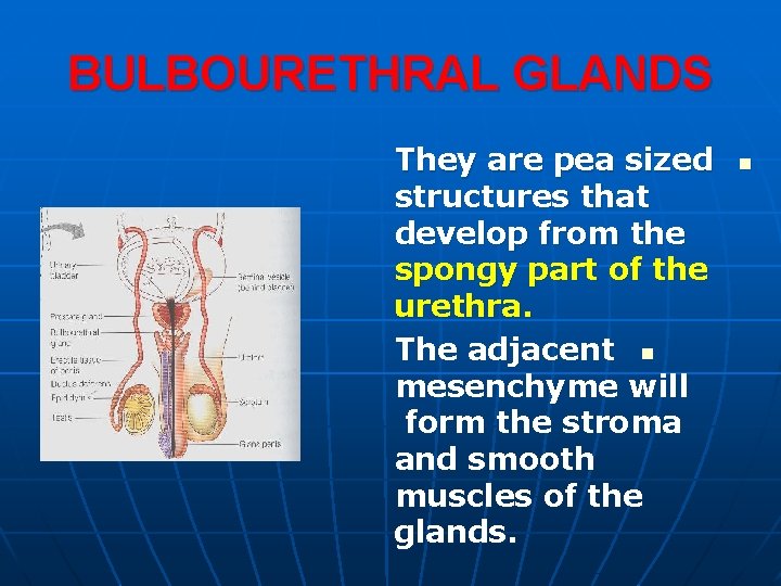
BULBOURETHRAL GLANDS They are pea sized structures that develop from the spongy part of the urethra. The adjacent n mesenchyme will form the stroma and smooth muscles of the glands. n
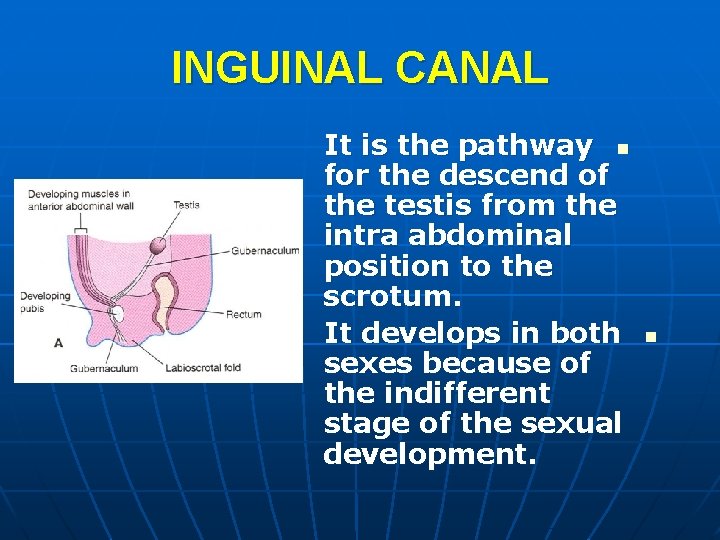
INGUINAL CANAL It is the pathway n for the descend of the testis from the intra abdominal position to the scrotum. It develops in both sexes because of the indifferent stage of the sexual development. n
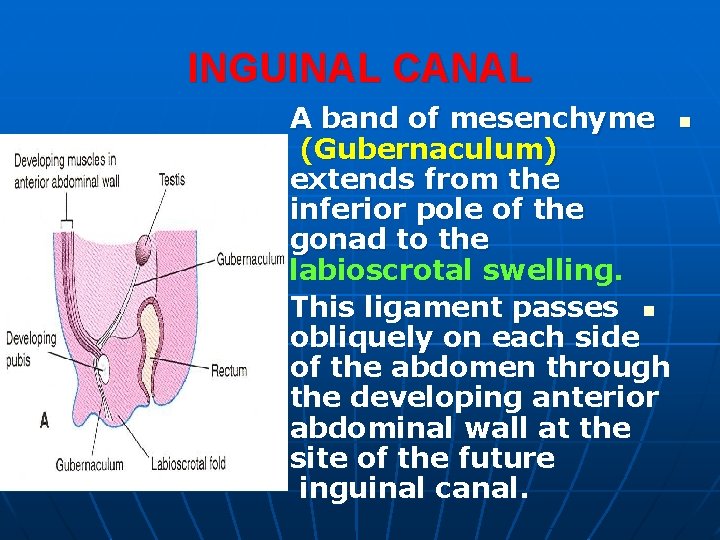
INGUINAL CANAL A band of mesenchyme n (Gubernaculum) extends from the inferior pole of the gonad to the labioscrotal swelling. This ligament passes n obliquely on each side of the abdomen through the developing anterior abdominal wall at the site of the future inguinal canal.
DESCENT OF THE TESTIS The descent of the n testes through the inguinal canals into the scrotum begins during the 26 th week. It takes (2 -3) days. n About (97%) of full n term new born boys have both testes in the scrotum.
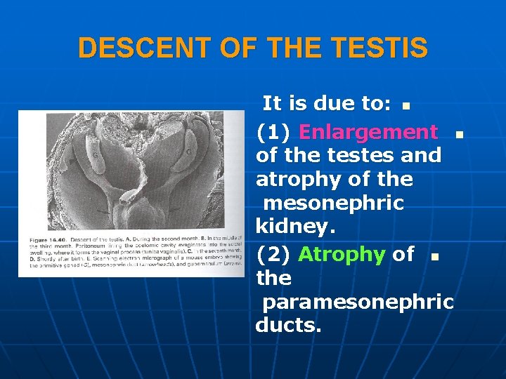
DESCENT OF THE TESTIS It is due to: n (1) Enlargement n of the testes and atrophy of the mesonephric kidney. (2) Atrophy of n the paramesonephric ducts.
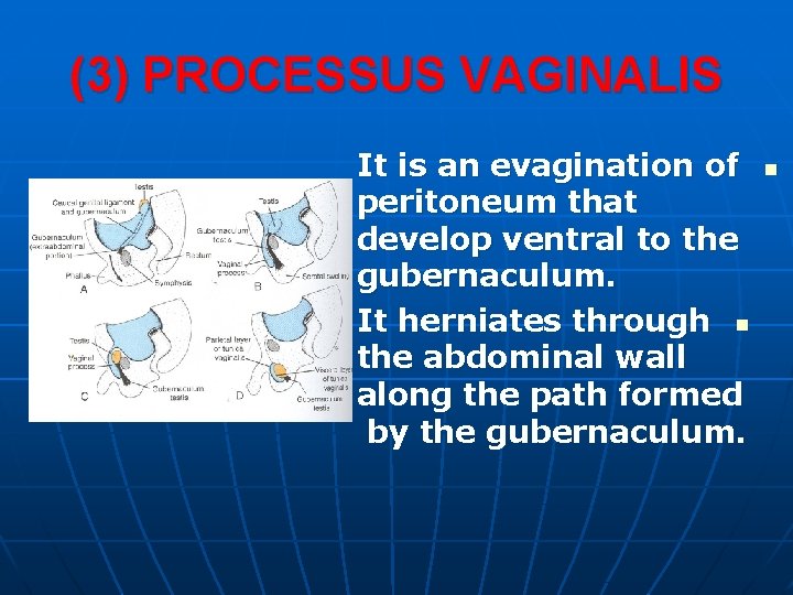
(3) PROCESSUS VAGINALIS It is an evagination of peritoneum that develop ventral to the gubernaculum. It herniates through n the abdominal wall along the path formed by the gubernaculum. n
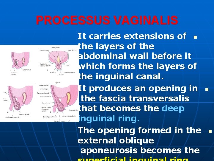
PROCESSUS VAGINALIS It carries extensions of n the layers of the abdominal wall before it which forms the layers of the inguinal canal. It produces an opening in the fascia transversalis that becomes the deep inguinal ring. The opening formed in the external oblique aponeurosis becomes the n n
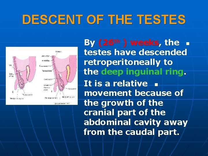
DESCENT OF THE TESTES By (26 th ) weeks, the n testes have descended retroperitoneally to the deep inguinal ring. It is a relative n movement because of the growth of the cranial part of the abdominal cavity away from the caudal part.
FACTORS CONTROLLING DESCENT OF TESTES 1. Androgens (e. g. n testosterone) produced by the fetal testes. 2. Gubernaculum : n Its role is uncertain. n It can guide the descent. The increase of the n abdominal pressure can produces passage through the inguinal n
DESCENT OF TESTES The descended testes carry their ductus deferens and vessels with them. Both the testes and n ductus deferens become ensheathed by the fascial extensions of the abdominal wall. n
TESTIS WITHIN SCROTUM Within the scrotum, the testis projects into the distal end of the processus vaginalis. During the intrauterine life the connecting stalk of the process is obliterated normally, so it isolates the tunica vaginalis. The inguinal canal n contracts around the spermatic cord. n n
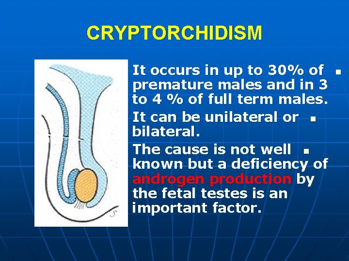
CRYPTORCHIDISM It occurs in up to 30% of premature males and in 3 to 4 % of full term males. It can be unilateral or n bilateral. The cause is not well n known but a deficiency of androgen production by the fetal testes is an important factor. n
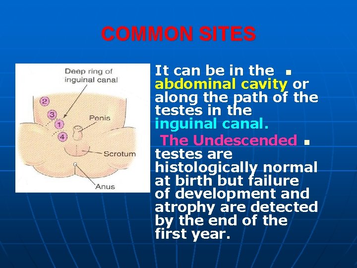
COMMON SITES It can be in the n abdominal cavity or along the path of the testes in the inguinal canal. The Undescended n testes are histologically normal at birth but failure of development and atrophy are detected by the end of the first year.
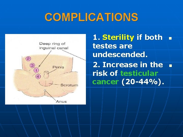
COMPLICATIONS 1. Sterility if both testes are undescended. 2. Increase in the risk of testicular cancer (20 -44%). n n