Female Reproductive System Essential Accessory Organs Gonads ovaries


 serve as duct for ovaries")


 period - small patches")
- Slides: 15
Female Reproductive System
Essential & Accessory Organs • Gonads - ovaries • Accessory Organs – Ducts - uterine tubes, uterus, vagina – Sex glands - bartholin’s and breasts – External genitals vulva
Ovaries • • Pair of them Uneven surface Weight: 3 g Attached to ligaments in pelvic cavity • Each side of uterus • 1 million ovarian follicles in baby girl
Follicle development • Each follicle contains an oocyte • By puberty 400, 000 develop into primary follicle • During lifetime, 350 - 500 will develop into mature follicles • These ovulate and release an ovum for pot’l fertilization - Graafian follicle
Primary follicle to Ovulation • Granulosa cell layer around oocyte increases • Forms hollow chamber (antrum) & secondary follicle is formed • Mature follicle (Graafian follicle) ruptures during ovulation • Remaining follicle is transformed into hormone secreting glandular structure (corpus luteum)
Oogenesis • Production of female sex cells via meiosis • Two meiotic divisions reduce chromosome number in half but cytoplasm is divided unequally b/t 4 cells • One large ovum & 3 small polar bodies
Estrogen and Progesterone • Granulosa cells around oocyte produce estrogen • Female sex characteristics • Stimulates growth of epithelial cells lining uterus • Initiates 1 st period • Secreted by Corpus luteum • Stimulated by pituitary gland to last for 11 days • Stimulates proliferation and vascularization of epithelial lining of uterus • Initiates menstrual cycle with estrogen
Reproductive Ducts • Uterine tubes (fallopian tubes or oviducts) serve as duct for ovaries • Uterus - small, but strong organ
Fallopian Tubes • Outer end has fringelike projections called fimbriae • Fimbriae produce wavelike movement to “catch” the ovum • Ovum travels toward uterus
Uterus • Size of a pear • Extremely strong • Holds baby during pregnancy • 2 parts - body & cervix • 3 layers - endometrium, myometrium, perimetrium • Functions in menstruation, pregnancy and labor
Vagina • Distensible tube about 10 cm long • Smooth muscle and lined with mucous membrane • Sperm enters on journey to reach ovum • Also called birth canal
Accessory and supportive sex glands • Bartholin’s glands secretes mucuslike fluid to genetalia • Breast - lie over pectoral muscles • 15 -20 lobes that contain milk-secreting glandular cells
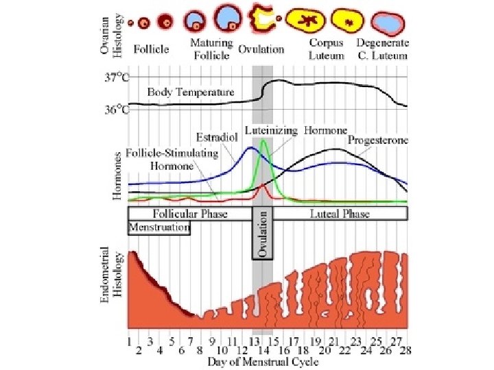
Menstrual Cycle • Days 1 - 5 - Menses (menstrual) period - small patches of dead cells of uterine lining slough off, leaving torn blood vessels • Days 6 - 13 Proliferative phase - epithelial cells reproduce, repairing uterine lining • Day 14 - Ovulation - ovum is released from ovary and moves into fallopian tube for possible fertiliaztion • Days 15 - 28 - Secretory phase - uterine lining prepares for pregnancy by growing thicker, secreting, more blood supply. On last day, blood supply goes down, causing cells to die.