The Axial Skeletal System Divisions of the Skeletal


 • Form the lateral walls and the superior portion of the")
 • Form the inferior lateral walls and a portion of the")


 • Forms the posterior wall and the floor of the cranium.")


 • Forms the anterior floor of the cranial cavity. – Also")


 • The small bone located anterior to the sphenoid bone in")


 • Form the prominence of the cheeks. – Also form part")


 • The smallest bones of the facial division. – Resemble the")
: “L” shaped bones that form the posterior portion of")


: a triangular bone that forms a portion of the posterior")


. • U shaped •")


. • Smaller bones")

. • Have facets")
. • The largest")


 What region of the vertebral column might be most affected by osteoporosis?")


- Slides: 100
The Axial Skeletal System
Divisions of the Skeletal System • Humans are born with approximately 300 bones which fuse to 206 bones as adults. • There are 2 main divisions of the skeletal system: axial skeleton and appendicular skeleton.
Divisions of the Skeletal System • Axial skeleton forms the vertical axis of the body. • 80 bones= skull (22), vertebral column (26), ribcage (25), auditory ossicles (6), and hyoid (1) • Appendicular skeleton forms the arms, legs and the girdles • Girdles attach the arms and legs to the axial skeleton • 126 bones= pectoral girdle (4), arms (60), legs (60) and pelvic girdle (2)
Skull • Superior end of the vertebral column • Composed of flat and irregular shaped bones • Large hollow space within the skull is called the cranial vault or cranial cavity. • Functions to: – Surround and protect the brain – Be points of attachment for the facial muscles (landmarks)
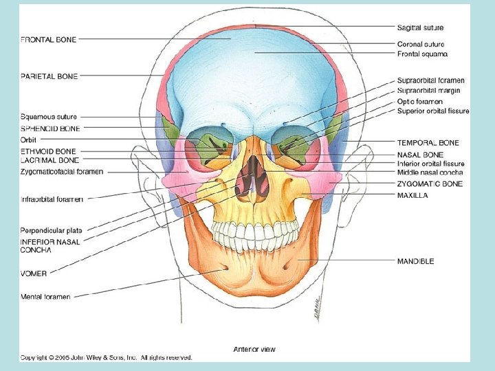
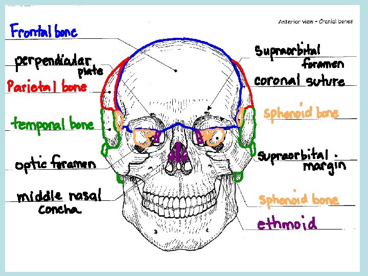
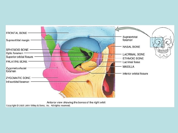
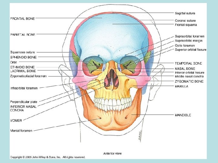
Divisions of the Skull • Cranial division consists of 8 flat bones that form a protective box around the brain. – Help to form the cranial vault (cavity) – Frontal (1): forms the anterior portion of the cranial cavity • Forms the superior orbits of the eyes and forms the forehead
Frontal Bone Landmarks • Supraorbital margin: a thickened ridge of bone found superior to the orbit of the eye. – Just deep to the eyebrow and more prominent on the lateral portion – Point for muscle attachment (PFMA) • Supraorbital foramen: a small opening found on the medial aspect of the supraorbital margin. – Can feel it best inferior to the margin – Allows blood vessels and nerves to enter the frontal bone
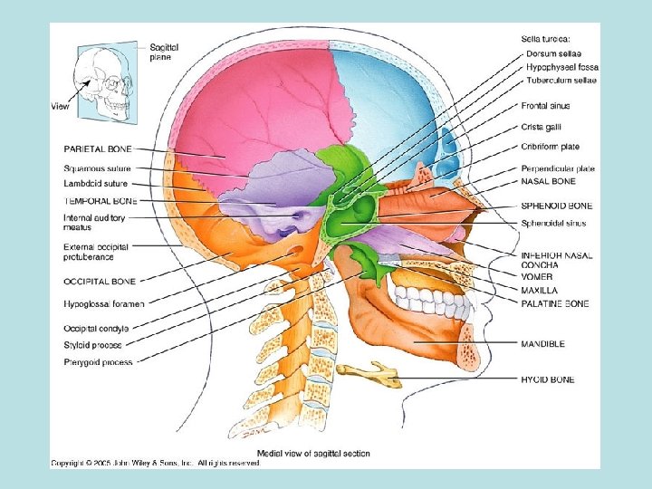
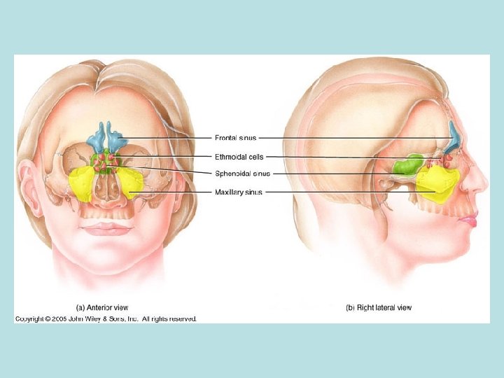
Frontal Bone Landmarks • Frontal sinus: A hollow space found within the frontal bone, superior and medial to the supraorbital margin. – Can only be seen with a sagittal cut – ¼ inch superior to the eyebrows – House mucus and macrophages for trapping and destroying foreign particles.
Frontal Bone Landmarks • Frontal sinus: A hollow space found within the frontal bone, superior and medial to the supraorbital margin. – Can only be seen with a sagittal cut – ¼ inch superior to the eyebrows – House mucus and macrophages for trapping and destroying foreign particles.
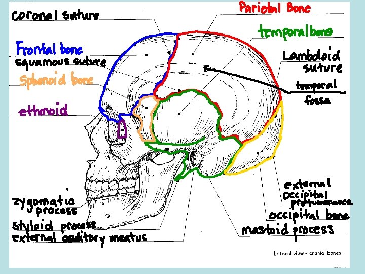
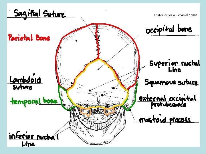
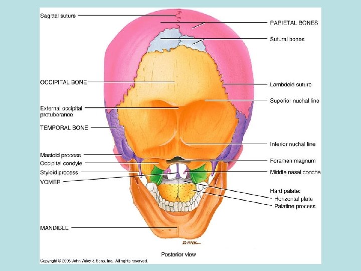
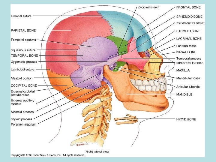
Parietal Bones (2) • Form the lateral walls and the superior portion of the cranium. • Landmarks: – Temporal fossa: A large, shallow depression that begins on the parietal bone and extends to the frontal bone. • PFMA
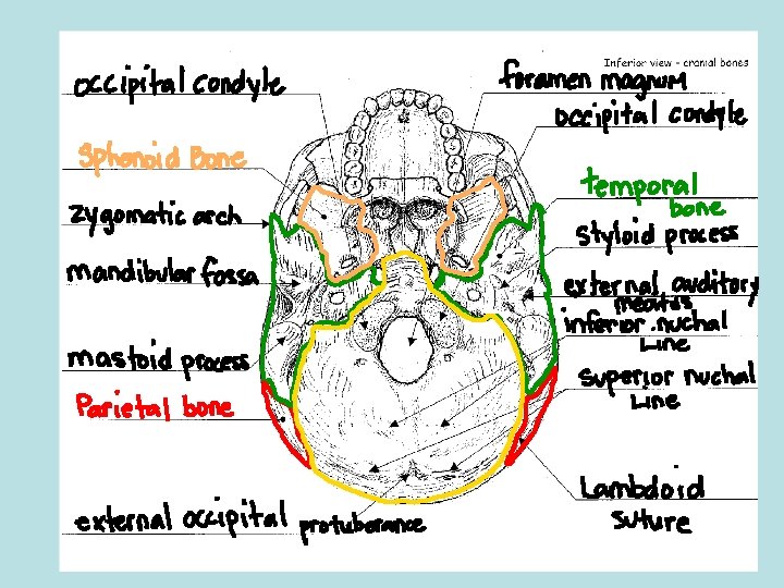
Temporal Bones (2) • Form the inferior lateral walls and a portion of the floor of the cranium. • Articulate with the mandible (lower jaw) to form the temporomandibular joint (TMJ) • Temporal bone landmarks: – Mastoid process: A large, blunt projection found posterior to the external auditory meatus. • Bump behind the ear • PFMA
Temporal Bone Landmarks cont. . – Styloid process: A thin, sharp projection found inferior and medial to the external auditory meatus. • Covered with muscle so it is more difficult to identify • PFMA – Zygomatic process: A thin, flat projection found anterior to the external auditory meatus. • PFMA – External auditory meatus: The external ear canal • Opening through which the auditory nerve runs.
Temporal Bone Landmarks cont. . – Mandibular fossa: A shallow depression found inferior and slightly anterior to the external auditory meatus • This forms an articulation with the mandible • Easy to see inferiorly if the mandible is removed
Occipital Bone (1) • Forms the posterior wall and the floor of the cranium. – The spinal cord passes through this as it exits the cranial vault. • Occipital bone landmarks: – External occipital protuberance: A prominent midline projection found on the superior surface. • Where the occipital bone turns to form the horizontal part.
Occipital Bone Landmarks cont. . – Superior nuchal line: Two curved ridges that extend laterally from the external occipital protuberance. • PFMA – Inferior nuchal line: Two curved ridges that extend laterally from the external occipital protuberance, inferior to the superior nuchal line. • PFMA – Foramen magnum: A large opening in the inferior surface of the occipital bone that allows the spinal cord to exit the cranial cavity. • Largest foramen in the body.
Occipital Bone Landmarks cont. . – Occipital condyles: Paired oval-shaped projections found lateral to the foramen magnum. • Form an articulation with the 1 st bone of the spine (atlas)
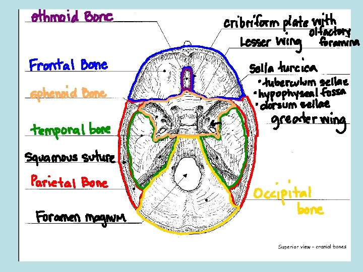
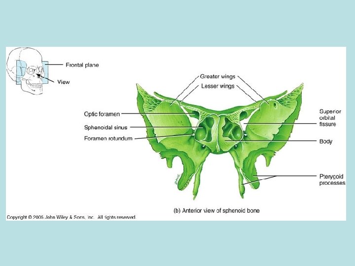
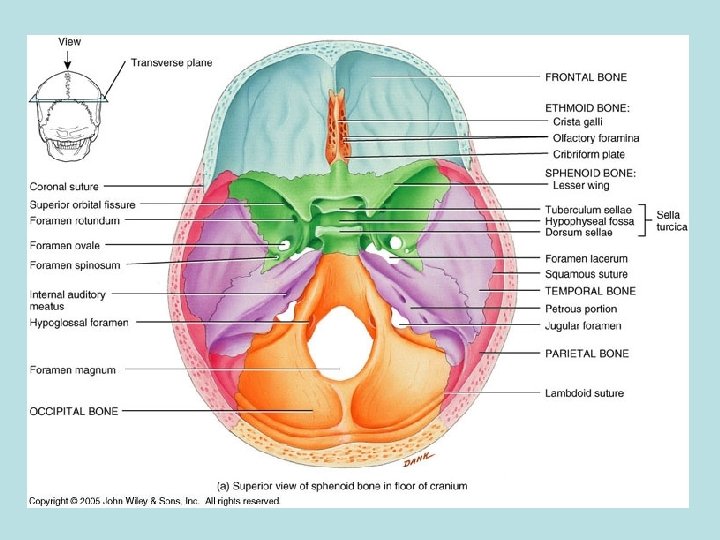
Sphenoid Bone (1) • Forms the anterior floor of the cranial cavity. – Also forms a portion of the lateral walls of the cranial cavity. – Forms the posterior wall of the orbits of the eyes. – The keystone bone for the cranium because it articulates with all other cranial bones. – Shape resembles a bat with outstretched wings when viewed superiorly.
Sphenoid Bone Landmarks • Greater wing: The larger, inferior projection of the sphenoid that forms a portion of the floor and the lateral walls of the cranium. • Also forms the posterior wall of the orbits of the eyes. • Lesser wing: The smaller, superior projection of the sphenoid bone located posterior to the frontal bone.
Sphenoid Bone Landmarks cont. . • Sella turcica: A small, saddle-like depression found between the greater and lesser wings that surrounds and protects the pituitary gland. • Pituitary is an important endocrine gland • 3 parts to the sella turcica: – Tuberculum sellae: The anterior portion of the sella turcica. • Closest to the lesser wing.
Sphenoid Bone Landmarks cont. . • 3 parts to the sella turcica continued: – Hypophyseal fossa: The seat of the saddle. • Where the pituitary gland resides – Dorsum sellae: The posterior portion of the sella turcica. • Closer to the greater wing
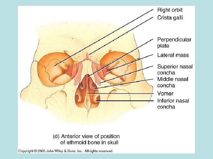
Ethmoid Bone (1) • The small bone located anterior to the sphenoid bone in the middle of the frontal bone. – Forms a small portion of the anterior floor of the cranium. – Also forms a small portion of the medial wall of the eye orbits. – Also forms the superior portion of the nasal septum.
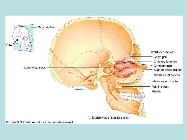
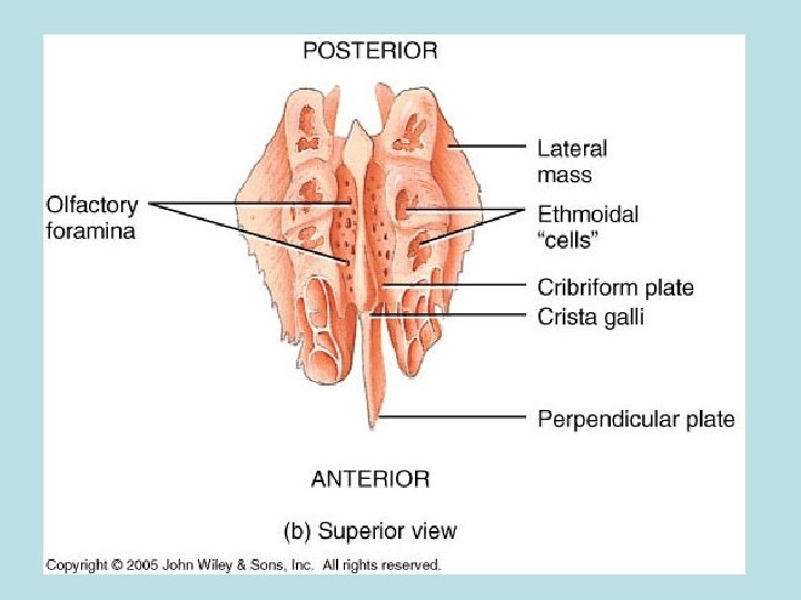
Ethmoid Bone Landmarks • Cribriform plate: Paired projections found lateral to the crista galli. – Has small openings called the olfactory foramina. • Olfactory foramina: A series of small openings found within the cribriform plate that allow nerves from the olfactory epithelium to pass directly into the brain. – These nerves give us our sense of smell.
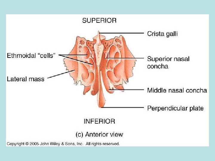
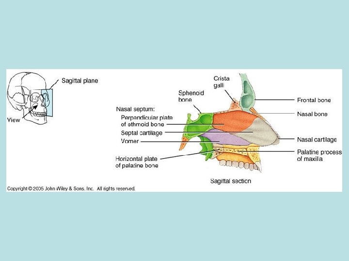
Ethmoid Bone Landmarks cont. . • Crista galli: A small triangular projection found in the center of the ethmoid bone. – Near the front of the cranial cavity. – Point of attachment for the meninges (protective coverings of the brain). • Perpendicular plate: A small vertical projection arising from the inferior surface of the ethmoid bone. – Forms the superior portion of the nasal septum. – Articulates with the vomer (facial bone).
Ethmoid Bone Landmarks cont. . • Superior and middle nasal conchae: Two thin, scroll-shaped projections found lateral to the perpendicular plate – The middle nasal conchae is inferior to the superior nasal conchae. – These increase surface area of the nasal passageways • Ethmoidal cells: Air spaces found within the lateral masses of the ethmoid bone. – Small sinuses
Sutures • Fibrous joints found between the bones of the cranium. • There are 4 major sutures: – Coronal: unites the frontal bone and both parietal bones – Sagittal: unites the two parietal bones on the superior midline of the skull – Lambdoid: unites the two parietal bones to the occipital bone. – Squamous (2): unite the parietal and temporal bones on the lateral sides of the skull
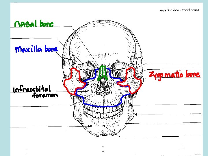
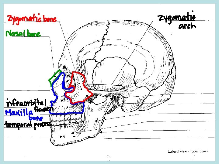
The Facial Division • A group of 14 irregular bones that serves as points of attachment for muscles of the face. • Nasal (2): form the bridge of the nose. – Rectangular shaped bones – PFMA • Maxillae (2): Form the upper jaw. – Articulate with every face bone except the lower jaw. – Form part of the floors of the orbits, lateral walls and floor of the nasal cavity, and most of the hard palate (bony roof of the mouth).
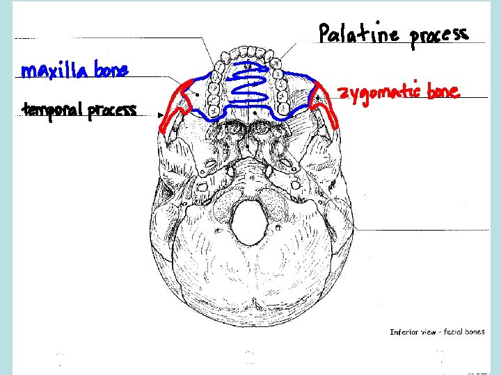
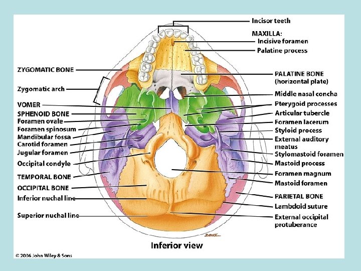
Maxillary Landmarks • Infraorbital foramen: Small openings found inferior to the orbits of the eyes. – Allows passage of blood vessels and nerves. • Palatine process: a lateral projection that forms one half of the anterior portion of the hard palate. – Typically the 2 processes unite during weeks 10 -12 of embryo development. If not, cleft palate will result. This negatively impacts speech and swallowing.
Maxillary Landmarks cont… • Maxillary sinuses: a series of small spaces within the maxillae. – Empty into the nasal cavity.
Zygomatic Bone (2) • Form the prominence of the cheeks. – Also form part of the lateral wall and floor of each orbit. – Articulate with the frontal, maxilla, sphenoid and temporal bones. Zygomatic bone landmarks: • Temporal process: a thin, flat projection arising from the lateral, posterior surface of the zygomatic bone. • Articulates with the zygomatic process of the temporal bone.
Zygomatic Bone Landmarks cont. . • Zygomatic arch: created by the articulation of the temporal process of the zygomatic bone and the zygomatic process of the temporal bone.
Lacrimal Bones (2) • The smallest bones of the facial division. – Resemble the shape and size of a fingernail – Posterior and lateral to the nasal bones and form part of the medial wall of each orbit. Lacrimal bone landmark: • Lacrimal fossa: a small vertical groove formed with the maxilla, that helps drain fluid away from the eye. • Houses a lacrimal sac that gathers tears and passes them into the nasal cavity.
• Palatine Bones (2): “L” shaped bones that form the posterior portion of the hard palate. – The parts that make-up the hard palate are called horizontal plates. • Inferior Nasal Conchae (2): scroll shaped bones that form a portion of the inferior, lateral walls of the nasal cavity. – Increase surface area and help filter air along with the superior and middle nasal conchae of the ethmoid bone.
• Vomer (1): a triangular bone that forms a portion of the posterior floor of the nasal cavity. – Articulates with the perpendicular plate of the ethmoid bone to form the inferior portion of the bony nasal septum. • Mandible (1): the largest bone of the facial division. – Except for the ossicles, it is the only moveable skull bone.
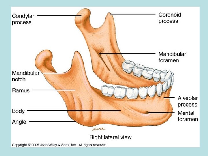
Mandibular Landmarks • Mandibular body: a triangular bone that forms a portion of the posterior floor of the nasal cavity. • Ramus: the short, vertical portion of the mandible. • Angle: the area where the ramus and the body of the mandible meet. • Coronoid process: a small triangular projection found on the superior anterior portion of the ramus.
Mandibular Landmarks cont… • Condylar process: a small rounded projection found on the superior posterior portion of the ramus. • Articulates with the mandibular fossa to create the temporomandibular joint (TMJ). • Mental foramen: small openings found in the anterior surface of the body of the mandible that allow blood vessels and nerves to enter the mandible. • Alveoli: sockets for teeth
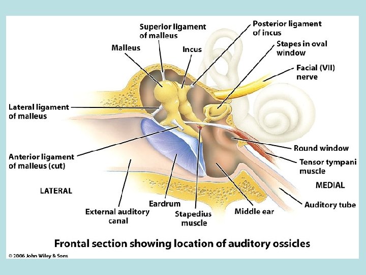
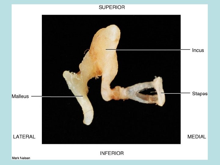
• • Auditory Ossicles The 6 smallest bones in the human body. Located medial to the eardrum. Connected by synovial joints. Function to transfer sound waves from the eardrum to the inner ear. • The bones are as follows: • Malleus- attaches to the eardrum and is commonly called the “hammer”. • Incus- middle bone that is commonly called the “anvil”. • Stapes- Smallest bone and is commonly called the “stirrup”.
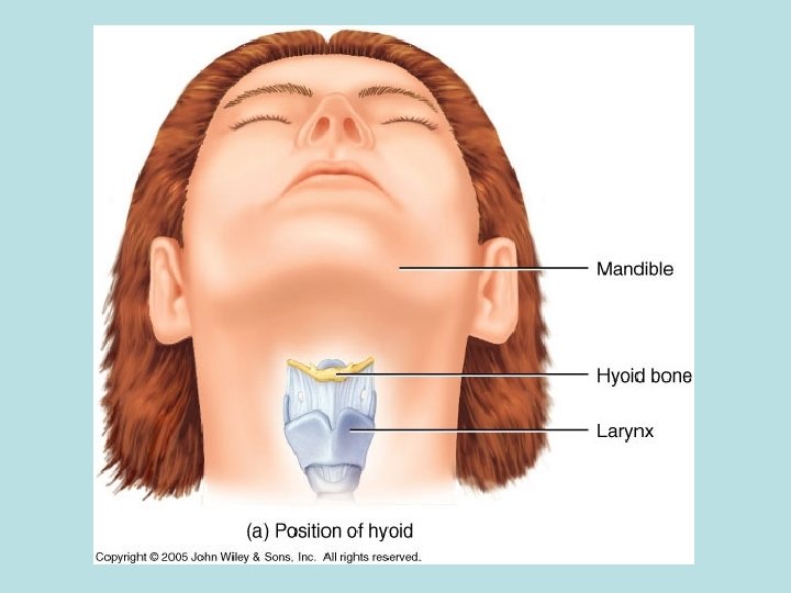
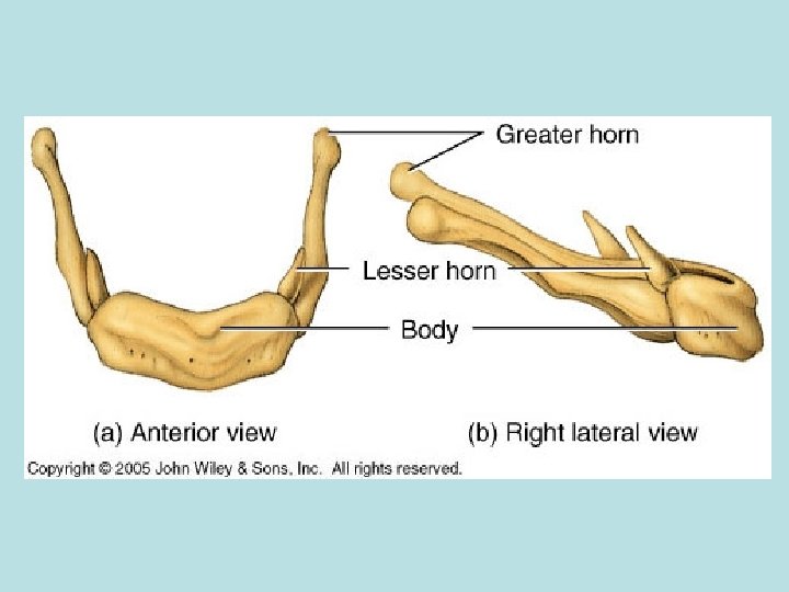
Hyoid Bone • Located superior to the larynx (voice box). • U shaped • The only bone that does not articulate with another bone. • Suspends from the styloid processes by ligaments and muscles. • Often fractured during strangulation. • Functions to support the tongue.
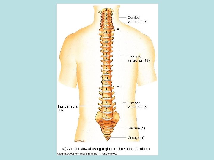
The Vertebral Column • Also called the spine, backbone or spinal column. • Consists of 33 (children) or 26 (adults) bones called vertebrae. • Functions to protect the spinal cord, support the head, and serve as attachment points for the ribs, pelvis, back muscles and arm muscles.
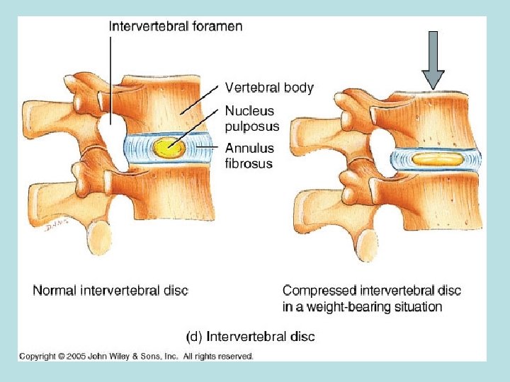
Vertebrae • Vary in size, shape and detail but have many similarities. • Consist of 3 main parts: vertebral body, vertebral arch and several processes. • Vertebral body: the thickened anterior portion of a vertebra. • Holds the intervertebral disc and contains foramina for the entrance of blood vessels. • Intervertebral discs: pads of fibrocartilage that help hold the vertebrae in place. • Compress throughout the day due to weight and water loss This compression does not change height as we age.
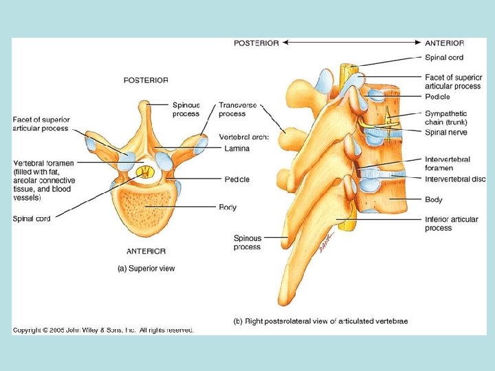
Vertebrae continued… • Vertebral arch: located posterior to the vertebral body. • Forms the vertebral foramen with the vertebral body. • The vertebral arch consists of the pedicles and the laminae. • Pedicles: the shorter anterior portions of the vertebral arch. • Laminae: the longer posterior portions of the vertebral arch.
Vertebrae continued… • Vertebral foramen: the opening formed by the vertebral body and the vertebral arch. – Contains the spinal cord, adipose tissue, areolar connective tissue and blood vessels. • Processes: bony projections that arise from the vertebral arch. – Transverse processes: paired lateral projections that arise from the vertebral arch. – Spinous process: the single posterior projection that arises from the vertebral arch. – Both of these are PFMA
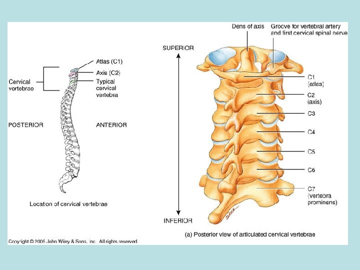
Cervical Vertebrae • The first 7 vertebrae (C 1 -C 7). • Smaller bones than other vertebrae but larger vertebral foramen. • Also have 2 transverse foramen through which the vertebral artery, vein and nerve fibers pass. • C 2 -C 6 have a branching spinous process. • C 1 is called the atlas. – Supports the skull. – It lacks a body and a spinous process.
Cervical Vertebrae cont… • C 2 is called the axis. – Has a body and a peglike process called the dens or the odontoid process. – The dens makes a pivot on which the atlas and head rotate. • C 7 is called the vertebra prominens. – Has a large spinous process that is not branched and can be felt at the back of the neck.
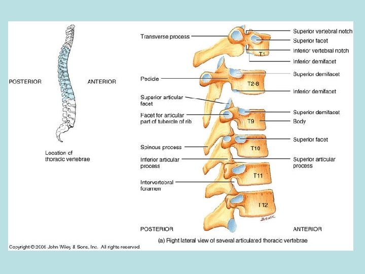
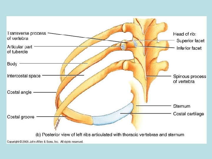
Thoracic Vertebrae • The next 12 vertebrae (T 1 -T 12). • Have facets (flat surfaces) where they articulate with the 12 rib pairs. • Movement of these vertebrae are most limited because the ribs attach to the sternum anteriorly.
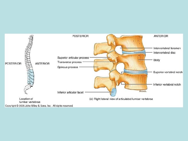
Lumbar Vertebrae • The next 5 vertebrae (L 1 -L 5). • The largest and strongest of the bones of the spine. • Spinous processes are thick and broad.
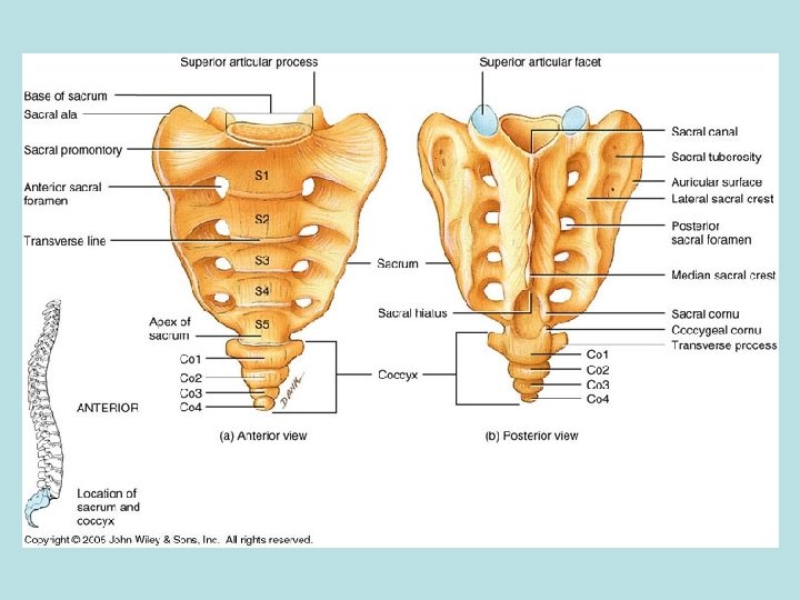
Sacrum • The next vertebra in the adult vertebral column. • A triangular shaped bone formed from the fusion of 5 bones. This starts around age 16 and is usually completed by age 30. • Auricular surfaces: large, ear-shaped roughened surfaces found on the lateral aspect of the sacrum. – This articulates with the hip bones of the pelvis.
Coccyx • The last vertebra in the adult vertebral column. • The tailbone that is formed by the fusion of 4 vertebrae that occurs between the age of 20 -30. • In males, it points anteriorly and in females it points inferiorly.
ACT-UP
ACT-UP 1) What region of the vertebral column might be most affected by osteoporosis? 2) Why? 3) What would we expect to see (on the outside) when looking at a person suffering from osteoporosis?
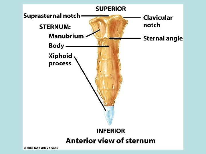
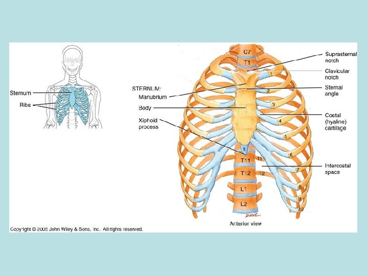
Thorax • Includes the sternum, ribs and the bodies of the thoracic vertebrae. • Sternum: located along the anterior midline of the thorax. – Also known as the breastbone – Consists of 3 parts that fuse by age 25 and the points of fusion can be seen as transverse ridges. – If thoracic surgery is necessary, the sternum may be cut along the midline.
Regions of the Sternum • Manubrium: the superior portion of the sternum – Articulates with the clavicles (collarbones) and the costal cartilages of the 1 st-2 nd rib pairs. • Sternal body: the intermediate portion of the sternum. – Articulates directly or indirectly with the costal cartilages of the 2 nd-10 th rib pairs. • Xiphoid process: the inferior portion of the sternum. – Where some abdominal muscles attach.
Ribs • 12 pairs of flat bones that form a protective cage around the heart and the lungs. – Increase in length from 1 -7 and then decrease from 7 -12. – Each rib pair articulates posteriorly with its corresponding thoracic vertebra. • Costal cartilage: elongated pads of hyaline cartilage used to attach the ribs to the sternum. – Allows the ribcage to be more elastic and limits fracturing from blows to the chest.
Types of Ribs • True ribs: Rib pairs 1 -7 – Their costal cartilages attach directly to the sternum. • False ribs: Rib pairs 8 -12 – Their costal cartilages do not attach directly to the sternum. Rib pairs 8 -10 have cartilages that attach to the cartilage of 7 (which attaches to the sternum. • Floating ribs: Rib pairs 11 -12 – The costal cartilages do not attach to the sternum at all.