The abdominal regions Nine regions R hypochondriac region

The abdominal regions 腹部分区 ——Nine regions R. hypochondriac region 右季肋区 Epigastric region 腹上区 R. lateral regions 左季肋区 右外区 R. inguinal region 右腹股沟区 Pubic region 耻区 L. hypochondriac region L. lateral regions 左外侧区 Umbilical region 脐区 L. inguinal region 左腹股沟区

The abdominal regions 腹部分区 ——Four quadrants n n Left and right upper quadrants Left and right lower quadrants

Composition Alimentary canal消化管 n n Mouth 口腔 Pharynx 咽 Esophagus 食管 Stomach 胃 Duodenum 十二指肠 n Small intestine小肠 Jejunum 空肠 Ileum 回肠 n Large intestine 大肠 Superior alimentary canal 上消化道 Inferior alimentary canal 下消化道 Alimentary glands 消化腺 n n n Major salivary glands 大唾液腺 Liver 肝 Pancreas 胰 Function: ingestion, digestion, absorption, egesting

Major salivary glands Mouth Pharynx Esophagus Liver Duodenum Ileum Stomach Pancreas Large intestine Jejunum

Alimentary canal 消化管

The Oral Cavity 口腔

The Oral Cavity 口腔 n Consists of two parts q Oral vestibule 口腔前庭: between cheeks and lip and teeth q Oral cavity proper 固有口腔: within arch of teeth n Boundaries q q n Anterior and lateral: gum and teeth Posterior: isthmus of fauces 咽峡 Roof: palate Floor: tongue, muscles and mucous membrane Oral vestibule leads, by the space behind the molar teeth, into the oral cavity proper

Palate 腭 n Two parts q Hard palate 硬腭: anterior 2/3, formed by the maxilla and palatine bone Soft palate 软腭: posterior 1/3 n n n Velum palatinum 腭帆 Uvula 腭垂 Palatoglossal arch 腭舌弓 Palatopharyngeal arch 腭咽弓 Isthmus of fauces 咽峡 formed by uvula, free border of velum palatinum, both side of palatoglossal arches, and root of tongue.

Teeth 牙

Teeth 牙 General features n n Two sets: q Deciduous 乳牙 q Permanent 恒牙 Classification: q Incisors 切牙 q Canine 尖牙 q Premolars 前磨牙 q Molars 磨牙

Deciduous teeth 乳牙 n n 20 in number, ten teeth in each mandibular and maxillary arch Deciduous central incisor 乳中切牙, deciduous lateral incisor 乳侧切牙, deciduous canine 乳尖牙, first deciduous molar 第一乳磨牙,second deciduous molar 第二乳磨牙 in each quadrant Upper jaw Lower jaw n n Ⅰ in. Ⅱ Ⅲ Ⅳ Ⅴ total 20 in. can. mol. Eruption: stars at about 6 mouth of age and continues to beginning of 3 rd year Shedding: occurs between 6 th and 12 th years with replacement by permanent teeth Ⅱ Ⅳ

Deciduous teeth 乳牙 Deciduous central incisor 乳中切牙 Deciduous lateral incisor 乳侧切牙 Deciduous canine 乳尖牙 First deciduous molar 第一乳磨牙 Second deciduous molar 第二乳磨牙
恒牙 n n 32 in number,sixteen in each mandibular and maxillary arch")
Permanent teeth (adult)恒牙 n n 32 in number,sixteen in each mandibular and maxillary arch Two incisors, one canine, two premolars, and three molars in each quadrant Upper jaw 1 2 3 4 5 6 7 8 total 32 Lower jaw n n First permanent molar- appears at about 6 years Third molars (wisdom teeth)-many erupt at any time after 12 years of age or not at all (impaction). 3 7

Permanent teeth 恒牙 3 rd molar 第三磨牙 2 nd molar 第二磨牙 1 st molar 第一磨牙 2 nd premolar 第二前磨牙 1 st premolar 第一前磨牙 Canine 尖牙 Lateral incisor 侧切牙 Central incisor 中切牙

Teeth 牙 Part and structure of the teeth n Each tooth consists of 3 parts: q q q n Crown 牙冠 Neck 牙颈 Root 牙根 Dental cavity 牙腔 q q Pulp chamber 牙冠腔 Root canal 牙根管 transmits the nerves and vessels to and from the dental cavity through the apical foramen 根尖孔

Teeth 牙 n Calcified tissues 牙组织 q q Dentine 牙质- is a yellowish white tissue, that forms the bulk of tooth. Enamel 釉质-is a hard, brittle white tissue that covers the crown of the tooth Cement 牙骨质-is an unusual form of bone that covers the root of the tooth Dental pulp 牙髓 formed connective tissue, blood vessels and nerves.

Teeth 牙 n Periodontal tissue 牙周组织 q Periodontal membrane 牙周膜 q Alveolar bone 牙槽骨 q Gum 牙龈

Teeth 牙

Tongue 舌 Two parts: divided two parts by v-shaped terminal sulcus 界沟 n Body of tongue 舌体-anterior 2/3 q q n Apex of tongue 舌尖-free rounded tip At the apex of terminal sulcus is a small median pit, the foramen cecum of tongue 舌盲孔 Root of tongue 舌根- posterior 1/3

Tongue 舌 Lingual mucous membrane n n Papillae of tongue 舌乳头 q Filiform papillae q Fungiform papillae 菌状乳头 q Foliate papillae 叶状乳头 q Vallate papillae 轮廓乳头 丝状乳头 Lingual tonsil 舌扁桃体 -masses of submucosal lymphoid tissue on the root of tongue contain taste buds 味蕾

Inferior surface of tongue Frenulum of tongue 舌系带 -a midline fold of mucous membrane connecting tongue to floor of mouth Sublingual caruncle 舌下阜 -small elevation Sublingual fold 舌下襞

Muscles of tongue 舌肌 Intrinsic muscles of tongue n n Involved in changing shape of tongue Include longitudinal, transverse and vertical muscles of tongue

Tongue 舌 n Extrinsic muscles of tongue q q q n Genioglossus 颏舌肌 n Arises from mental spine of mandible and inserts into either side of midline of tongue n Action: acting together draw tongue forward and downward (depresses and protrudes tongue ); acting along making apex of tongue to opposite side Hyoglossus 舌骨舌肌 Tyloglossus 茎突舌肌 Involved in determining shape and position of tongue

Major salivary glands 大唾液腺 Parotid gland 腮腺 n n n Superficial part: triangular in shape, lies below and in front of the external acoustic meatus, and partially covers the masseter. Deep part: lies deep to medial pterygoid. Parotid duct: arises front anterior border of gland, runs over the masseter a finger’s breadth below the zygomatic arch to pierce the buccinator and opens into the mouth cavity, opposite the upper second molar tooth

Major salivary glands 大唾液腺 Submandibular gland 下颌下腺 n Position: lies in submandibular triangle, between anterior and posterior bellies of digastric n Duct opens on to sublingual caruncle

Major salivary glands 大唾液腺 Sublingual gland 舌下腺 n Position: situated beneath the mucous membrane of the floor of mouth n Ducts q q Major sublingual duct-opens onto the sublingual caruncle Minor sublingual ducts-open onto the sublingual fold

The Pharynx 咽 General features n n A –fibromuscular tube, part of digestive and respiratory systems Extends from base of skull to the inferior border of cricoid cartilage (lower border of C 6 level) Three segments n n n Nasopharynx 鼻咽 Oropharynx 口咽 Laryngopharynx 喉咽

The Pharynx 咽 Nasopharynx 鼻咽—posterior to nasal cavities n Extends from the base of skull to level of soft palate, below n Features q Pharyngeal opening of auditory tube 咽鼓管咽口 q Tubal torus 咽鼓管圆枕 q Pharyngeal recess 咽隐窝 q Tubal tonsil 咽鼓管扁桃体 q Pharyngeal tonsil 咽扁桃体

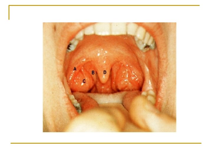
The Pharynx 咽 Oropharynx 口咽-posterior to oral cavity n n Lies below soft palate, extends to upper border of epiglottis Features q Median glossoepiglottic fold 舌会厌正中襞 q q Epiglottic vallecula 会厌谷 Palatine tonsil 腭扁桃体 -lies within tonsillar fossa

The Pharynx 咽 Laryngopharynx 喉咽-posterior n n to larynx Extends from upper border of epiglottis to the level of lower border of C 6 Piriform recess 梨状隐窝 -a deep depression on each side of aperture of larynx, common side for lodgement of foreign bodies (for example, fish bones)

The Pharynx 咽 Lymphatic ring n n Consists of q Pharyngeal tonsil q tubal tonsil q Palatine tonsil q lingual tonsil Forming a circular band of lymphoid tissue at oropharyngeal isthmus

The Esophagus 食管 n General features - a muscular tuber about 25 cm long, connecting the pharynx at level of C 6 vertebra, passes through the diaphragm at level of T 10 vertebra and after 1~2 cm enters the stomach n Division q q q Cervical part Thoracic part Abdominal part

The Esophagus 食管 ★Three constrictions 1. Where it beginning, 15 cm from incisors, lies at level of C 6, is the narrowest part of the esophagus 2. Where it is crossed by left principal bronchus, 25 cm from incisors, lies at level of intervertebral disc between T 4 and T 5. 3. Where it passes through the esophageal hiatus of diaphragm, 40 cm from incisors, at level of T 10

The Stomach 胃 Shape n n n Two surface: anterior and posterior Two curvatures q Lesser curvature 胃小弯 : short, concave and directed to the right and upward, near its lower part is angular incisure 角切迹 q Greater curvature 胃大弯: long, convex and directed to the left and downward, at the junction of left margin of esophagus and greater curvature is cardiac incisure 贲门切迹 Two openings q Cardia 贲门 q Pylorus 幽门

The Stomach 胃 n Four parts q q Cardiac part 贲门部 Fundus of stomach 胃底 Body of stomach 胃体 Pyloric part 幽门部 n n Pyloric antrum 幽门窦 Pyloric canal 幽门管

Fundus of stomach 胃底 Cardiac part 贲门部 Body of stomach 胃体 Pyloric canal 幽门管 Pyloric antrum 幽门窦 Pyloric part 幽门部

The Stomach 胃 Structure of stomach wall n n n -consists of four usual layers Mucous membrane Submucous ( loose areolar tissue connecting the mucous and muscular layer) Muscular layer contains: q The most superficial longitudinal fibers q Inner circular fibres n n q n Sphincter of pylorus Pyloric valve 幽门瓣 幽门括约肌 Innermost oblique fibres Serous (visceral peritoneum)

The Stomach 胃 Location n Mainly parts is situated in the left hypochondriac region Small in the epigastric region The cardia is situated to the left of T 11, the pylorus lies to the right of L 1

Variations in position of stomach

The Small Intestine小肠 Duodenum n About 5 -7 m long n Division: q q q Duodenum 十二指肠 Jejunum 空肠 Ileum 回肠 Jejunum Ileum

Duodenum 十二指肠 Four parts n Superior part 上部 q Duodenal cap q Superior duodednal flexure 十二指肠球 十二指肠上曲 n Descending part 降部 q Inferior duodenal flexure 十二指肠下曲 n Horizontal part 水平部 n Ascending part 升部 q Duodenojejunal flexure 十二指肠空肠曲

Duodenum 十二指肠 Descending part 降部 n Longitudinal fold of duodenum 十二指肠纵襞 n Major duodenal papilla 十二指肠大乳头 the common opening of the common bile duct and pancreatic duct, 75 cm from incisors n Minor duodenal papilla 十二指肠小乳头

n Contrast radiographic appearance of the duodenum showing a distended duodenal cap and the remainder of the duodenum up to the duodenojejunal flexure
, a surgical landmark,")
Duodenum 十二指肠 n Suspensory muscle of duodenum 十二指肠悬肌 (ligament of Treitz), a surgical landmark, descends from the right crus of diaphragm to duodenal termination.

Jejunum and ileum Characteristic Jejunum Ileum Position Upper 2/5, upper left Lower 3/5, lower right part of abdominal cavity Diameter Greater Less Wall Thicker Thin Circular folds Larger, numerous and large villi Fewer,smaller and less abundant villi Vascularity Greater Less Vasa recta Long Short Color Deeper red Paler pink Lymphatic follicles Solitary Aggregated Fat in mesentery Less More

Jejunum and ileum
. The")
n n Barium studies of the jejunum and ileum. Small bowel enema (enteroclysis). The plicae circulares are clearly demonstrated by this technique.

Jejunum and ileum Meckel’s diverticulum n n Persistence of proximal portion of yolk sac (vitelline duct, omphalomesenteric duct) Common malformation of digestive tract (2-4%) - more prevalent in males About 2-5 cm long and located 30-100 cm from ileocecal valve Usually asymptomatic but: q q May become inflamed (mimicking appendcitis) or bleed May be attached to umbilicus by a fibrous cord (distal end of yolk stalk) and cause intestinal obstruction by compressing adjacent intestinal loops

Large Intestine 大肠 n n Approximately 1. 5 m long, Five parts: q q q Cecum 盲肠 Vermiform appendix 阑尾 Colon 结肠 Rectum 直肠 Canal 肛管

Large Intestine 大肠 Features n n n Colic bands 结肠带 Haustra of colon 结肠袋 Epiploic appendices 肠脂垂

Cecum 盲肠 n n Position: lies in right iliac fossa Shape: q q Blind sac, first part of large intestine, with largest diameter and thinnest wall The ileum enters the cecum obliquely, and partially invaginates into it, forming the ileocecal valve 回盲瓣 n Consists of two folds n Probably delays flow of ileal contents into large intestine

Vermiform appendix 阑尾 n n n Blind worm-like tube, 6-8 cm long, about 0. 5 cm in diameter Opens into posteromedial aspect of cecum,about 2 cm below ileoceal orifice The base of the appendix lies at the point of convergence of three colic bands (used as a guide to find the appendix during operation)

Vermiform appendix 阑尾 n Surface marking of the base is at the so-called Mc. Burney’s point which is at junction of lateral and middle thirds of line joining right anterior superior iliac spine and umbilicus

Vermiform appendix 阑尾 n n n Tip variable in position Preileal -7% Pelvic-41% Retrocecal -29% Retroileal-4% Subcecal-17%

Vermiform appendix 阑尾 Mesoappendix 阑尾系膜 n n Triangular mesentery- extends from terminal part of ileum to appendix Appendicular a. runs in free margin of the meseoappendix then along wall of appendix

Double contrast barium enema appearance of the caecum and ileocaecal valve.

Colon 结肠 n Ascending colon 升结肠 q n Transverse colon 横结肠 q n right colic flexure 结肠右曲 left colic flexure 结肠左曲 Descending colon 降结肠 descends almost vertically from left colic flexure to sigmoid colon at left iliac crest. n Sigmoid colon乙状结肠 -extends from descending colon to rectum at level of S 3.

Appearance of the abdominal colon on double contrast barium enema examination demonstrating the transverse colon, hepatic and splenic flexures.

n n Barium studies of the jejunum and ileum. Barium follow-through. The feathery appearance of the small intestine is due to the plicae circulares, this is most prominent in the jejunum. The constrictions are the result of peristalsis.

Rectum 直肠 n Position: within pelvic cavity, extends from S 3 to pelvic diaphragm. n Curves q Sagittal plane n n q Coronal plane n n n Sacral flexure 直肠骶曲: convex backward Perineal flexure 直肠会阴曲: convex forward. Upper and lower part-convex to the right. Middle part-convex to the left. Lower part of rectum dilated, to from ampulla of rectum 直肠壶腹 q Three transverse folds of rectum 直肠横襞

Sagittal T 2 -weighted MRI of the rectum in the male. Sagittal T 2 -weighted MRI of the rectum in the female.

Anal canal 肛管 n n n Anal columns 肛柱: 6 - 11 in number Anal valves 肛瓣 Anal sinuses 肛窦 Anorectal line 肛直肠线 Dentate line 齿状线 Above line, of endodermal origin q Below line, of ectodermal origin Anal pecten 肛梳 White line 白线 (Hilton’s line) Anus 肛门 Anal sphincters 肛门括约肌 q Sphincter ani internus 肛门内括约肌 q Sphincter ani externus 肛门外括约肌 q n n
 joining")
Anterior coronal MRI endocoil section in a woman showing the transverse perineii (TP) joining the external anal sphincter anteriorly (between arrows). EAS, external anal sphincter; IAS, internal anal sphincter; PR, puborectalis.
- Slides: 65