THE SKELETAL SYSTEM 8646 C Introduction The skeletal


 which are in contact with")


 epiphysial")


 – ribs that are attached to the sternum by cartilage.")
 that fuse together as animal ages to form")
 are the")

 by muscles and connective")
 – a triangular-shaped flat bone that attaches to the humerus. Humerus")


 – a complex region of small, cube-shaped bones (sliding joints) that function")

 – the bones that form the toes (fingers in humans) on the")
 is made up of three phalanges (small bones). The first phalanx")
 are the")
 that are fused together to")


 – two rows of tarsal bones in the hind leg that correspond")
 – bones that are similar to the metacarpals of the")
- Slides: 95
THE SKELETAL SYSTEM #8646 -C
Introduction The skeletal system is all the bony tissues in an animal’s body. Animal’s that have an internal skeleton, or endoskeleton, include humans and domestic animals. The skeletons are similar in most species, but may vary in lengths and sizes of bones.
Functions of the skeleton include: • Giving the body shape and form, • Protecting vital organs, • Allowing for body movement, • Storing minerals, and • Serving as a site formation of blood cells.
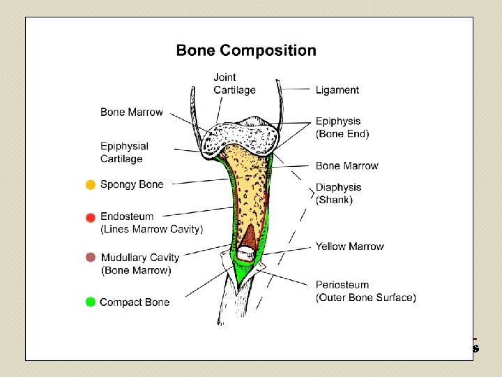
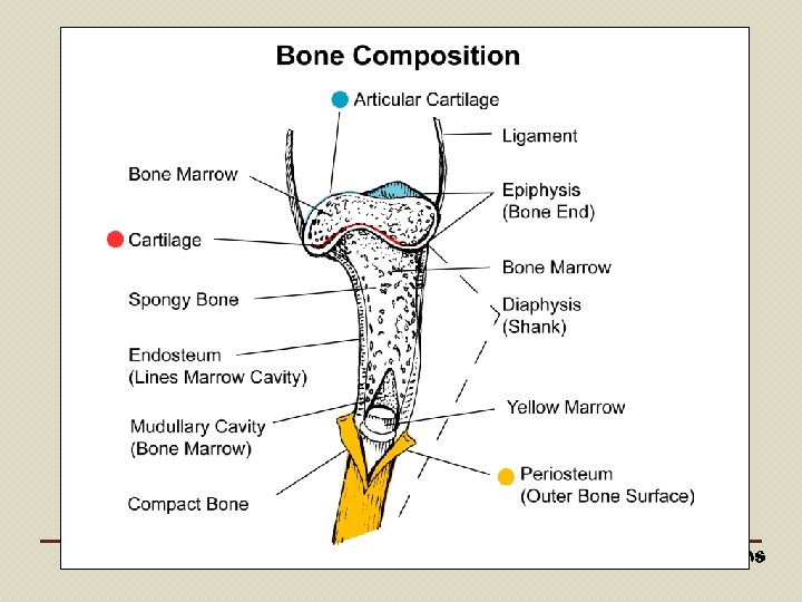
Anatomy of Bones and Bone Tissue The outer portion of the bone is hard, dense bone and forms the cortex. The inner portion of the bone is spongy, porous bone that forms a network called the medulla cavity. The medulla cavity has a membrane lining called the endosteum.
Bone marrow is a thick, red mass of cells inside the medulla cavity, which makes essential blood cells. Blood cells created in the bone marrow include the following: • Leukocytes – fight infection, • Erythrocytes – carry oxygen, and • Platelets – help the blood to clot.
As the bone ages, the red bone marrow gradually changes into yellow fatty marrow.
Parts of the Bone Epiphysis – refers to either end or extremity of a long bone. Proximal epiphysis – end closest to the main body of the animal. Distal epiphysis – end farthest from the main body of the animal. Diaphysis – the long bone shaft between the two joint ends.
Epiphysial cartilage – layer of cartilage between the joint ends and the shaft that allows the bone to increase in length. Periosteum – fibrous membrane that covers the exterior of the bone, excluding the joint ends. Articular cartilage – thin layer of cartilage that covers each joint end.
Total Bone Mass Of the total bone mass, 26% is mineral matter; the other chemical compositions are 20% protein, 4% fat, and 50% water.
The outer layer of a bone is composed of mineral deposits, which makes the bone hard and inflexible. Calcium phosphate makes up almost 85% of the mineral matter and the remaining 15% is calcium carbonate and magnesium phosphate.
A bone is the body’s primary mineral reservoir, which is constantly being depleted or replenished.
One-third of the bone’s total weight is comprised of living tissues that contain replicating cells, blood vessels, lymphatic vessels, and nerves.
Because a bone is made up of living organic matter, composed of fibrous tissue and cells, it is vulnerable to disease. A bone can repair itself if injured and reacts to changes caused by stress.
Classification of Bones are classified based on function and shape. Classifications include long bones, short bones, flat bones, sesamoid bones, pneumatic bones, and irregular bones.
Long bones – bones found in limbs that serve as supporting columns and levers for the skeleton, assisting in body support, locomotion, and eating.
A long bone is an elongated, round shaft with two ends. Examples of long bones include the femur and the humerus.
Short bones – short bones are cube-shaped bones that contain a spongy substance filled with marrow spaces surrounded by a thin layer of compact bone. Short bones function to reduce friction and change the direction of tendons in the joint of a limb.
Examples of short bones can be found in the knee and hock.
Flat bones – relatively thin, long, and wide bones that contain two plates of compact bone surrounded by spongy bone. Flat bones function to protect vital organs, such as the brain, heart, lungs, and pelvic viscera, and serve as areas of muscle attachment.
Examples of flat bones are the ribs, sternum, and scapula.
Sesamoid bones – flat and round bones that are located along the course of tendons. Sesamoid bones reduce friction and change the direction of tendons or the angle of muscle pull.
The kneecap, or patella, is an example of a sesamoid bone.
Pneumatic bones – bones that contain air spaces (sinuses) which are in contact with the atmosphere. Frontal and maxillary bones are examples of pneumatic bones that can be found in the face.
Irregular bones – bones that protect and support the central nervous system and are points some muscle attachments. of The bones in the vertebral column and some unpaired bones of the skull are examples of irregular bones.
The surfaces of bones have projections and depressions that differ in size and shape, depending on the function.
Articular projections and depressions, located in the joint, are covered with articular cartilage.
Non-articular projections and depressions, serve as points of attachment for tendons and ligaments.
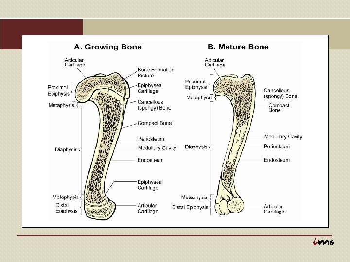
Physiology of Bones grow in the region of the Section of Bone (Red) epiphysial cartilage, from Growth Plate Area which is located between the epiphysis (end) and diaphysis (shaft). This bone growth is an increase in both diameter and length. Epiphysial Cartilage Photo by Rob Flynn courtesy of USDA Agricultural Research Service.
The periosteum produces new boney tissue that increases the diameter of the bone. The periosteum, which is the outside covering of the bone, is also involved with repairing bone fractures.
As the bone increases in diameter, the bone marrow cavity increases. This is accomplished by the removal or re-absorption of portions of the inner bone.
As an animal matures, bone growth stops. Ossification occurs; that is, the epiphysial cartilage becomes calcified, bony material. Although bone continues to be reabsorbed and replaced, there is no net bone growth.
Osteogenesis is the process of bone formation. Osteoblasts, which are the parent cells of connective tissue, accomplish this process by multiplying and secreting an enzyme called phosphatase.
Phosphatase causes some of the cells to mature and secrete calcium salts for ossification. Osteocytes (mature bone cells) are surrounded by calcified osteoid material.
This osteoid material appears as small cavities, which are connected by tiny canals that transmit tissue fluid, the life support for osteocytes. Some of the new cells stay in the periosteum, where they reproduce and are stored until needed.
Re-absorption of bone will occur due to the following reasons: • bone growth or repair of fractures, • aging, • hormonal imbalances, • inflamation, • pressure, and • certain bone diseases.
Osteoclasts, cells that secrete phosphatase which dissolves bone tissue, and increasing blood supply are responsible for bone re-absorption.
Bone growth is affected by hormones, vitamins, and other nutrients. Nutritional deficiencies can make bones fragile and distorted. Because of their rigidity, especially in older animals, bones can break easily. Bone tissue can also repair itself.
When a fracture occurs, it becomes filled with blood and connective tissue cells. The bone-forming cells, osteoblasts, replicate rapidly, forming a fibrin clot or callus, which becomes calcified into true bone tissue with a marrow cavity. Osteoclasts then reabsorb excess bone tissue from the callus.
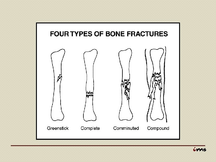
Common Types of Bone Fractures Simple Fracture – a broken bone that does not puncture the skin. Compound Fracture – a broken bone that results in the bone protruding through the skin, making infections possible.
Greenstick Fracture – one side of the bone is fractured and the other side is bent. Epiphysial Fracture – a break in the bone that occurs at the juncture of the epiphysis (end) and diaphysis (shaft). Greenstick and epiphysial fractures occur in young animals only.
Complete Fracture – the bone is broken completely across. Comminuted Fracture – the bone is broken into fragments due to crushing or splintering.
When a broken bone occurs, both ends of the fracture should be held together and immobilized by a splint or bone pin.
Anatomy of Bone Joints There are three main types of joints or articulations that join bones together: • Immovable joints, • Slightly movable joints, and • Freely movable joints.
Immoveable Joints – joints that are filled with fibrous tissue early in life and ossify as the animal matures, making them immobile. Example: skull
Slightly Movable Joints – joints that allow limited movement forward, backward, and sideways. These joints have flattened discs of cartilage and are sometimes called gliding joints. Example: joints of vertebral column (backbone) and those adjacent to the pelvic bone.
Freely Movable Joints – also called synovial joints, allow friction-free movement. These joints consist of the following • Articular surfaces – bone surfaces are shaped to operate smoothly with the bones to which they connect;
• Articular cartilage – cartilage that covers the articular surface and absorbs concussions; • Joint capsule – capsule that contains synovial fluid, which lubricates the joint and allows for the friction-free movement; and • Ligaments – connective tissue bands that connect bone to bone.
Types of freely movable joints: • Hinge joints – moves in two directions, flexion and extension. Example: knee joint. • Plane joints – move in slight gliding motions between flat surfaces. Example: carpals or small cubeshaped bones in knee joint.
• Pivot joint – allows rotary motion. Example: in the neck between the axis and atlas vertebrae; and • Ball-and-Socket Joint – allows movement in almost all directions and is characterized by a spherical head on one bone fitting into a cup-shaped socket of the other bone. Example: hip joint
Parts of the Skeleton There are three general areas of the vertebrate’s skeletal system: • Axial skeleton, • Pectoral limb, and • Pelvic limb.
Anatomy of the Axial Skeleton The axial skeleton consists of the bones that are either on the midline of the back or are attached to the bones of the midline, including the skull, vertebral column, ribs, and sternum.
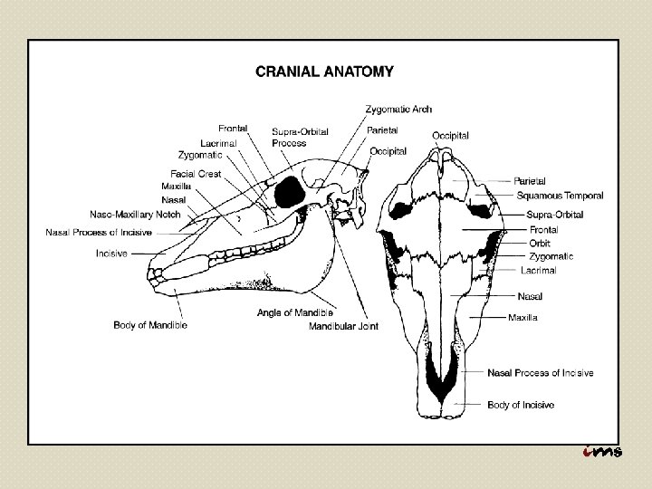
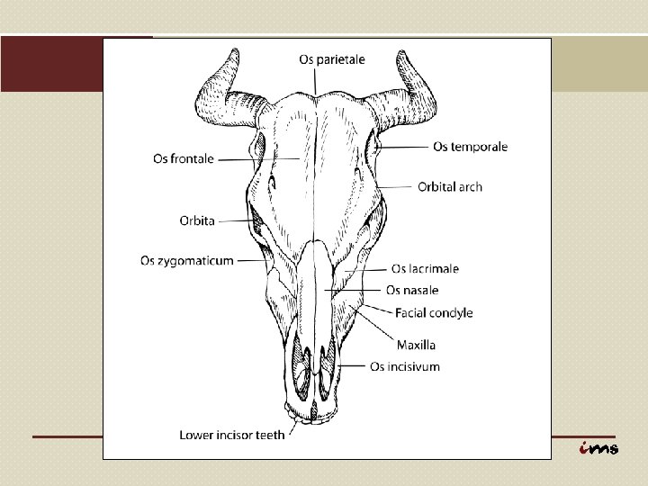
Skull – protects the brain, houses many of the sense organs, and contains the beginnings of the digestive and respiratory systems. It is composed of the cranial part and the facial part. Many cranial and facial bones occur in pairs, one on each side of the head and are connected at joints called sutures.
Major bones in the cranium: • Occipital bones – situated at the back and lower part of the cranium; • Parietal bones – form the sides and roof of the cranium; • Frontal bones – serve as the origin of horns in horned animals.
• Ethmoid bone – contains openings for olfactory nerves that are responsible for the sense of smell; and • Sphenoid bone – supports the brain and pituitary gland.
Facial parts of the skull: • Orbital section – contains eye sockets that house and protect the eyes; • Nasal section – two small, oblong bones that form the “bridge” of the nose. • Oral section – “mouth” bones that support the teeth and provide muscle attachment for chewing and swallowing.
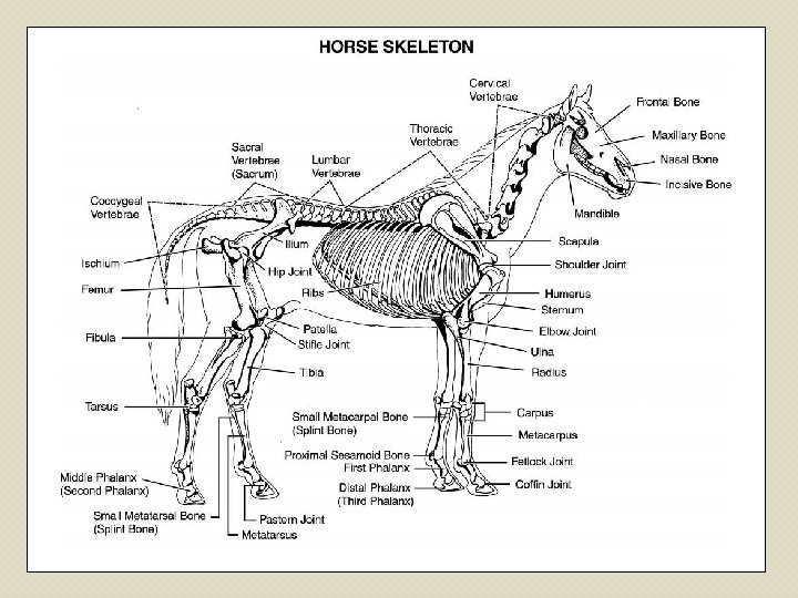
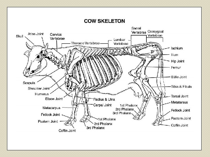
Vertebral Column – the vertebral column, or backbone, is divided into 5 anatomical regions including the cervical, thoracic, lumbar, sacral, and coccygeal vertebrae. The number of vertebrae in each region varies among the different species.
Cervical vertebrae – the cervical vertebrae are in the neck area and allow for movement of the head.
Atlas – first cervical vertebrae that forms a hinge joint with the occipital bone of the skull and allows the head to move up and down. Axis – second cervical vertebrae that forms a pivotal joint with the atlas and allows the head to turn from side to side.
Thoracic Vertebrae – the thoracic vertebrae are in the chest area and are the attachment sites of the ribs.
Lumbar vertebrae – the lumbar vertebrae are in the loin area and are slightly more mobile than the thoracic vertebrae. The large, flat projections of the lumbar vertebrae, that extend to either side of the midline, are the long bones seen in a T-bone cut.
Sacral vertebrae – the sacral vertebrae are in the pelvic region and are usually fused to form a single wedge-shaped bone, sacrum, to which the pelvic limb is attached.
Coccygeal Vertebrae – the coccygeal vertebrae are in the tail area and vary greatly in number depending on the species.
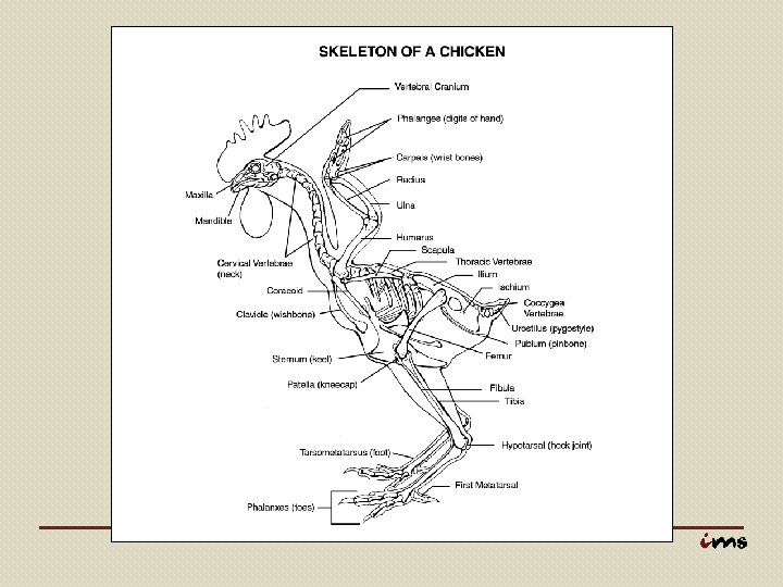
Number of Vertebrae in Selected Animal Species Cattle Swine Sheep Horse Humans Poultry Cervical 7 Thoracic 13 Lumbar 6 5 -7 Sacral 5 4 Coccygeal 18 -20 7 7 18 12 6 -7 6 5 4 5 5 (fused) 13 -17 13 -14 20 -23 16 -18 4 (fused) 14 6
Ribs – thin, flat, curved bones that protect the heart, lungs, stomach, spleen, and kidneys. Ribs also assist in respiration by lifting up and allowing the lungs to expand during inhalation and by moving down and squeezing air out during exhalation.
The number of rib pairs usually corresponds to the number of thoracic vertebrae, but sometimes extra ribs can occur in front of or behind the thoracic vertebrae.
True ribs (sternal ribs) – ribs that are attached to the sternum by cartilage. False ribs – ribs that do not connect directly to the sternum, but may connect to the last sternal rib by cartilage. Floating ribs – ribs that have no connection to other ribs in the sternum area.
Sternum – several small bones (sternebrae) that fuse together as animal ages to form the floor of the thoracic cavity. The sternum, as previously mentioned, is the site of attachment for the sternal (true) ribs. Number of Sternebrae Swine and sheep ……………. 6 Cattle ……………. 7 Horses and dogs ……………. 8
Anatomy of the Pectoral Limbs The front legs of four-legged animals (quadrupeds) are the pectoral limbs. The bones included in the pectoral limb are the scapula, humerus, radius and ulna, carpus, metacarpus, and phalanges.
The pectoral limbs are connected to the axial skeleton (body) by muscles and connective tissues. The joints formed by the scapula and humerus are ball-and-socket joints, but function as hinge joints. The remaining joints in the pectoral limb also function as hinge joints.
Scapula (shoulder blade) – a triangular-shaped flat bone that attaches to the humerus. Humerus (arm) – a long bone that extends toward the front of the animal, forming the point of the shoulder.
The articular angle, the angle formed by the scapula and humerus, is important to the soundness of the front legs. In poultry, the wishbone or coracoid, connects to the scapula.
Radius – the larger, well-developed bone in the forearm, located on the inside of the foreleg. The radius connects to the humerus forming the elbow joint. Ulna – the smaller of the two bones in the forearm.
Together, the radius and ulna make up the bones in the forearm. In horses, the radius and ulna are fused, allowing no movement between the bones. In cattle, sheep, and swine, the ulna is more developed and limited movement occurs between the radius and ulna.
Carpus (knee) – a complex region of small, cube-shaped bones (sliding joints) that function together as a hinge joint. Metacarpal (cannon) bones – bones that form the lower part of the foreleg.
The number of metacarpal bones vary with the species.
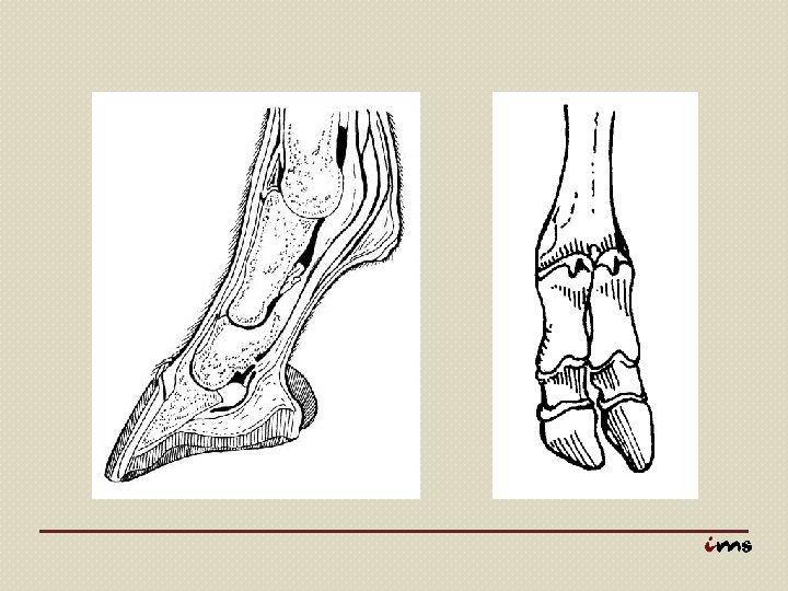
Phalanges (digits) – the bones that form the toes (fingers in humans) on the pectoral limb. The number of digits vary with the species: horses (1), cattle and sheep (2), swine (4), and dogs (5). Dewclaws – second and fifth digits in swine (more developed), cattle and sheep (non-functional).
Each digit (toe) is made up of three phalanges (small bones). The first phalanx is the long pastern bone, the second phalanx is the short pastern bone, and the third phalanx is the coffin bone.
Anatomy of the Pelvic Limbs The hind legs of quadrupeds (fourlegged animals) are the pelvic limbs. The pelvic limbs are connected to the axial skeleton by the pelvic girdle. The femur, tibia, fibula, tarsals, metatarsals, and phalanges are the bones that form the hind leg.
Pelvic girdle – three bones (ilium, ischium, and pubis) that are fused together to form an irregular bone called the os coxae or pelvis. The pelvis is connected to the sacrum at the sacroiliac joint.
Ilium bones – front, dorsal bones in the pelvis; in cattle, the front points of the ilium bones are called the hook bones. Ischium bones – rear, dorsal bones in the pelvis; in cattle, the rear points of the ischium bones are called the pin bones. Pubis – ventral bones in the pelvis that form the floor of the pelvic girdle.
Femur – a long bone that extends from the hip joint to the stifle joint and is the site of several hip and thigh muscle attachments. Patella (kneecap in humans) – the largest sesamoid bone in quadrupeds.
Tibia – the larger, thicker of the two long bones in the hind leg, the tibia is located on the inside of the hind leg and extends from the stifle joint to the hock joint. Fibula – the thinner bone in the hind leg, the fibula extends from the upper end of the tibia to lengths that vary depending on the species.
Tarsus (hock) – two rows of tarsal bones in the hind leg that correspond to the ankle in humans and are similar to the carpus in the front leg. Fibular tarsal – bone that forms the point of the hock and serves as a lever for muscles that extend the hock.
Metatarsus (rear cannon bone) – bones that are similar to the metacarpals of the foreleg, but are slightly longer. Phalanges – bones that make up the digits of the hind leg, which are similar to those in the front leg.